. 2025 Jul 17:16:1610422.
doi: 10.3389/fimmu.2025.1610422. eCollection 2025.

Adjuvant combination and antigen multimerization shape neutralizing antibody and T cell responses to a SARS-CoV-2 RBD subunit vaccine

Affiliations

Adjuvant combination and antigen multimerization shape neutralizing antibody and T cell responses to a SARS-CoV-2 RBD subunit vaccine

João Pedro da Silva Nunes et al. Front Immunol. .

Abstract

Introduction: The rapid development and deployment of multiple safe and effective COVID-19 vaccines were critical cornerstones of pandemic control. However, vaccine inequity and the emergence of new variants of concern (VOCs) highlighted major gaps in the global strategy to control SARS-CoV-2 infection. Despite the use of distinct platforms, most approved vaccines utilize the Spike protein as the main antigen due to its pivotal role in virus entry, mediated by the receptor binding domain (RBD). In this context, RBD stands out as a promising antigen for a subunit vaccine candidate, as it is the main target of neutralizing antibodies, has a well-established scalable production pipeline, and has proven safety. Approaches to enhance RBD immunogenicity encompass the addition of adjuvants and antigen multimerization.

Methods: In this study, we compared the immunogenic properties of the Wuhan RBD monomer and homodimer with an RBD heterotrimer formulation composed of the Delta, Beta and Gamma variants. We also screened different adjuvants to optimize both humoral and cellular immunity.

Results: Our results showed that immunization with the RBD dimer and trimer, in the presence of the adjuvant AddaS03, elicited a higher humoral response and a broader neutralization profile. Additionally, RBD-trimer immunization more efficiently inhibited viral replication in the lungs of mice challenged with the ancestral Wuhan strain compared to the monomer. We further optimized our vaccine formulation by combining the adjuvants AddaS03 and Poly I:C, which demonstrated a synergistic effect, integrating the potent humoral response induced by AddaS03 with the cellular Th1 skewing capacity of Poly I:C. The AddaS03+ Poly I:C mixture induced antibodies with higher affinity and an increased frequency of RBD-specific IgG2c-producing bone marrow plasma cells, highlighting the potential of this adjuvant combination to generate long-lived memory plasma cells. Additionally, we identified sequences within the RBD that induced specific IFNγ T cell responses. Peptide 12 (393-TNVYADSFVIRGDEVRQ-409) emerged as the immunodominant CD4 T cell epitope, whereas peptides 28 (505-YQPYRVVVLSFELLHAP-521) and 29 (512-VLSFELLHAPATVCGPK-528) successfully activated CD8 T cells.

Conclusions: These findings underscore that antigen multimerization and the strategic combination of adjuvants can significantly improve vaccine immunogenicity.

Keywords: RBD; SARS-CoV-2; adjuvant; recombinant protein; vaccine.


Conflict of interest statement

The authors declare that the research was conducted in the absence of any commercial or financial relationships that could be construed as a potential conflict of interest.

Figures

Figure 1
Figure 1
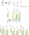
AddaS03 is the most potent adjuvant to induce humoral response. (A) Study design. C57BL/6 mice (n=4) were immunized subcutaneously with two doses of the RBD monomer together with AddaS03, AddaVax, Poly I:C or Alum + CpG 15 days apart. The control groups received only adjuvants (Alum+ CpG, AddaS03, Poly I:C). (B) Anti-RBD total IgG titers 15 days after two doses. Data represent the mean ± SD of antibody titers in log10 scale. (C) NT50 pseudovirus neutralization assay (PNA) against Wuhan pseudovirus after two doses. Serum neutralizing antibody responses after two doses as measured by live virus neutralization assay (VNT100) against (D) Delta, (E) Gamma and (F) Omicron BA.1 variants of SARS-CoV-2. For ELISA and PNA, serum of each animal was assayed individually. For the VNT100, serum of each group was pooled and tested in triplicate. Data represent the mean ± SD. b, c: Statistical analysis was determined by one-way ANOVA followed by Tukey post-hoc test.; d, e, f: Statistical analysis was determined by Kruskal-Wallis followed by Dunn’s post hoc test. *p < 0.05, **p < 0.01, ***p < 0.001, and ****p < 0.0001. Only statistically significant comparisons are depicted.
Figure 2
Figure 2
The RBD dimer and trimer are superior immunogens to induce humoral responses. (A) Study design. C57BL/6 mice were immunized subcutaneously with two doses of RBD monomer (n=18), dimer (n=34) or trimer (n=21) combined with AddaS03. (B) Anti-RBD total IgG titers 15 days after one and two doses. Data represent the mean ± SEM. (C) NT50 PNA against Wuhan and Omicron BA.2–15 days after two doses. Data represent the median ± interquartile range. (D) VNT100 against Wuhan, (E) Gamma, (F) Delta and (G) Omicron BA.1–15 days after two doses. For ELISA and PNA, serum of each animal was assayed individually. Data are from 7 independent experiments. For VNT, the samples from monomer RBD were derived from two independent experiments and assayed in triplicate (n=6). For dimer and trimer RBDs, samples came from three independent experiments—two performed in triplicate and one in duplicate (n=8 each). Data represent the mean ± SEM. b, c: Statistical analysis was determined by two-way ANOVA followed by Tukey post-hoc test.; d, e, f, g: Statistical analysis was determined by Kruskal-Wallis followed by Dunn’s post hoc test. *p < 0.05, **p < 0.01, ***p < 0.001, and ****p < 0.0001. Wu/Om = Wuhan/Omicron fold of decay. Only statistically significant comparisons are depicted.
Figure 3
Figure 3
Lethal SARS-CoV-2 challenge in K18-hACE2 mice following immunization with RBD monomer, dimer and trimer in the presence of AddaS03. (A) Study design. K18-hACE2 mice were immunized subcutaneously with two doses of RBD monomer (n=8), dimer (n=8) or trimer (n=8) together with AddaS03. Control groups received AddaS03 only (n=11) or saline (n=6). Fifteen days after the second dose, mice were intranasally challenged with SARS-CoV-2 (2.8x10⁵ TCID50/mL) and accompanied for 6 days followed by euthanasia. (B) Total anti-RBD IgG titers 15 days after two doses. Full circles represent pre-challenge titers while empty circles describe post-challenge titers. Data represent the mean ± SD. (C) NT50 PNA against Wuhan 15 days after two doses. Data represent the median ± interquartile range. (D) VNT100–15 days after two doses against Wuhan. For ELISA, PNA and qPCR, samples from each animal were assayed individually. For VNT, serum of each group was pooled and used in triplicate. Data represent the mean ± SD. (E) Weight loss. (F) Survival rate. (G) Lung and (H) Brain viral load after challenge. Dashed line represents the threshold of detection. Data represent the mean ± SD. (B) Statistical analysis was determined by two-way and (C, G, H) by one-way ANOVA followed by Tukey post-hoc test; (D) Statistical analysis was determined by Kruskal-Wallis followed by Dunn’s post hoc test; (F) Statistical analysis was determined by Log-Rank test. *p < 0.05, **p < 0.01, ***p < 0.001, and ****p < 0.0001. Only statistically significant comparisons are depicted.
Figure 4
Figure 4
Humoral analysis after two or three doses of RBD dimer with AddaS03, Poly I:C or AddaS03 + Poly I:C mixture. (A) Study design. C57BL/6 mice (n=3) were immunized subcutaneously with two or three doses of RBD dimer together with AddaS03, Poly I:C or AddaS03 + Poly I:C mixture. Control group received AddaS03 + Poly I:C only. (B) Total anti-RBD IgG titers 15 days after each dose. (C) Anti-RBD IgG1 (D) IgG2c and (E) IgG2b subtypes after two or three doses. For ELISA and PNA, the serum of each animal was assayed individually. Data represent the mean ± SD. Statistical analysis was determined by two-way ANOVA followed by Tukey post-hoc test. *p < 0.05, **p < 0.01, ***p < 0.001, and ****p <0.0001. Only statistically significant comparisons are depicted.
Figure 5
Figure 5
Affinity and neutralization after two or three doses of RBD dimer together with AddaS03, Poly I:C or AddaS03 + Poly I:C mixture. C57BL/6 mice were immunized subcutaneously with two or three doses of RBD dimer together with AddaS03, Poly I:C or AddaS03 + Poly I:C mixture. (A) Antibody affinity of pooled mouse sera after incubation with increasing concentrations of ammonium thiocyanate. (B) Area under the curve of the affinity assay. (C) NT50 PNA against Wuhan and (D) Omicron BA.2–15 days after two or three doses. For PNA, serum of each animal was assayed individually. For affinity ELISA, serum of each group was pooled and used in triplicate. Data represent the mean ± SD. Statistical analysis was determined by two-way ANOVA followed by Tukey post-hoc test. *p < 0.05, ***p < 0.001, and ****p < 0.0001. Only statistically significant comparisons are depicted.
Figure 6
Figure 6
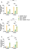
Bone marrow plasma cell frequency after two or three doses of dimer together with AddaS03, Poly I:C or AddaS03 + Poly I:C mixture. C57BL/6 mice were immunized subcutaneously with two or three doses of RBD dimer together with AddaS03, Poly I:C or AddaS03 + Poly I:C mixture. After the last dose, mice were euthanized, and bone marrow cells were cultured for 12 hours in dimer pre-coated plates. (A) Total IgG, (B) IgG1, and (C) IgG2c-producing bone marrow plasma cells were quantified by dimer-specific B cell ELISpot. Bone marrow cells were pooled and tested in quadruplicate. Data represent the mean ± SD. Statistical analysis was determined by two-way ANOVA followed by the Tukey post-hoc test. *p < 0.05, **p < 0.01, and ****p < 0.0001. ASC: antibody-secreting cells. Only statistically significant comparisons are depicted.
Figure 7
Figure 7
T cell response after two or three doses of RBD dimer together with AddaS03, Poly I:C or AddaS03 + Poly I:C mixture. C57BL/6 mice were immunized subcutaneously with two or three doses of RBD dimer, together with AddaS03, Poly I:C or AddaS03 + Poly I:C. After the last dose, splenocytes were harvested and cultured with dimer and peptides for 18 hours. Comparison of T cell responses after two or three-dose regimens against (A) pool 3, (B) pool 8, and (C) peptide 12. Cut-off=mean of control group + 3 SD. Splenocytes from each group were pooled and tested in triplicate. Data represent the mean ± SD. Statistical analysis was determined two-way ANOVA followed by the Tukey post-hoc test. *p < 0.05 and ***p < 0.001. Only statistically significant comparisons are depicted.
Figure 8
Figure 8
CD4+ and CD8+ T cell epitope mapping. Pooled spleens from groups that received three doses of dimer in the presence of AddaS03 + Poly I:C and control group (AddaS03 + Poly I:C) were sorted and utilized for (A) IFNγ ELISpot of CD4 and (B) CD8 T cells stimulated with peptide pools and RBD dimer protein. (C) IFNγ ELISpot of CD4+ and (D) CD8+ T cells stimulated with individual peptides (12, 28 and 29). Splenocytes from each group were pooled and tested in triplicate. Cut-off= mean of control group + 3 SD. Data represent the mean ± SD. (E) RBD amino acids sequence from Wuhan strain aligned with Delta, Beta, Gamma, Omicron BA.1, Omicron BA.2, Omicron BA.5, XBB 1.5 and JN.1. Amino acids highlighted in gray represent point mutations in variants compared to the Wuhan strain. In silico predictions using the IEDB MHC binding prediction tool. The top responders’ core peptides (highlighted in white) “in silico – Class II” (score = 0.5702) and “in silico – Class I” (score = 0.8929) aligned with the single peptides 12 (highlighted in blue), 28 (highlighted in green), and 29 (highlighted in green), respectively.

Similar articles

References

    1. Harvey WT, Carabelli AM, Jackson B, Gupta RK, Thomson EC, Harrison EM, et al. SARS-CoV-2 variants, spike mutations and immune escape. Nat Rev Microbiol. (2021) 19:409–24. doi: 10.1038/s41579-021-00573-0, PMID: - DOI - PMC - PubMed
    1. Van Kerkhove MD. COVID-19 in 2022: controlling the pandemic is within our grasp. Nat Med. (2021) 27:2070. doi: 10.1038/s41591-021-01616-y, PMID: - DOI - PubMed
    1. Hotez PJ, Bottazzi ME. Developing a low-cost and accessible covid-19 vaccine for global health. PloS Negl Trop Dis. (2020) 14:1–6. doi: 10.1371/journal.pntd.0008548, PMID: - DOI - PMC - PubMed
    1. Hare J, Hesselink R, Bongers A, Blakeley P, Riggall G. Improving vaccine equity by increasing vaccine thermostability. Sci Transl Med. (2024) 16(757):eadm7471. doi: 10.1126/scitranslmed.adm7471, PMID: - DOI - PubMed
    1. Li M, Wang H, Tian L, Pang Z, Yang Q, Huang T, et al. COVID-19 vaccine development: milestones, lessons and prospects. Signal Transduction Targeted Ther. (2022) 7. doi: 10.1038/s41392-022-00996-y, PMID: - DOI - PMC - PubMed