. 2025 Jun:54:197-223.
doi: 10.3114/persoonia.2025.54.06. Epub 2025 May 14.

Taxonomic revision of Bisifusarium (Nectriaceae)

Affiliations

Taxonomic revision of Bisifusarium (Nectriaceae)

K Zhang et al. Persoonia. 2025 Jun.

Abstract

Species of Bisifusarium (previously the Fusarium dimerum species complex) have been associated with cheese fermentation and a wide range of opportunistic human infections, but they are generally regarded as saprotrophs. Bisifusarium spp. are also commonly isolated from soils and tissues of plants growing in arid climates. The genus is typically characterized by its distinct pionnotal growth in culture, and typically very short, 0-2(-3)-septate macroconidia, produced in sporodochia or on lateral phialidic hyphal pegs. Only 16 species of Bisifusarium have been described to date, and this study sought to re-evaluate these taxa by examining 116 Bisifusarium isolates from the culture collection of the Westerdijk Fungal Biodiversity Institute in Utrecht, The Netherlands. A multi-gene phylogenetic analysis using partial nucleotide sequences of the translation elongation factor 1-alpha (tef1), partial RNA polymerase II second largest subunit (rpb2), the 5.8S nrDNA with its flanking intergenic spacer regions (ITS), and partial β-tubulin (tub2) genes resolved 25 phylogenetic lineages. Further evaluation of culture and morphological characters, and host-substrates, confirmed eight of these clades as novel taxa that are formally described here. In addition, two putative novel species were identified but not described due to limited available data. We provide the morphological descriptions and photographic illustrations for B. hedylamarriae and B. lovelliae, which were formerly known only from their DNA data. This study significantly increases the number of species in Bisifusarium and provides a crucial foundation for future studies to elucidate the ecology and evolutionary relationships within this expanding genus. Citation: Zhang K, Sandoval-Denis M, Kandemir H, Yilmaz N, Groenewald JZ, Roets F, Yáñez-Morales M de J, Wingfield MJ, Crous PW (2025). Taxonomic revision of Bifusarium (Nectriaceae). Persoonia 54: 197-223. doi: 10.3114/persoonia.2025.54.06.

Keywords: cheese fermentation; multi-locus; new taxa; opportunistic human infections; systematics.


Conflict of interest statement

The authors declare that there is no conflict of interest.

Figures
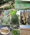
Fig. 1
Fig. 1
Substrates associated with Bisifusarium spp. A. B. africanum from Encephalartos leaf. B. B. hedylamarriae on leaves of Aloe sp. C. B. cacticola on Opuntia ficus-indica. D. B. sechiicola on Sechium edule. E. B. colchici on Trachyandra sp. F. B. allantoides on Saint Nectaire cheese (photo credit: Monica Coton). G. B. solicola from soil adjacent to Agapanthus praecox. H. B. namibense from desert soil in Namibia.
Fig. 2
Fig. 2
Phylogenetic tree inferred from a Maximum Likelihood (RAxML) analysis based on aligned, concatenated ITS, rpb2, tef1 and tub2 sequences of 116 strains representing Bisifusarium and outgroups. Numbers at branches indicate support values (RAxML-BS / IQ-TREE-BS / BI-PP) above 50 % / 90 % / 0.9. New species are printed in bold font. “T” indicates ex-type, “NT” indicates neotype, “ET” indicates epitype and “LT” indicates lectotype strains, all of which are labelled in bold. The tree is rooted with two species of Rectifusarium. Scale bar represents expected number of changes per site.
Fig. 3
Fig. 3
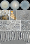
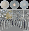
Bisifusarium africanum (ex-type CBS 153387). A. Colony surface on OA after 7 d at 25 °C. B. Colony on PDA. C. Colony on SNA. D, E. Sporodochia on carnation leaf (D) and adjacent agar (E).. F–H. Sporodochial conidiophores and conidiogenous cells. I. Chlamydospores. J. Sporodochial macroconidia. Scale bars: F, G = 20 µm; all others = 10 µm.
Fig. 4
Fig. 4
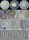
Bisifusarium australianum (ex-type CBS 366.73). A. Colony surface on OA after 7 d at 25 °C. B. Colony on PDA. C. Colony on SNA. D–F. Sporodochia on carnation leaf. G–I. Sporodochial conidiophores and conidiogenous cells. J. Sporodochial conidia. Scale bars = 10 µm.
Fig. 5
Fig. 5
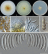
Bisifusarium cacticola (ex-type CBS 101615). A. Colony surface on OA after 7 d at 25 °C. B. Colony on PDA. C. Colony on SNA. D. Sporodochia on carnation leaf. E, F. Sporodochial conidiophores and conidiogenous cells. G. Chlamydospores. H. Sporodochial macroconidia. Scale bars = 10 µm.
Fig. 6
Fig. 6
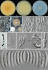
Bisifusarium colchici (ex-type CBS 153389). A. Colony surface on OA after 7 d at 25 °C. B. Colony on PDA. C. Colony on SNA. D, E. Sporodochia on carnation leaf. F–H. Sporodochial conidiophores and conidiogenous cells. I. Microconidia. J, K. Chlamydospores. L. Sporodochial macroconidia. Scale bars = 10 µm.
Fig. 7
Fig. 7
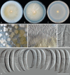
Bisifusarium hedylamarriae (ex-type CBS 150895). A. Colony surface on OA after 14 d at 25 °C. B. Colony on PDA. C. Colony on SNA. D, E. Sporodochia on carnation leaf. F, G. Sporodochial conidiophores and conidiogenous cells. H–J. Aerial conidiophores and conidiogenous cells. K. Microconidia. L. Sporodochial macroconidia. Scale bars = 10 µm.
Fig. 8
Fig. 8
Bisifusarium lovelliae (ex-type CBS 150777). A. Colony surface on OA after 14 d at 25 °C. B. Colony on PDA. C. Colony on SNA. D, E. Sporodochia on carnation leaf. F, G. Sporodochial conidiophores and conidiogenous cells. H, I. Aerial conidiophores and conidiogenous cells. J. Microconidia. K, L. Chlamydospores. M. Sporodochial macroconidia. Scale bars: G, L = 5 µm; H = 20 µm; all others = 10 µm.
Fig. 9
Fig. 9
Bisifusarium namibense (ex-type CBS 153393). A. Surface of colony on OA after 7 d at 25 °C. B. Colony on PDA. C. Colony on SNA. D. Sporodochia on carnation leaf. E, F. Sporodochial conidiophores and conidiogenous cells. G, H. Aerial conidiophores and conidiogenous cells. I. Microconidia. J. Chlamydospores. K. Sporodochial macroconidia. Scale bars: J = 5 µm; all others = 10 µm.
Fig. 10
Fig. 10
Bisifusarium phloginum (ex-type CBS 110137). A. Surface of colony on OA after 7 d at 25 °C. B. Colony on PDA. C. Colony on SNA. D, E. Sporodochia on carnation leaf. F–H. Sporodochial conidiophores and conidiogenous cells. I, J. Chlamydospores. K. Sporodochial macroconidia. Scale bars = 10 µm.
Fig. 11
Fig. 11
Bisifusarium salinasense (ex-type CBS 110139). A. Surface of colony on OA after 7 d at 25 °C. B. Colony on PDA. C. Colony on SNA. D, E. Sporodochia on carnation leaf. F, G. Sporodochial conidiophores and conidiogenous cells. H. Sporodochial macroconidia. Scale bars = 10 µm.
Fig. 12
Fig. 12
Bisifusarium sechiicola (ex-type CBS 153536). A. Surface of colony on OA after 7 d at 25 °C. B. Colony on PDA. C. Colony on SNA. D, E. Sporodochia on carnation leaf. F. Sporodochial conidiophores and conidiogenous cells. G. Sporodochial macroconidia. Scale bars: F = 10 µm; G = 5 µm.
Fig. 13
Fig. 13
Bisifusarium sp. 1 (CBS 110318). A. Surface of colony on OA after 7 d at 25 °C. B. Colony on PDA. C. Colony on SNA. D. Sporodochia on carnation leaf. E. Sporodochial conidiophores and conidiogenous cells. F. Chlamydospores. G. Sporodochial macroconidia. Scale bars: E = 20 µm; all others = 10 µm.
Fig. 14
Fig. 14
Bisifusarium sp. 2 (CBS 135686). A. Surface of colony on OA after 7 d at 25 °C. B. Colony on PDA. C. Colony on SNA. D. Sporodochia on carnation leaf. E, F. Sporodochial conidiophores and conidiogenous cells. G. Sporodochial macroconidia. Scale bars = 10 µm.

References

    1. Bachmann H-P, Bobst C, Bütikofer U, et al. (2003). Sticky cheese smear and natural white mould. Milchwissenschaft 58: 117–232.
    1. Bachmann HP, Bobst C, Bütikofer U, et al. (2005). Occurrence and significance of Fusarium domesticum alias anticollanti on smear-ripened cheeses. LWT - Food Science and Technology 38: 399–407. 10.1016/j.lwt.2004.05.018 - DOI
    1. Bhunjun CS, Chen YJ, Phukhamsakda C, et al. (2024). What are the 100 most cited fungal genera? Studies in Mycology, 108: 1–412. 10.3114/sim.2024.108.01 - DOI - PMC - PubMed
    1. Brent G, Abdul-Wahab A, Borman AM, et al. (2023). Disseminated Bisifusarium infection following toxic epidermal necrolysis in a child with B-cell acute lymphoblastic leukemia. Pediatric Dermatology 40: 503–506. 10.1111/pde.15179 - DOI - PubMed
    1. Collado C, Medina L, Zorraquino A, et al. (2013). Cutaneous fusariosis by a species of the Fusarium dimerum species complex in a patient with acute myeloblastic leukemia. Revista Iberoamericana de Micologia 30: 119–121. 10.1016/j.riam.2012.11.001 - DOI - PubMed

LinkOut - more resources