. 2025 Jun 24;5(7):3311-3327.
doi: 10.1021/jacsau.5c00441. eCollection 2025 Jul 28.

Structure of the Lipopolysaccharide from Paenalcaligenes hominis: A Chemical Perspective on Immune Recognition

Affiliations

Structure of the Lipopolysaccharide from Paenalcaligenes hominis: A Chemical Perspective on Immune Recognition

Ferran Nieto-Fabregat et al. JACS Au. .

Abstract

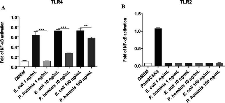
Gram-negative bacterium Paenalcaligenes hominis, which is increasingly prevalent in elderly individuals, is associated with cognitive decline and gut-brain axis dysfunction. Here, we present a comprehensive structural characterization of P. hominis lipopolysaccharide (LPS), a key modulator of immune recognition and the main component of its outer membrane. Using a multidisciplinary approach combining chemical, spectroscopic, spectrometric, biophysical and computational methods, we unveil a unique O-antigen characterized by a trisaccharide repeating unit containing rhamnose and glucosamine, displaying nonstoichiometric O-acetylation and a terminal methylated rhamnose capping the saccharide chain. Furthermore, we disclose a short core oligosaccharide and a Lipid A composed of penta- to tetra-acylated species. Notably, this LPS exhibits reduced activation of Toll-Like Receptor-dependent signaling compared to the highly immunostimulatory Escherichia coli LPS and elicits a poor pro-inflammatory cytokine response. Moreover, P. hominis LPS exhibits selective binding to immune lectins such as Ficolin-3 and Galectin-4, as shown by the microarray assays. This raises the possibility that lectin-mediated recognition may represent an alternative route of immune engagement, which could help explain altered immune responses observed in elderly individuals. These findings provide a molecular basis for further exploring the role of P. hominis LPS in microbiota-induced immune modulation and its possible impact on age-related inflammatory and neurodegenerative conditions.

Keywords: NMR spectroscopy; Paenalcaligenes hominis; innate immunity; lipopolysaccharide; mass spectrometry.


Figures

1
1
(A) 1H NMR spectra of O-deacylated LPS (LPS OdeAc , top) and of O-antigen (OS, bottom) form P. hominis LPS (schematic representation with SNFG). Anomeric signals are expanded in the inset and attributed in Table S1 (B) Zoom-in of the multiplicity edited heteronuclear single-quantum coherence (HSQC) spectrum (blue and red), highlighting the assignment of proton–carbon correlations for individual sugar residues. (C) Overlay of NOESY (red) and TOCSY (blue) P. hominis LPS OdeAc spectra, used to identify both intraresidue proton connectivities and inter-residue NOEs crucial for establishing the monosaccharide sequence and linkage positions. Key intra- and inter-residue cross-peaks are labeled. (D) Superimposition of HSQC (blue/red) and heteronuclear multiple–bond correlation (HMBC) (black) from P. hominis LPS OdeAc spectra, providing short- and long-range heteronuclear correlations, respectively, essential for confirming anomeric configurations and interglycosidic linkages. Sugar residue assignments correspond to letters defined in Table S1.
2
2
Structure of P. hominis LPS O-antigen.
3
3
(A) Negative-ion MALDI-TOF MS spectrum, recorded in reflectron mode, of lipid A fraction from P. hominis LPS. (B) Proposed structures of main mono- and bis-phosphorylated lipid A species.
4
4
(A) Negative-ion ESI mass spectrum of the P. hominis O-deacylated LPS (LPS deOAc ). LPS preparation yielded quadruply and quintuple charged deprotonated molecular ions for the major LPS, while mainly singly and triply charged species were detected for lipid A and core OS species. Key ions and their proposed structures are listed in Table S2. OAgAc and OAg stand for a repeating unit of the O-Antigen bearing or not an additional acetyl group, respectively. LipA stands for lipid A comprising the bis-phosphorylated glucosamine disaccharide backbone carrying two 14:0 (3-OH), whereas LipA2 and LipA3 bear an additional 12:0 and 12:0 and 14:0 (3-OH), respectively. A cartoon describing the O-deacylated positions of lipid A resulting in LipA1–3 is reported in the inset. (B,C) Enlargements of the negative-ion ESI-MS/MS spectrum of the core OS species detected at m/z 1230.42. Main singly charged ions involving cleavage of the glycoside linkage (i.e., Y, B, Z, and C type ions) were indicated in the spectrum. Unassigned peaks are related to cross-ring fragmentations in combination or not with linkage cleavage. Schematic structures of the core OS identified using MS-based characterization strategy are reported in the insets. Symbols legend: green triangle: rhamnose; green circle: mannose; yellow circle: galactose; blue square: N-acetyl-glucosamine; green hexagon: heptose; yellow hexagon: 3-deoxy-d-manno-octulosonic acid, Kdo. The structure is tentatively given according to compositional analyses.
5
5
Torsion angle distributions around the glycosidic linkages throughout the MD simulation of P. hominis LPS OdeAc octasaccharide. The torsion angles (Φ and Ψ) were monitored for each glycosidic bond along the entire MD simulation. The two-dimensional plots display the distribution of torsion angles for each linkage, with the color gradient representing the frequency of each angle occurrence. The LPS OdeAc octasaccharide is shown in its most representative conformation, with each sugar residue labeled according to Table S1.
6
6
Computational study of the MD of P. hominis O-antigen and LPS OdeAc tetradecasaccharides. (A) Superposition of all poses obtained during the MD simulation of the O-antigen tetradecasaccharide. (B) Most representative pose of the O-antigen tetradecasaccharide (white), highlighting acetyl groups in teal. The terminal sugar (B residue) is highlighted in pink, with its terminal OMe group shown in cyan, displayed both with and without the molecular surface representation. (C) Most representative pose of LPS OdeAc tetradecasaccharide (green), also highlighting the terminal sugar (pink) and its OMe group (cyan), shown with and without surface. (D) Front view of O-antigen (white) and LPS OdeAc (green) tetradecasaccharides, emphasizing their similar helical conformations.
7
7
TLR activation by P. hominis LPS. (A) Stimulation of HEK Blue hTLR4 and (B) HEK Blue hTLR2 cells. Secreted embryonic alkaline phosphatase (SEAP) levels (OD) upon stimulation with LPS from E. coli and P. hominis at the indicated concentrations (1, 10, 100 ng/mL). Ultrapure LPS from E. coli 0111:B4, a potent TLR4 agonist, was used as a positive control in (A), whereas in (B), it was used as a negative control. The positive control in (B) was Pam3CSK4 (500 ng/mL), a synthetic triacylated lipopeptide agonist for TLR2. Significant difference between P. hominis LPS values and the corresponding E. coli LPS (P. hominis vs E. coli, *** p < 0.001, ** p < 0.01) by the Student t-test are indicated. Data are expressed as mean ± SD of three independent experiments.
8
8
Microarray analysis of P. hominis LPS recognition by various lectins and immune-related proteins. Two different LPS preparations, Mix1 (containing both long and short O-antigen forms) and Mix2 (enriched in the shorter O-antigen form), were printed as duplicates at different concentrations and the binding of the different proteins was assayed using AF647-streptavidin for final detection. Data shown correspond to the mean of the fluorescence signals obtained for samples printed at 1, 0.3, 0.1, and 0.03 mg/mL (depicted in dark to light blue color scale), and error bars indicate the standard deviation of the mean. HG, human galectin; FIC, ficolin; COL, collectin; INTL, intelectin; HS, human Siglec.
9
9
(A) Hydrodynamic radius distribution of P. hominis LPS, (B) Zimm plot of P. hominis LPS, (C) NR data with the corresponding fits, (D) scattering length density (SLD) profiles, and (E) volume fraction occupancy of P. hominis LPS moieties: silicon (black), silicon dioxide (gray), inner head groups (light green), PC tails (orange), LPS tails (purple), core region (midgreen), O-Antigen region (dark green).

References

    1. de Vos W. M., Tilg H., Van Hul M., Cani P. D.. Gut microbiome and health: Mechanistic insights. Gut. 2022;71(5):1020–1032. doi: 10.1136/gutjnl-2021-326789. - DOI - PMC - PubMed
    1. Thursby E., Juge N.. Introduction to the human gut microbiota. Biochem. J. 2017;474(11):1823–1836. doi: 10.1042/BCJ20160510. - DOI - PMC - PubMed
    1. Miyauchi E., Shimokawa C., Steimle A., Desai M. S., Ohno H.. The impact of the gut microbiome on extra-intestinal autoimmune diseases. Nat. Rev. Immunol. 2022;23:9–23. doi: 10.1038/s41577-022-00727-y. - DOI - PubMed
    1. Cho I., Blaser M. J.. The human microbiome: at the interface of health and disease. Nature reviews. Genetics. 2012;13(4):260–270. doi: 10.1038/nrg3182. - DOI - PMC - PubMed
    1. Di Lorenzo F., De Castro C., Silipo A., Molinaro A.. Lipopolysaccharide structures of Gram-negative populations in the gut microbiota and effects on host interactions. FEMS Microbiol Rev. 2019;43(3):257–272. doi: 10.1093/femsre/fuz002. - DOI - PubMed

LinkOut - more resources