. 2025 Jul;15(7):101246.
doi: 10.1016/j.jpha.2025.101246. Epub 2025 Feb 27.

E3 ubiquitin ligase FBXW11-mediated downregulation of S100A11 promotes sensitivity to PARP inhibitor in ovarian cancer

Affiliations

E3 ubiquitin ligase FBXW11-mediated downregulation of S100A11 promotes sensitivity to PARP inhibitor in ovarian cancer

Ligang Chen et al. J Pharm Anal. 2025 Jul.

Abstract

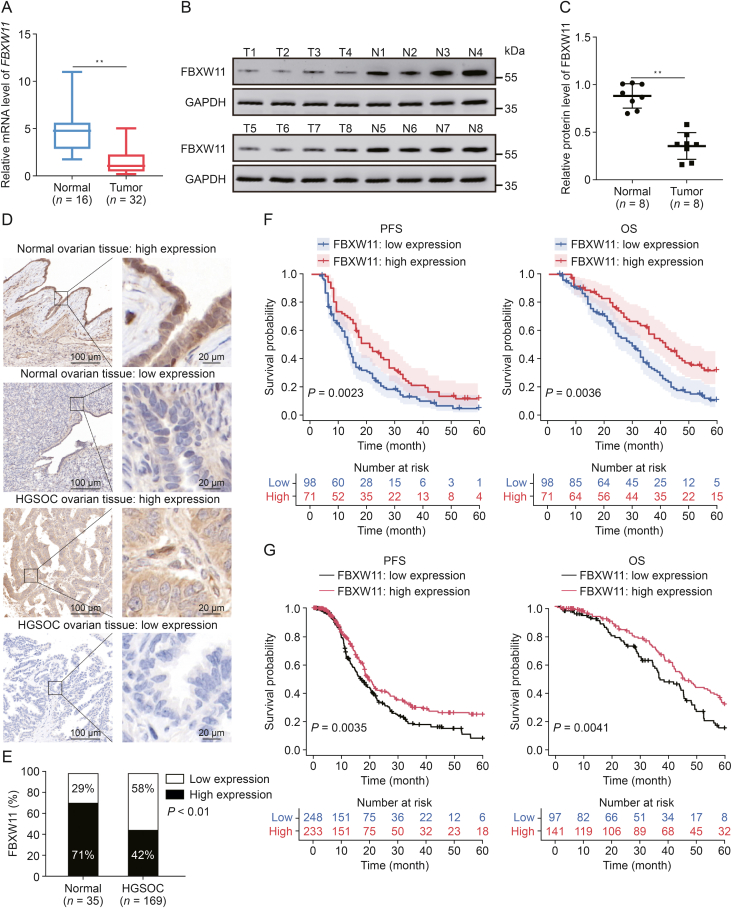
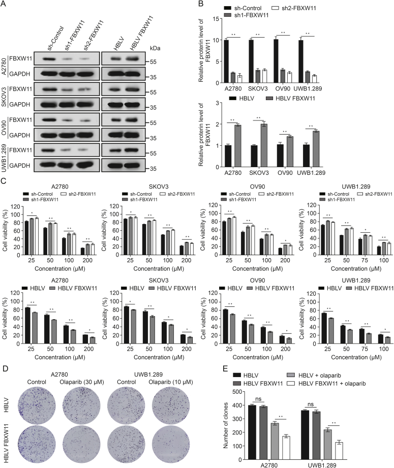
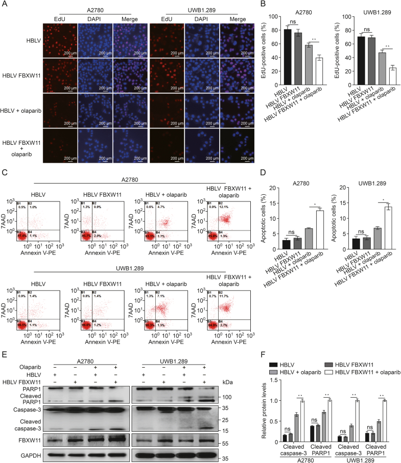
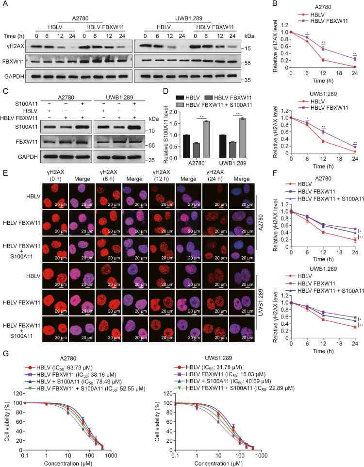
Resistance to poly adenosine diphosphate (ADP)-ribose polymerase inhibitor (PARPi) presents a considerable obstacle in the treatment of ovarian cancer. F-box and tryptophan-aspartic (WD) repeat domain containing 11 (FBXW11) modulates the ubiquitination of growth-and invasion-related factors in lung cancer, colorectal cancer, and osteosarcoma. The function of FBXW11 in PARPi therapy is still ambiguous. In this study, RNA sequencing (RNA-seq) showed that FBXW11 expression was raised in ovarian cancer cells that had been treated with PARPi. FBXW11 was abnormally expressed at low levels in high-grade serous ovarian cancer (HGSOC) tissues, and low levels of FBXW11 were associated with shorter overall survival (OS) and progression-free survival (PFS) in HGSOC patients. Overexpressing FBXW11 made ovarian cancer more sensitive to PARPi, while knocking down FBXW11 made it less sensitive. The four-dimensional (4D) label-free quantitative proteomic analysis revealed that FBXW11 targeted S100 calcium binding protein A11 (S100A11) and promoted its degradation through ubiquitination. The increased degradation of S100A11 led to less efficient DNA damage repair, which in turn contributed to increased PARPi-induced DNA damage. The role of FBXW11 in promoting PARPi sensitivity was also confirmed in xenograft mouse models. In summary, our study confirms that FBXW11 promotes the susceptibility of ovarian cancer cells to PARPi via affecting S100A11-mediated DNA damage repair.

Keywords: FBXW11; Ovarian cancer; PARPi resistance; S100A11.


Conflict of interest statement

The authors declare that there are no conflicts of interest.

Figures

Image 1
Graphical abstract
Fig. 1
Fig. 1
F-box and tryptophan-aspartic (WD) repeat domain containing 11 (FBXW11) is upregulated in ovarian cancer cells subjected to olaparib treatment. (A) RNA sequencing (RNA-seq) was performed to detect genetic differences in three kinds of cell lines treated with olaparib (A2780, 60 μM; SKOV3 100 μM; and OV90 50 μM) for 72 h. In the 18 cell samples from the three cell lines subjected to olaparib therapy, 989 genes were found to be upregulated and 1030 genes downregulated. The heat map shows the 50 upregulated and 50 downregulated genes with the largest fold change. (B) The results of Kyoto Encyclopedia of Genes and Genomes (KEGG) enrichment analysis for the gene expression differences across the three cell lines. (C) FBXW11 messenger RNA (mRNA) levels before and after 72 h of olaparib treatment were determined by quantitative real-time reverse transcription polymerase chain reaction (qRT-PCR) (A2780, 60 μM; SKOV3 100 μM; and OV90 50 μM). (D, E) Western blot demonstrated the expression of FBXW11 in small interfering (si)-FBXW11 and si-control (D) and quantification of FBXW11 protein levels (E). (F) The Cell Counting Kit-8 (CCK-8) assay was employed to ascertain the half maximal inhibitory concentration (IC50) values of the three ovarian cell types following 72 h of olaparib treatment. Fig. S1 displays the statistical analysis's findings. ∗∗P < 0.01. GAPDH: glyceraldehyde-3-phosphate dehydrogenase.
Fig. 2
Fig. 2
F-box and tryptophan-aspartic (WD) repeat domain containing 11 (FBXW11) is expressed at low levels and is associated with worse prognosis in high-grade serous ovarian cancer (HGSOC). (A) The transcript levels of FBXW11 in fresh tissue samples were determined by quantitative real-time reverse transcription polymerase chain reaction (qRT-PCR). (B, C) The protein levels of FBXW11 in fresh tissue samples determined by Western blot (B) and quantification of FBXW11 protein levels (C). (D) Representative images of immunohistochemistry (IHC) staining of FBXW11 in normal ovarian tissue and HGSOC tissue. (E) The ratio of low and high expression of FBXW11 in the two tissues. (F) Kaplan-Meier curves for progression-free survival (PFS) and overall survival (OS) in HGSOC patients with low versus high FBXW11 expression levels. Data were obtained from clinical collections. (G) Kaplan-Meier curves for PFS and OS in HGSOC patients with low versus high FBXW11 expression levels. Data were obtained from The Cancer Genome Atlas (TCGA). ∗∗P < 0.01. T: tumor; N: normal; GAPDH: glyceraldehyde-3-phosphate dehydrogenase.
Fig. 3
Fig. 3
F-box and tryptophan-aspartic (WD) repeat domain containing 11 (FBXW11) augmented the inhibition of ovarian cancer cell proliferation by olaparib. (A, B) Western blot was employed to assess the knockdown and overexpression efficacy of FBXW11 in the four ovarian cancer cell lines (A) and quantification of FBXW11 protein levels (B). (C) The vitality of these four ovarian cancer cells subjected to olaparib treatment for 72 h was evaluated utilizing the Cell Counting Kit-8 (CCK-8) assay. (D, E) Colony formation test was utilized to measure the colony formation rate of cells subjected to olaparib treatment for 7–14 days (D) and measurement of the clone count (E). P < 0.05 and ∗∗P < 0.01. ns: not significant. GAPDH: glyceraldehyde-3-phosphate dehydrogenase; sh: short hairpin; HBLV: HanBio lentivirus.
Fig. 4
Fig. 4
F-box and tryptophan-aspartic (WD) repeat domain containing 11 (FBXW11) enhances olaparib-induced apoptosis. (A, B) 5-Ethynyl-20-deoxyuridine (EdU) method was employed to evaluate cell proliferation in the HanBio lentivirus (HBLV) and HBLV FBXW11 groups. The drug-treatment groups underwent olaparib (A2780, 30 μM and UWB1.289, 10 μM) processing 72 h prior to testing (A) and quantification of the percentage of EdU-positive cells (B). (C, D) The HBLV and HBLV FBXW11 groups were tested for cell apoptosis using a flow cytometry technique. The drug-treatment groups underwent olaparib (A2780, 30 μM and UWB1.289, 10 μM) processing 72 h prior to testing (C) and quantification of the proportion of apoptotic cells (D). (E, F) The two ovarian cancer cell lines, transfected with HBLV and HBLV FBXW11, were subjected to olaparib treatment (A2780 at 30 μM and UWB1.289 at 10 μM) for 72 h. Levels of cleaved poly adenosine diphosphate (ADP)-ribose polymerase 1 (PARP1), cleaved caspase-3, and FBXW11 were measured using Western blotting (E) and quantification of protein levels (F). P < 0.05 and ∗∗P < 0.01. ns: not significant. DAPI: 4′,6-diamidino-2-phenylindole; 7AAD:7-aminoactinomycin D; PE: phycoerythrin; GAPDH: glyceralde-hyde -3-phosphate dehydrogenase.
Fig. 5
Fig. 5
F-box and tryptophan-aspartic (WD) repeat domain containing 11 (FBXW11) specifically targets S100 calcium binding protein A11 (S100A11). (A) The volcano plot was drawn by the expression difference fold and P value of ubiquitination sites between HanBio lentivirus (HBLV) FBXW11 group and HBLV group in A2780 cells. (B) The intrinsic relationship between FBXW11 and S100A11 in ovarian cancer cells was validated via co-immunoprecipitation (Co-IP). (C) Immunofluorescence confocal analysis revealed the co-localization of FBXW11 and S100A11 within ovarian cancer cells. (D) The cells were subjected to a 4 h treatment with 20 μM MG132, and then the magnetic beads coated with the S100A11 antibody were utilized to incubate the cell lysates, and S100A11 ubiquitination was detected in anti-S100A11 pull-down precipitation. The two HBLV samples in the upper panel are the sample immunoprecipitated with IgG antibody on the left and the sample immunoprecipitated with S100A11 antibody on the right. DAPI: 4′,6-diamidino-2-phenylindole; sh: short hairpin; IB: immunoblotting; Ubi: ubiquitin; GAPDH: glyceraldehyde-3-phosphate dehydrogenase.
Fig. 6
Fig. 6
Effect of F-box and tryptophan-aspartic (WD) repeat domain containing 11 (FBXW11) on the stability of S100 calcium binding protein A11 (S100A11). (A, B) FBXW11 and S100A11 protein levels were assessed using Western blotting (A) and the relative levels of FBXW11 and S100A11 (B). (C) Correlation analysis between FBXW11 and S100A11 in 169 clinical tissue samples. (D, E) Transfected HanBio lentivirus (HBLV) and HBLV FBXW11 cells underwent treatment with 50 μM of cycloheximide (CHX). Protein expression of S100A11 was assessed using Western blotting after treatment for 0, 12, 24, and 36 h (D) and quantification of S100A11 relative protein levels (E). (F, G) S100A11 protein levels were measured by Western blotting following treatment of the cells with 20 μM MG132 for 8 h (F) and quantification of S100A11 protein levels (G). P < 0.05 and ∗∗P < 0.01. sh: short hairpin; GAPDH: glyceraldehyde-3-phosphate dehydrogenase.
Fig. 7
Fig. 7
F-box and tryptophan-aspartic (WD) repeat domain containing 11 (FBXW11) enhances olaparib-induced DNA damage through S100 calcium binding protein A11 (S100A11). (A, B) A2780 and UWB1.289 cells were cultured in medium containing 30 μM and 10 μM olaparib for a duration of 24 h, respectively, and then transferred to fresh medium without olaparib. Protein was extracted and phosphorylation of H2A histone family member X at serine 139 (γH2AX) levels measured at various time points after drug withdrawal (A) and quantification of γH2AX relative protein levels (B). (C, D) Western blot was utilized to quantify S100A11 levels after import S100A11 overexpression plasmid (C) and quantification of S100A11 protein levels (D). (E) A2780 and UWB1.289 cells were cultured in medium containing 30 and10 μM olaparib for a duration of 24 h. Immunofluorescence analysis was employed to detect γH2AX foci at various time intervals following drug withdrawal. The proportion of γH2AX-positive cells is quantified in Figs. S5A and B. (F) Following the discontinuation of olaparib, the recovery rate of γH2AX foci in Fig. 7E was evaluated. (G) Cell Counting Kit-8 (CCK-8) method was utilized to determine the half maximal inhibitory concentration (IC50) values of the two ovarian cell types following 72 h of olaparib treatment. P < 0.05 and ∗∗P < 0.01. HBLV: HanBio lentivirus; GAPDH: glyceraldehyde-3-phosphate dehydrogenase.
Fig. 8
Fig. 8
F-box and tryptophan-aspartic (WD) repeat domain containing 11 (FBXW11) increased the killing effect of olaparib on ovarian tumors in mice. (A–C) Each mouse received a subcutaneous injection of 5 × 10⁶ SKOV3 cells transfected with HanBio lentivirus (HBLV) or HBLV FBXW11 in the left axilla. When the tumor volumes attained roughly 50 mm3, the mice were distributed at random into four groups (HBLV, HBLV FBXW11, HBLV + olaparib, and HBLV FBXW11 + olaparib) and administered an intraperitoneal dose of olaparib (50 mg/kg) or phosphate-buffered saline (PBS) daily. Mice were euthanized 20 days post-injection, and tumor volume were assessed (A) and quantitative analyses of tumor volume (B) and body weight of the nude mice (C). (D, E) Typical images of immunohistochemistry (IHC) staining for FBXW11, S100 calcium binding protein A11 (S100A11), phosphorylation of H2A histone family member X at serine 139 (γH2AX), cleaved caspase-3, and terminal deoxynucleotidyl transferase (TdT) dUTP nick-end labeling (TUNEL) were observed in tumor tissues (D) and quantifying protein expression levels and TUNEL-positive cell rates (E). ∗∗P < 0.01. ns: not significant.

Similar articles

References

    1. Siegel R.L., Miller K.D., Wagle N.S., et al. Cancer statistics, 2023. CA Cancer J. Clin. 2023;73:17–48. - PubMed
    1. Torre L.A., Trabert B., DeSantis C.E., et al. Ovarian cancer statistics, 2018. CA Cancer J. Clin. 2018;68:284–296. - PMC - PubMed
    1. Gorski J.W., Ueland F.R., Kolesar J.M. CCNE1 amplification as a predictive biomarker of chemotherapy resistance in epithelial ovarian cancer. Diagnostics (Basel) 2020;10:279. - PMC - PubMed
    1. Kuroki L., Guntupalli S.R. Treatment of epithelial ovarian cancer. BMJ. 2020;371 - PubMed
    1. Kurnit K.C., Fleming G.F., Lengyel E. Updates and new options in advanced epithelial ovarian cancer treatment. Obstet. Gynecol. 2021;137:108–121. - PMC - PubMed

LinkOut - more resources