. 2025 Jul 25:53:459-479.
doi: 10.1016/j.bioactmat.2025.07.001. eCollection 2025 Nov.

Metformin carbon dots-based osteogenic and protein delivery system to promote bone regeneration in periodontitis

Affiliations

Metformin carbon dots-based osteogenic and protein delivery system to promote bone regeneration in periodontitis

Jingjing Wei et al. Bioact Mater. .

Abstract

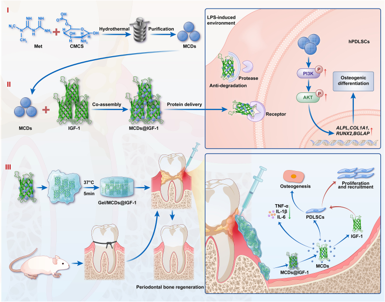
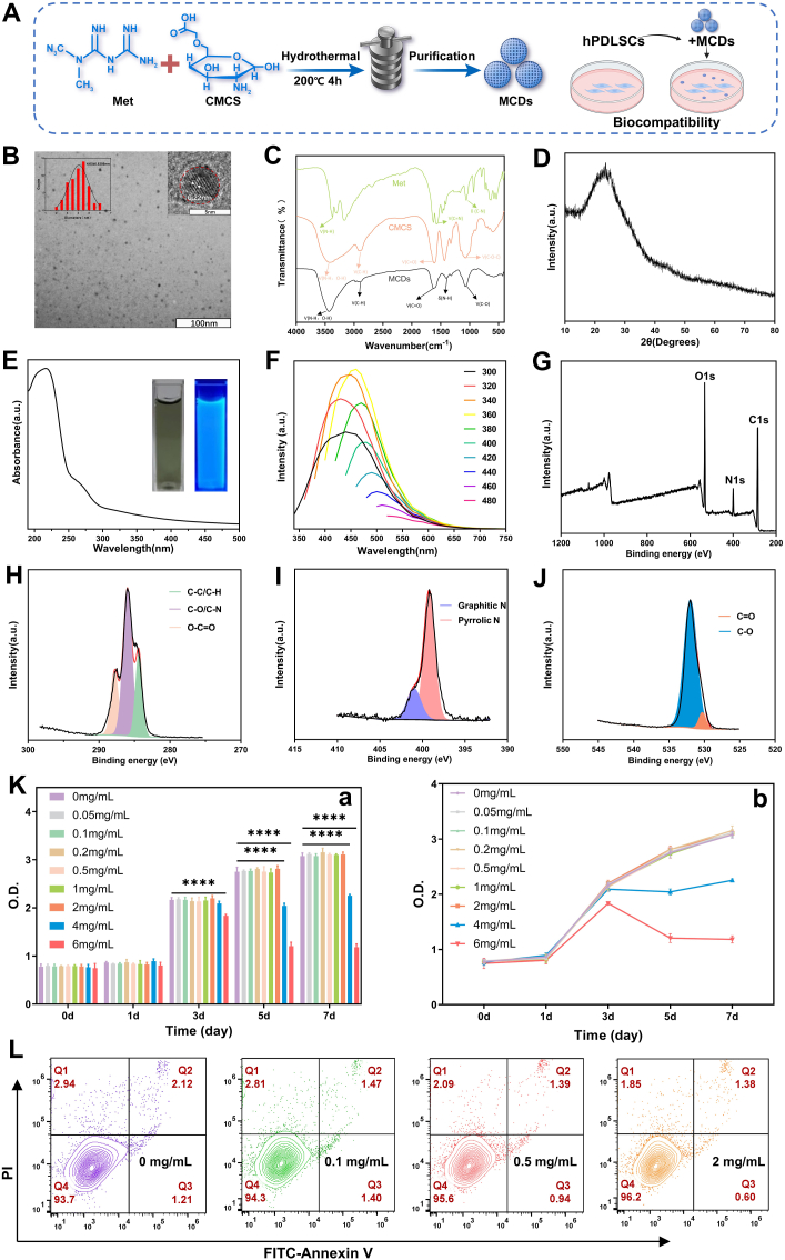
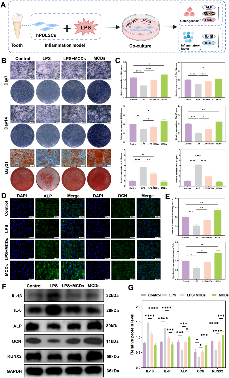
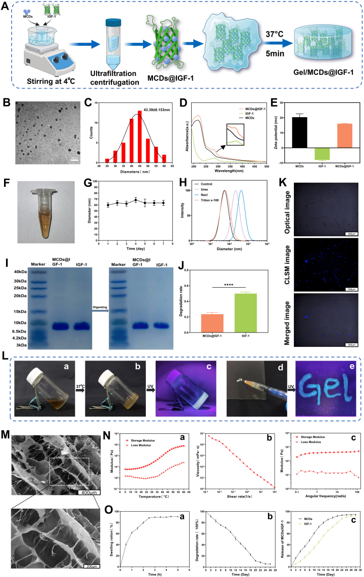
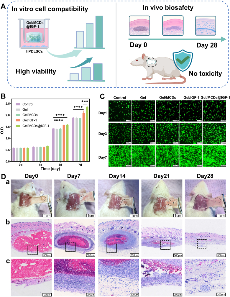
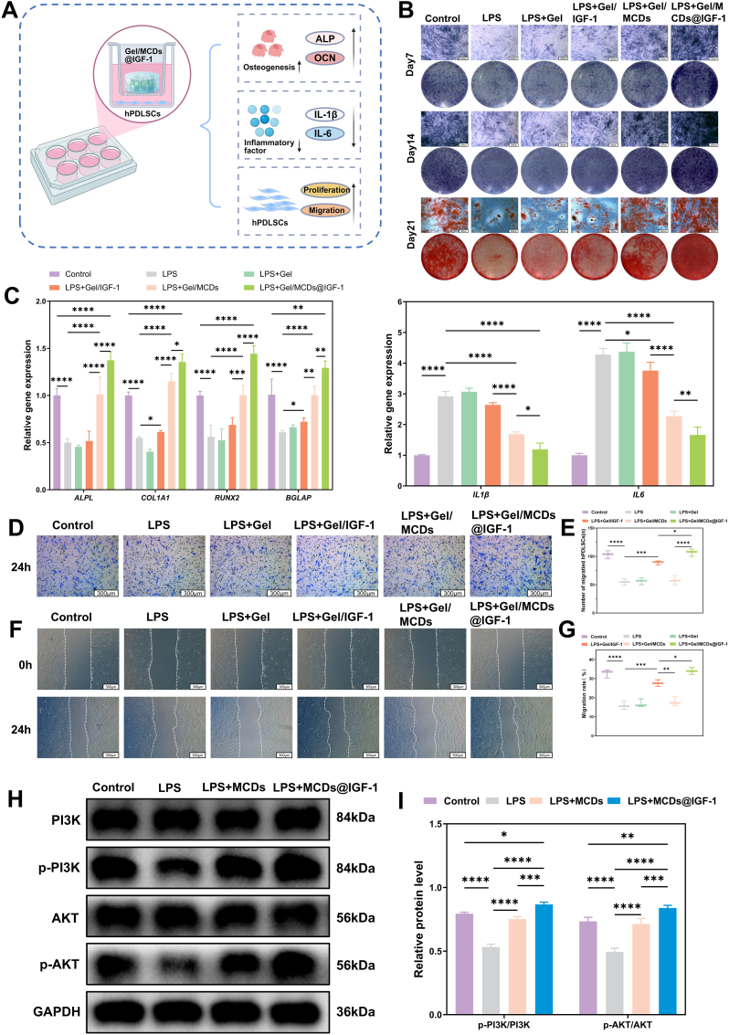
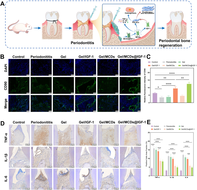
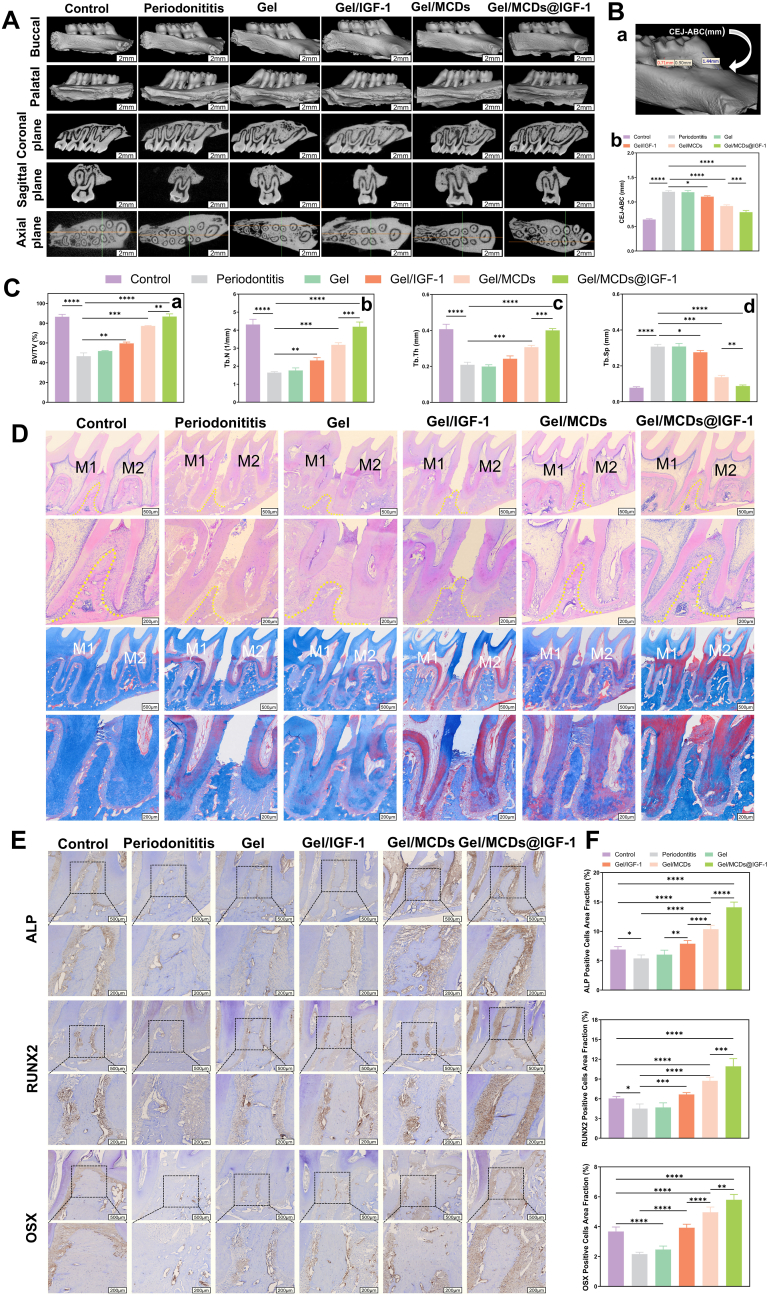
The chronic inflammation in periodontitis suppresses the osteogenic potential of human periodontal ligament stem cells (hPDLSCs), posing a significant challenge to endogenous bone regeneration. To address this, we developed an osteogenic and protein-delivery composite hydrogel system based on metformin carbon dots (MCDs) to enhance the osteogenic potential of hPDLSCs under inflammatory conditions. We successfully synthesized a novel Gel/MCDs@IGF-1 composite hydrogel (Gel) that exhibited excellent biocompatibility and sequentially released MCDs and insulin-like growth factor 1 (IGF-1). First, MCDs were synthesized using a one-step hydrothermal method. MCDs promote the osteogenic differentiation of hPDLSCs under lipopolysaccharide (LPS)-induced inflammatory conditions by activating the PI3K/AKT signaling pathway, and alleviate inflammation. Next, MCDs and IGF-1 were assembled into MCDs@IGF-1 complexes through supramolecular interactions, facilitating efficient IGF-1 delivery and reducing its degradation by trypsin. Furthermore, in vitro and in vivo studies demonstrated that the Gel/MCDs@IGF-1 composite hydrogel effectively recruited stem cells, exerted early anti-inflammatory effects, increased the osteogenesis of hPDLSCs under inflammatory conditions, and significantly promoted alveolar bone regeneration in a Sprague-Dawley (SD) rat model of periodontitis. In conclusion, MCDs, with their dual roles in promoting osteogenesis and protein delivery, are a promising candidate nanoplatform for periodontitis therapy. Additionally, the MCDs-based sequential release hydrogel system presents a novel material strategy for the treatment of periodontitis.

Keywords: Carbon dots; IGF-1; Metformin; Osteogenic differentiation; Protein delivery.


Conflict of interest statement

The authors declare that they have no known competing financial interests or personal relationships that could have appeared to influence the work reported in this paper.

Figures

Image 1
Graphical abstract
Scheme 1
Scheme 1
Schematic diagram illustrating the dual functions of MCDs in promoting osteogenesis and protein delivery, along with the design principle and working mechanism of the Gel/MCDs@IGF-1 composite hydrogel system.
Fig. 1
Fig. 1
Characterization and cytotoxicity assays of MCDs. A) Schematic illustration of the synthesis of MCDs and their excellent biocompatibility. B) TEM and HRTEM images of MCDs. The inset shows the particle size distribution of the MCDs. C) FTIR spectra of carboxymethyl chitosan (red), metformin (green), and MCDs (black). D) XRD pattern of MCDs. E) UV–Vis absorption spectrum of MCDs. Insets: photographs of MCDs solution under sunlight (left) and UV light (right). F) Photoluminescence (PL) spectra of MCDs. G) XPS survey scan spectrum of MCDs. H) C 1s spectrum of MCDs. I) N 1s spectrum of MCDs. J) O 1s spectrum of MCDs. K) a: CCK8 assays of MCDs; b: Line graph corresponding to a. L) Apoptosis assay of hPDLSCs treated with MCDs at different concentrations. The data are presented as the means ± SDs (n ≥ 3). ∗∗∗∗p < 0.0001. ∗p < 0.05 was considered to be statistically significant.
Fig. 2
Fig. 2
Osteogenesis of hPDLSCs induced by MCDs under both inflammatory and normal conditions. A) Schematic showing that the coculture of hPDLSCs with MCDs under LPS-induced inflammatory conditions reduced the levels of inflammatory factors and promoted the osteogenic differentiation of hPDLSCs. B) ALP staining on days 7 and 14 and ARS staining on day 21. C) Validation of osteogenic gene and inflammatory factor expression via qRT‒PCR. D-E) Immunofluorescence staining and semiquantitative analysis of osteogenesis-related proteins (ALP and OCN). (Nuclei: blue; ALP/OCN: green). F-G) Western blot and semiquantitative analysis of osteogenesis-related proteins (RUNX2, ALP, and OCN) and inflammatory factors (IL-1β and IL-6). Data are presented as the means ± SDs (n ≥ 3). ∗p < 0.05, ∗∗p < 0.01, ∗∗∗p < 0.001 and ∗∗∗∗p < 0.0001. ∗p < 0.05 was considered to be statistically significant.
Fig. 3
Fig. 3
MCDs promote the osteogenic differentiation of hPDLSCs under the inflammatory conditions by activating the PI3K/AKT signaling pathway. A) Overview of the procedure. Transcriptomic sequencing was performed on hPDLSCs treated with MCDs. B) Venn diagram showing the number of DEGs in hPDLSCs cultured under Control, LPS, or LPS + MCDs conditions after a 7-day osteogenic induction. C) Heatmap showing that 49 DEGs were upregulated in the Control and LPS + MCDs groups but downregulated in the LPS group; and 14 DEGs with the opposite pattern. D) Chord diagram of the enrichment of osteogenesis-related signaling pathways. E) GO analysis of the 63 DEGs. F) KEGG analysis of the top 15 enriched pathways among the 63 DEGs. G) Enrichment plot of the PI3K/AKT signaling pathway from GSEA analysis comparing LPS and LPS + MCDs groups. H) Heatmap showing genes related to the PI3K/AKT signaling pathway and osteogenic differentiation. I) Statistical analysis of gene expression levels related to the PI3K/AKT signaling pathway and osteogenesis in hPDLSCs cultured under Control, LPS, or LPS + MCDs conditions after a 7-day of osteogenic induction. J) Effect of LY294002 on MCDs-induced osteogenic differentiation in an inflammatory environment, as assessed by ALP staining on days 7 and 14 and ARS staining on day 21. K-L) Western blot and semiquantitative analysis of osteogenesis-related proteins (RUNX2, ALP, and OCN) and key proteins of the PI3K/AKT signaling pathway (PI3K, p-PI3K, AKT, and p-AKT). Data are presented as the means ± SDs. (n ≥ 3). ∗p < 0.05, ∗∗p < 0.01, ∗∗∗p < 0.001 and ∗∗∗∗p < 0.0001. ∗p < 0.05 was considered to be statistically significant.
Fig. 4
Fig. 4
Characterization of MCDs@IGF-1 and Gel/MCDs@IGF-1. A) Schematic illustration of the synthesis route of the MCDs@IGF-1 complex and the Gel/MCDs@IGF-1 composite hydrogel. B) TEM image of MCDs@IGF-1. C) Particle size distribution of MCDs@IGF-1. D) UV–vis absorption spectra of MCDs, IGF-1, and MCDs@IGF-1. E) Zeta potentials of MCDs, IGF-1, and MCDs@IGF-1. F) Photographs of MCDs@IGF-1 solution on the 7th day. G) Changes in the diameter of MCDs@IGF-1 determined by DLS for 7 days. H) DLS results of mixtures formed by MCDs@IGF-1 in the presence of inhibitors (urea, NaCl, and Triton X-100; 25 mM). I) SDS‒PAGE analysis of IGF-1 and MCDs@IGF-1. J) Comparison of degradation rates of IGF-1 and MCDs@IGF-1 after pancreatic enzyme digestion. K) In vitro CLSM images of hPDLSCs treated with MCDs@IGF-1 (0.2 mg/mL) at 37 °C for 12 h. L) a: Photograph of Gel/MCDs@IGF-1 mixture solution at room temperature; b: Photograph of the as-prepared Gel/MCDs@IGF-1 hydrogel formed within 5 min at 37 °C; c: Photograph of Gel/MCDs@IGF-1 hydrogel under UV light; d: Photographs demonstrating injectability of Gel/MCDs@IGF-1 hydrogel; e: Photographs showing fluorescent properties of Gel/MCDs@IGF-1 hydrogel. M) Representative SEM image of Gel/MCDs@IGF-1 hydrogel. N) a: Variations in storage and loss moduli (G′ and G″, respectively) versus temperature; b: Viscosity of the hydrogel versus shear frequency; c: Variations in storage and loss moduli (G′ and G″, respectively) versus angular frequency. O) a–c: Swelling rates, degradation ratios and release profiles of Gel/MCDs@IGF-1 hydrogel. Data are presented as the means ± SDs. (n ≥ 3). ∗∗∗∗p < 0.0001. ∗p < 0.05 was considered to be statistically significant.
Fig. 5
Fig. 5
Toxicity and degradation of hydrogel systems in vitro and in vivo. A) Schematic illustration of the in vitro and in vivo biocompatibility evaluation of the Gel/MCDs@IGF-1 composite hydrogel. B) CCK-8 viability assay of hPDLSCs cultured with hydrogel components after 1, 3, and 7 days of culture. C) Live/dead cell staining images of hPDLSCs cultured with hydrogel components after 1, 3, and 7 days. Live cells were stained green, and dead cells were stained red. D) a: Rat dorsal skin was stripped at different time intervals, and the locally formed hydrogel is circled with a black dotted line. b: Histology of subcutaneous tissue from SD rats at different time points. c: Magnified images of b. Data are presented as the means ± SDs. (n ≥ 3). ∗∗∗p < 0.001, ∗∗∗∗p < 0.0001. ∗p < 0.05 was considered to be statistically significant.
Fig. 6
Fig. 6
Evaluation of the in vitro osteoinductive and promigratory capabilities of the hydrogel system and MCDs@IGF-1 synergistically activating the PI3K/AKT signaling pathway. A) Schematic illustration showing the in vitro osteogenic, anti-inflammatory, and promigratory capabilities of the Gel/MCDs@IGF-1 composite hydrogel. B) hPDLSCs incubated with hydrogels under different conditions were subjected to ALP staining on days 7 and 14, and ARS staining on day 21. C) Relative gene expression in hPDLSCs incubated with hydrogels under different conditions. D-E) Optical images of hPDLSCs recruited by the hydrogel system using a Transwell migration assay and quantification of the number of migrated hPDLSCs. F-G) Optical images of wound healing assays and their quantitative analysis. H-I) Western blot and semiquantitative analysis of key proteins of the PI3K/AKT signaling pathway (PI3K, p-PI3K, AKT, and p-AKT). Data are presented as the means ± SDs. (n ≥ 3). ∗p < 0.05, ∗∗p < 0.01, ∗∗∗p < 0.001 and ∗∗∗∗p < 0.0001. ∗p < 0.05 was considered to be statistically significant.
Fig. 7
Fig. 7
The recruitment of stem cells and early anti-inflammatory effects of composite hydrogels. A) Schematic illustration of the construction of the rat periodontitis model and the treatment procedure with Gel/MCDs@IGF-1 composite hydrogel. B-C) Immunofluorescence staining and relative quantitative analysis of CD90 in periodontal defects at day 7. D-E) Immunohistochemical staining images and relative quantitative analysis of TNF-α, IL-1β and IL-6 in periodontal defects at day 7. Data are presented as the means ± SDs. (n ≥ 3). ∗p < 0.05, ∗∗p < 0.01, ∗∗∗p < 0.001 and ∗∗∗∗p < 0.0001. ∗p < 0.05 was considered to be statistically significant.
Fig. 8
Fig. 8
Composite hydrogel promotes bone regeneration in rat periodontitis models. A) 3D micro-CT reconstruction images of maxillary alveolar bone after 4 weeks of treatment. B) a: Scheme of the vertical distance between the cementoenamel junction (CEJ) and the apex of the alveolar bone crest (ABC); b: Quantitative analysis of the distance from CEJ to ABC. C) a–d: Quantitative analysis of BV/TV, Tb.N, Tb.Th, and Tb.Sp determined from micro-CT images. D) HE staining images of periodontal bone tissue sections. The area enclosed by the yellow dotted line indicates the alveolar bone. E) Masson's trichrome staining images of periodontal bone tissue sections. M1 represents the first molar, and M2 represents the second molar. E-F) Immunohistochemical staining of ALP, RUNX2, and OSX after 4 weeks of treatment and the corresponding quantitative analysis. Data are presented as the means ± SDs. (n ≥ 3). ∗p < 0.05, ∗∗p < 0.01, ∗∗∗p < 0.001 and ∗∗∗∗p < 0.0001. ∗p < 0.05 was considered statistically significant.

Similar articles

References

    1. Kinane D.F., Stathopoulou P.G., Papapanou P.N. Periodontal diseases. Nat. Rev. Dis. Primers. 2017;3 doi: 10.1038/nrdp.2017.38. - DOI - PubMed
    1. Ding T., Kang W., Li J., Yu L., Ge S. An in situ tissue engineering scaffold with growth factors combining angiogenesis and osteoimmunomodulatory functions for advanced periodontal bone regeneration. J. Nanobiotechnol. 2021;19:247. doi: 10.1186/s12951-021-00992-4. - DOI - PMC - PubMed
    1. Zhao Z., Sun Y., Qiao Q., Weir M.D., Schneider A., Masri R., Lynch C.D., Zhang N., Zhang K., Bai Y., Xu H. Calvaria defect regeneration via human periodontal ligament stem cells and prevascularized scaffolds in athymic rats. J. Dent. 2023;138 doi: 10.1016/j.jdent.2023.104690. - DOI - PubMed
    1. Gu F., Zhang K., Li J., Xie X., Wen Q., Sui Z., Su Z., Yu T. Changes of migration, immunoregulation and osteogenic differentiation of mesenchymal stem cells in different stages of inflammation. Int. J. Med. Sci. 2022;19:25–33. doi: 10.7150/ijms.58428. - DOI - PMC - PubMed
    1. Đorđević I.O., Kukolj T., Krstić J., Trivanović D., Obradović H., Santibañez J.F., Mojsilović S., Ilić V., Bugarski D., Jauković A. The inhibition of periodontal ligament stem cells osteogenic differentiation by IL-17 is mediated via MAPKs. Int. J. Biochem. Cell Biol. 2016;71:92–101. doi: 10.1016/j.biocel.2015.12.007. - DOI - PubMed

LinkOut - more resources