. 2025 Mar 24;12(2):e21200036.
doi: 10.14440/bladder.2024.0060. eCollection 2025.

Pentosan polysulfate alleviates interstitial cystitis/bladder pain syndrome by modulating bile acid metabolism and activating the TGR5 receptor through gut microbiota regulation

Affiliations

Pentosan polysulfate alleviates interstitial cystitis/bladder pain syndrome by modulating bile acid metabolism and activating the TGR5 receptor through gut microbiota regulation

Zhangrui Zhu et al. Bladder (San Franc). .

Abstract

Background: The disrupted gut microbiome has been found to be implicated in the development of interstitial cystitis/bladder pain syndrome (IC/BPS). Pentosan polysulfate (PPS) is an oral medication used for treating IC/BPS, acting as both an anti-inflammatory agent and a bladder barrier protector. However, the precise mechanisms by which the PPS-mediated modulation of the gut microbiome alleviates IC/BPS are not fully understood.

Objective: This study aimed to identify the key gut microbiota species and metabolites involved in PPS's protective effects against IC/BPS.

Methods: We employed a multifaceted approach, including 16S rDNA gene sequencing, antibiotic treatment, and fecal microbiota transplantation, to validate the dependency of PPS's protective effects on the gut microbiome. Furthermore, we performed a comprehensive metabolomic profiling using non-targeted metabolomics and liquid chromatography-tandem mass spectrometry.

Results: PPS significantly elevated the abundance of the xylan-degrading bacteria, Eubacterium xylanophilum group, which, through its interaction with the gut microbiome, markedly reduced inflammation and barrier damage induced by cyclophosphamide in IC/BPS. In addition, PPS significantly increased the level of ursodeoxycholic acid (UDCA), a secondary bile acid, demonstrating a strong correlation with the abundance of the E. xylanophilum group. Ex vivo supplementation with UDCA mitigated lipopolysaccharide-induced inflammation and barrier disruption in SV-HUC-1 cells by activating the TGR5 receptor.

Conclusion: PPS exerts its protective effects against IC/BPS by modulating the gut microbiome and its metabolites.

Keywords: Bladder barrier; Cyclophosphamide; Gut microbiota; Interstitial cystitis/bladder pain syndrome; Pentosan polysulfate; Ursodeoxycholic acid.


Conflict of interest statement

The authors declare no conflicts of interest.

Figures
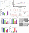
Figure 1
Figure 1
Pentosan polysulfate protects against bladder dysfunction in mice with CYP-induced interstitial cystitis/bladder pain syndrome. (A) Experimental workflow for assessing the protective effects of PPS on CYP-induced IC/BPS in mice. (B) Weight change curves of mice in four groups, n = 6. (C) Von Frey test results showed changes in mechanical pain thresholds in four groups of mice after PPS gavage and CYP modeling, n = 6. (D-F) Urodynamic plots and quantitative parameters of bladder function in four groups, n = 6. (G-I) Voiding stains and quantitative parameters of voiding behavior in four groups, n = 6. Note: Results are presented as mean ± standard error of the mean, **p < 0.01, ****p < 0.0001, analyzed by one-way analysis of variance. Abbreviations: C + P: Cyclophosphamide + pentosan polysulfate (CYP + PPS); CYP: Cyclophosphamide; IC/BPS: Interstitial cystitis/bladder pain syndrome; ns: Not significant; PPS: Pentosan polysulfate.
Figure 2
Figure 2
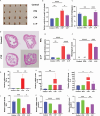
Pentosan polysulfate protects against bladder inflammation and barrier damage in CYP-induced interstitial cystitis/bladder pain syndrome mice. (A) Gross appearance of bladders from four groups of mice. (B) Bladder-to-body weight ratios in four groups of mice, n = 6. (C) GAG concentrations in bladder tissues, detected by ELISA, n = 6. (D-F) Hematoxylin and eosin staining, quantitative scores, and pathological damage scores of bladder tissues, scale bar: 50 μm, magnification: 20×, n = 5. (G-I) Expression levels of inflammatory cytokines TNF-α, IL-1β, and IL-6 in bladder tissues, n = 6. (J-L) mRNA expression levels of tight junction proteins ZO-1, occludin, and claudin-1 in bladder tissues, n = 6. Note: Results are presented as mean ± SEM, *p < 0.05, **p < 0.01, ***p < 0.001, ****p < 0.0001, analyzed by one-way analysis of variance. Abbreviations: C + P: Cyclophosphamide + pentosan polysulfate (CYP + PPS); CYP: Cyclophosphamide; GAG: Glycosaminoglycan; IC/BPS: Interstitial cystitis/bladder pain syndrome; ns: Not significant; PPS: Pentosan polysulfate.
Figure 3
Figure 3
16S rDNA sequencing revealed Pentosan polysulfate-mediated alterations in gut microbiota composition in CYP-treated mice. (A) Bar plot showing differences in gut microbiota composition at the genus level between CYP and C + P groups, n = 5. (B) Venn diagram of gut microbiota composition between the CYP group and the C + P group. (C) Heatmap of differences in gut microbiota composition at the genus level between CYP and C + P groups, n = 5. (D) Differences in gut microbiota composition between CYP and C + P groups at the genus level, n = 5. (E) LEfSe analysis for identifying differentially abundant taxa. (F) Cladogram representation of taxonomic differences, n = 5 for both CYP and C + P groups. Abbreviations: C + P: Cyclophosphamide + Pentosan polysulfate (CYP + PPS); CYP: Cyclophosphamide; PPS: Pentosan polysulfate.
Figure 4
Figure 4
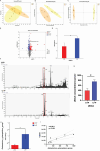
Pentosan polysulfate intervention alters the composition of intestinal and serum metabolites in CYP-treated mice. (A-C) Principal component analysis (PCA), partial least squares discriminant analysis (PLS-DA), and OPLS-DA of untargeted metabolomic data. (D) Volcano plot showing differential metabolites between CYP and C + P groups. (E) UDCA concentrations in cecal contents of two groups. (F-G) LC-MS targeted the detection of UDCA concentrations in the serum of two groups of mice. (H) Relative abundance of Eubacterium xylanophilum group in two groups, n = 5. (I) Scatter plot analysis of the correlation between UDCA concentrations in cecal contents and relative abundance of E. xylanophilum group, n = 7. Note: Statistical significance determined at *p < 0.05, **p < 0.01, analyzed by Student’s t-test. Abbreviations: C + P: Cyclophosphamide + Pentosan polysulfate (CYP + PPS); CYP: Cyclophosphamide; LC-MS: Liquid chromatography–mass spectrometry; PPS: Pentosan polysulfate; UDCA: Ursodeoxycholic acid.
Figure 5
Figure 5
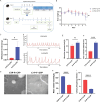
FMT from CYP + Pentosan polysulfate mice alleviates CYP-induced bladder dysfunction. (A) Experimental workflow for assessing the effects of FMT from C + P mice on CYP-induced IC/BPS in mice. (B) Weight change curves of two groups of mice, n = 6. (C) Von Frey test results showed changes in mechanical pain thresholds in two groups, n = 6. (D-F) Urodynamic plots and quantitative parameters of bladder function in two groups, n = 6. (G-I) Voiding stains and quantitative parameters of voiding behavior in two groups, n = 6. Note: Results are presented as mean ± SEM, **p < 0.05, ***p < 0.001, ****p < 0.0001, analyzed by Student’s t-test. Abbreviations: C + P: Cyclophosphamide + Pentosan polysulfate (CYP + PPS); CYP: Cyclophosphamide; FMT: Fecal microbiota transplantation; IC/BPS: Interstitial cystitis/bladder pain syndrome; ns: Not significant; PPS: Pentosan polysulfate.
Figure 6
Figure 6
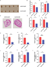
FMT from CYP + Pentosan polysulfate mice alleviates CYP-induced bladder inflammation and barrier damage. (A) Gross appearance of bladders from two groups. (B) Bladder-to-body weight ratios in two groups, n = 6. (C) GAG concentrations in bladder tissues detected by ELISA, n = 6. (D-F) HE staining, quantitative scores, and pathological damage scores of bladder tissues, scale bar: 50 μM, magnification: 20×, n = 5. (G-I) Expression levels of inflammatory cytokines TNF-α, IL-1β, and IL-6 in bladder tissues, n = 6. (J-I) mRNA expression levels of tight junction proteins ZO-1, occludin, and claudin-1 in bladder tissues, n = 6. Note: Results are presented as mean ± SEM, *p < 0.05, ***p < 0.01, ***p < 0.001, analyzed by Student’s t-test. Abbreviations: C + P: Cyclophosphamide + Pentosan polysulfate (CYP + PPS); CYP: Cyclophosphamide; FMT: Fecal microbiota transplantation; GAG: Glycosaminoglycan; IC/BPS: Interstitial cystitis/bladder pain syndrome; ns: Not significant; PPS: Pentosan polysulfate.
Figure 7
Figure 7
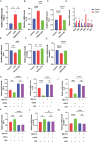
Inhibition of TGR5 by SBI-115 confirmed that UDCA alleviated lipopolysaccharide-induced inflammation and barrier damage in SV-HUC-1 cells by activating TGR5. (A-C) mRNA expression levels of inflammatory cytokines TNF-α, IL-1β, and IL-6 after SV-HUC-1 cells were pretreated with UDCA for 2 h and incubated with LPS for 24 h. (D) mRNA expression levels of TGR5, VDR, FXR, PXR, GR, and α5β1 after UDCA incubation for 24 h. (E-G) mRNA expression levels of tight junction proteins ZO-1, occludin, and claudin-1 after SV-HUC-1 cells were pretreated with UDCA for 2 h and incubated with LPS for 24 h. (H-J) mRNA expression levels of inflammatory cytokines TNF-α, IL-1β, and IL-6 and (K-M) mRNA expression levels of tight junction proteins ZO-1, occludin, and claudin-1 after SV-HUC-1 cells were pretreated with SBI-115 for 2 h and incubated with LPS for 24 h. Note: Results are presented as mean ± SEM, *p < 0.05, ***p < 0.01, ***p < 0.001, ****p < 0.0001, analyzed by one-way analysis of variance (A-C, E-M) or Student’s t-test (D). Abbreviations: LPS: Lipopolysaccharide; ns: Not significant; UDCA: Ursodeoxycholic acid.
Figure 8
Figure 8
shRNA-mediated knockdown confirmed that UDCA alleviated lipopolysaccharide-induced inflammation and barrier damage in SV-HUC-1 cells by activating TGR5. (A) qPCR validation of shRNA-mediated knockdown of TGR5. (B-D) mRNA expression levels of inflammatory cytokines TNF-α, IL-1β, and IL-6. (E-G) mRNA expression levels of tight junction proteins ZO-1, occludin, and claudin-1 after constructing stable SV-HUC-1 cell lines with knockdown of TGR5 and pretreating them with UDCA for 2 h, followed by LPS incubation for 24 h. Results are presented as mean ± SEM, *p < 0.05, **p < 0.01, ***p < 0.001, ****p < 0.0001, analyzed by one-way analysis of variance. Abbreviations: LPS: Lipopolysaccharide; ns: Not significant; qPCR: Quantitative polymerase chain reaction; UDCA: Ursodeoxycholic acid; SEM: Scanning electron microscopy.
Figure S1
Figure S1
Standard curve of voiding spot on paper (VSOP). (A) Urine samples were collected to construct a standard curve and were dispensed onto filter paper in various volumes (1, 2, 4, 10, 20, 40, 80, 100, and 150 μL). (B) The formula y = 6.6579x-1.0468 (R² = 0.9966) was utilized to calculate individual void areas on the filter paper.
Figure S2
Figure S2
Analysis of microbial diversity through 16S rDNA sequencing. Microbial α-diversity was assessed using (A) Shannon index, (B) Simpson index, (C) Chao1 index, and (D) Ace index. Microbial β-diversity was evaluated through (E) principal component analysis (PCA), (F) principal coordinates analysis (PCoA), and (G) non-metric multidimensional scaling (NMDS). Abbreviations: C + P: Cyclophosphamide + Pentosan polysulfate; CYP: Cyclophosphamide; OTU: Operational taxonomic unit.
Figure S3
Figure S3
Successful establishment of an IC/BPS cell model in SV-HUC-1 cells induced by lipopolysaccharide. (A–C) Changes in mRNA levels of inflammatory cytokines TNF-α, IL-1β, and IL-6 after incubation with different concentrations of LPS for 24 h in SV-HUC-1 cells, n = 3. (D–F) Changes in mRNA levels of bladder epithelial barrier tight junction proteins ZO-1, occludin, and claudin-1 after incubation with different concentrations of LPS for 24 h in SV-HUC-1 cells, n = 3. Note: Results are presented as mean ± SEM, with statistical significance indicated by *p < 0.05, **p < 0.01, ****p < 0.0001 (one-way analysis of variance). Abbreviations: LPS: Lipopolysaccharide; ns: Not significant.
Figure S4
Figure S4
The toxic effects of ursodeoxycholic acid on SV-HUC-1 cells at different concentrations using CCK8. Optical density (OD) values of SV-HUC-1 cells treated with UDCA at concentrations of 10 μM, 20 μM, 30 μM, 40 μM, and 50μM for durations of (A) 2 h, (B) 4 h, (C) 8 h, (D) 16 h, (E) 24 h, and (F) 32 h. Note: Results are presented as mean ± SE, with statistical significance indicated by *p < 0.05, **p < 0.01, ***p < 0.001, ****p < 0.0001 (one-way analysis of variance). Abbreviations: ns: Not significant; UDCA: Ursodeoxycholic acid.

Similar articles

References

    1. Clemens JQ, Erickson DR, Varela NP, Lai HH.Diagnosis and treatment of interstitial cystitis/bladder pain syndrome. J Urol 2022;208(1):34–42. doi: 10.1097/JU.0000000000002756 - PubMed
    1. Di XP, Peng L, Xiang LY, Wang MH, Zhang J, Luo DY.A bibliometric analysis of top-cited journal articles in interstitial cystitis and bladder pain syndrome. Int Urogynecol J 2022;33(9):2557–2563. doi: 10.1007/s00192-022-05298-z - PubMed
    1. Mohammad A, Laboulaye MA, Shenhar C, Dobberfuhl AD.Mechanisms of oxidative stress in interstitial cystitis/bladder pain syndrome. Nat Rev Urol 2024;21(7):433–449. doi: 10.1038/s41585-023-00850-y - PubMed
    1. Dalghi MG, Montalbetti N, Carattino MD, Apodaca G.The urothelium:Life in a liquid environment. Physiol Rev 2020;100(4):1621–1705. doi: 10.1152/physrev.00041.2019 - PMC - PubMed
    1. Kayama H, Okumura R, Takeda K.Interaction between the microbiota, epithelia, and immune cells in the intestine. Annu Rev Immunol 2020;38:23–48. doi: 10.1146/annurev-immunol-070119-115104 - PubMed

LinkOut - more resources