Review
. 2025 Oct;56(4):160.
doi: 10.3892/ijmm.2025.5601. Epub 2025 Aug 1.

Research progress on mesenchymal stem cell‑derived exosomes in the treatment of osteoporosis induced by knee osteoarthritis (Review)

Affiliations
Review

Research progress on mesenchymal stem cell‑derived exosomes in the treatment of osteoporosis induced by knee osteoarthritis (Review)

Hai-Yan Xue et al. Int J Mol Med. 2025 Oct.

Abstract

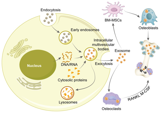
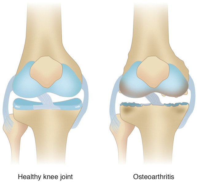
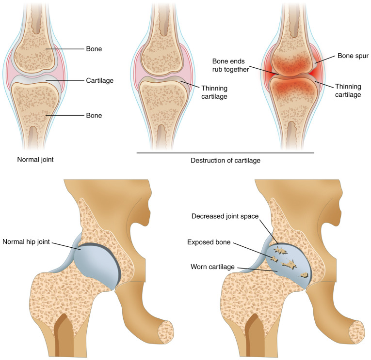
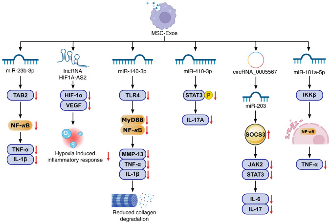
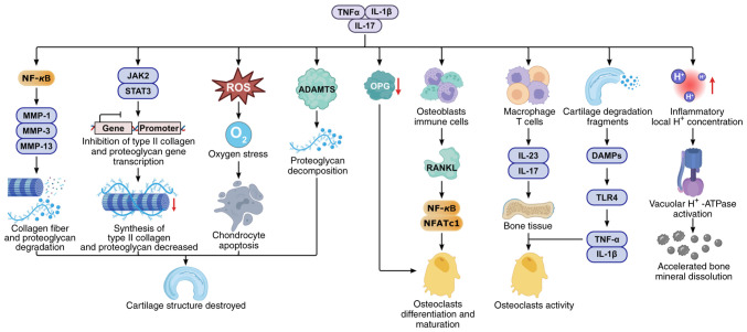
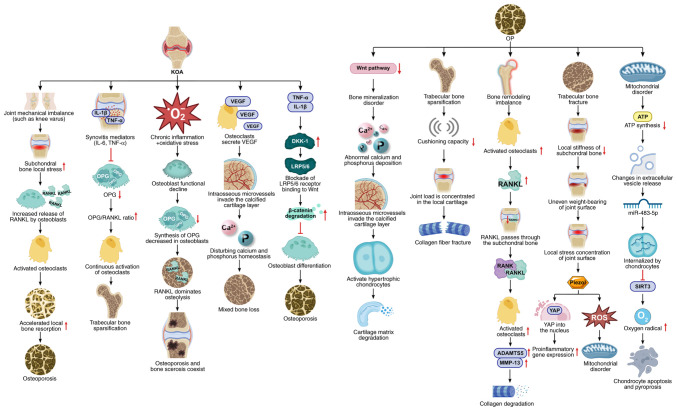
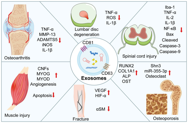
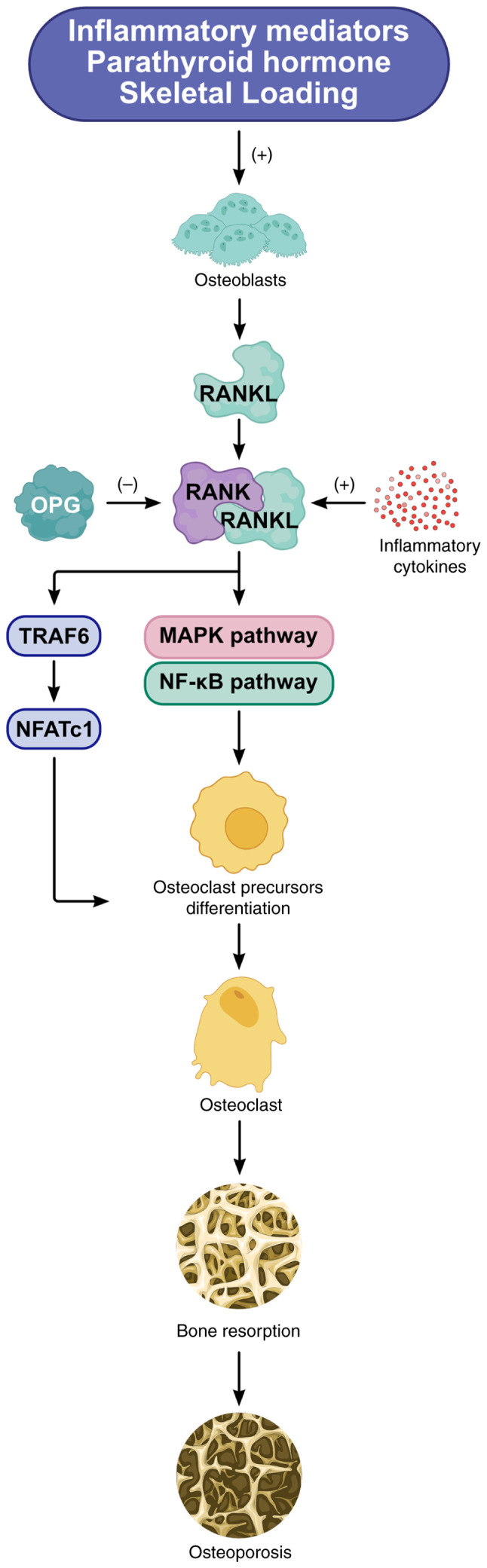
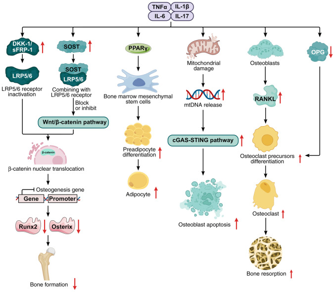
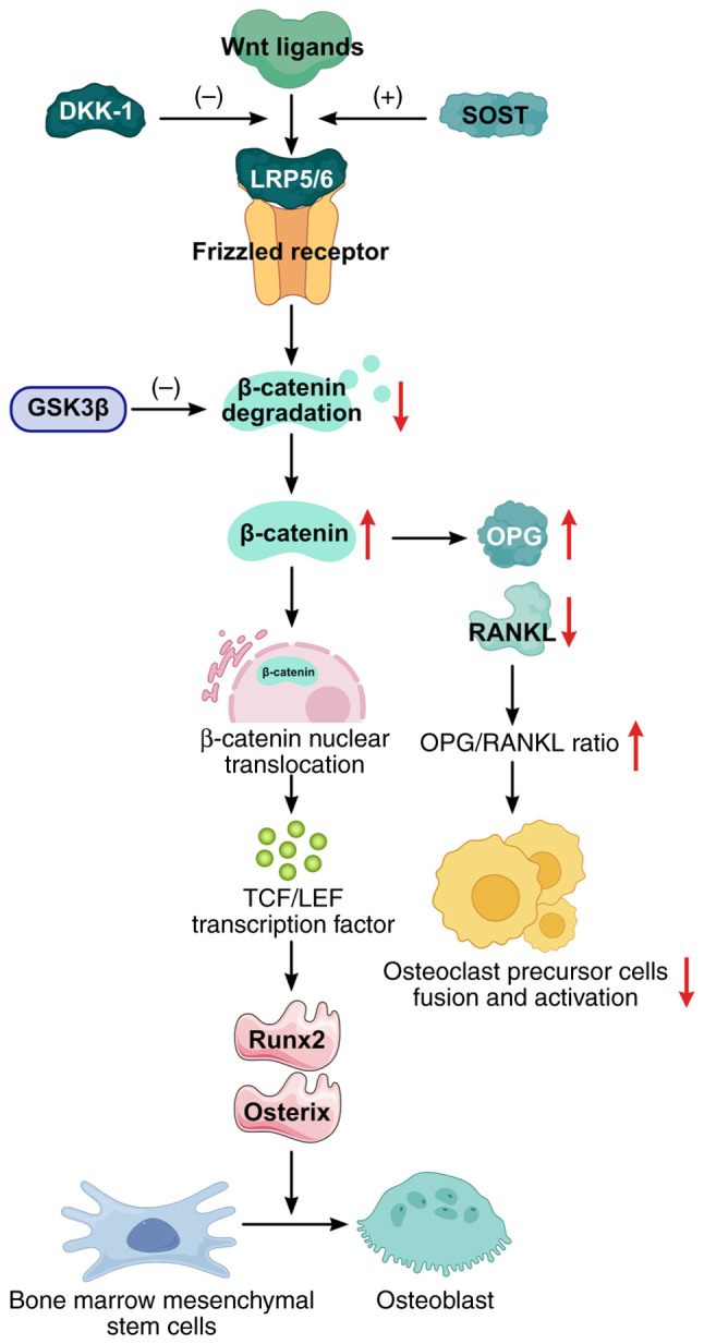
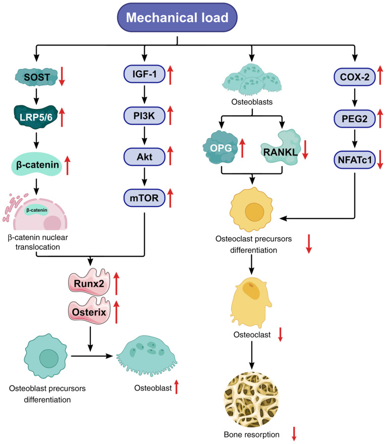
Knee osteoarthritis (KOA) and osteoporosis (OP) are closely related, age‑related, degenerative orthopedic conditions. Elderly patients with OP frequently develop concurrent KOA, with high co‑occurrence rates. Studies indicate that OP significantly increases KOA risk and that these conditions mutually exacerbate each other. Anti‑OP therapies show significant efficacy in KOA management, substantially delaying disease progression. Mesenchymal stem cell‑derived exosomes (MSC‑Exos) have significant therapeutic potential for both KOA and OP. These exosomes enhance chondrocyte proliferation, modulate cartilage matrix synthesis and degradation, and suppress synovial inflammation, suggesting a novel therapeutic approach for KOA. However, their OP mechanisms remain unclear but may involve disrupted bone metabolic signaling, amplified inflammation, and dysregulated intercellular communication in the bone microenvironment. The present review summarizes MSC‑Exos research advances in KOA and OP, providing a foundation for future studies and clinical applications.

Keywords: exosomes; knee osteoarthritis; mesenchymal stem cells; osteoporosis.


Conflict of interest statement

The authors declare that they have no competing interests.

Figures

Figure 1
Figure 1
Relationship between exosomes and bone formation. BM-MSCs, bone marrow-mesenchymal stem cells; RANKL, receptor activator of nuclear factor-kappaB ligand; M-CSF, macrophage colony-stimulating factor.
Figure 2
Figure 2
Source, isolation and application of extracellular vesicles from MSCs. MSCs, mesenchymal stem cells.
Figure 3
Figure 3
Articular cartilage degeneration. Healthy cartilage: Smooth, lubricin-rich, intact collagen fibers; Knee osteoarhtritis cartilage: Fibrillated, eroded, with fissures/ulcers; reduced lubricin.
Figure 4
Figure 4
Cartilage degeneration in osteoarthritis.
Figure 5
Figure 5
MSC-Exos from mesenchymal stem cells regulate multiple inflammatory pathways. MSC-Exos ameliorate osteoporosis and osteoarthritis by regulating the following pathways: miR-23b-3p/TAB2/NF-κB, lncRNA HIF1A-AS2/HIF-1α/VEGF, miR-140-3p/TLR4/MyD88/NF-κB, miR-410-3p/STAT3, circRNA_0005567/miR-203/SOCS3, and miR-181a/IKKβ/NF-κB. MSC-Exos, mesenchymal stem cell-derived exosomes; TAB2, TGF-β-activated kinase 1 binding protein 2; NF-κB, nuclear factor kappa B; HIF-1α, hypoxia-inducible factor 1-alpha; VEGF, vascular endothelial growth factor; TLR4, Toll-like receptor 4; MyD88, myeloid differentiation primary response 88; STAT3, signal transducer and activator of transcription 3; SOCS3, suppressor of cytokine signaling 3; IKKβ, IκB kinase beta; MMP-13, matrix metalloproteinase-13; JAK2, Janus kinase 2; miRNAs, microRNAs; lncRNAs, long non-coding RNAs; circRNAs, circular RNAs. The arrows indicate the direction of change: ↑, increased expression/activity; ↓, decreased expression/inhibition.
Figure 6
Figure 6
Inflammatory regulatory mechanisms in OP and KOA. In KOA, inflammatory mediators (IL-6, TNF-α and IL-1β) activate NF-κB, upregulating MMPs (MMP-1/3/13) to degrade collagen/proteoglycans; simultaneously, they activate JAK-STAT signaling, inhibiting COL2A1/proteoglycan gene transcription, reducing collagen/proteoglycan synthesis and causing chondrocyte structural damage; ADAMTS upregulation mediates collagen breakdown; ROS overproduction induces chondrocyte apoptosis; inflammation elevates osteoblast/immune cell-derived RANKL while suppressing OPG, promoting osteoclast differentiation/maturation and bone resorption; macrophage/T-cell-derived IL-17/IL-23 enhances osteoclast activity; cartilage fragments act as DAMPs, activating TLR4 signaling; and activity and leading to increased bone resorption. OP, osteoarthritis; KOA knee OA; MMPs, matrix metalloproteinases; ADAMTS, a disintegrin and metalloproteinase with thrombospondin motifs; ROS, reactive oxygen species; JAK, Janus kinase; STAT, signal transducer and activator of transcription; RANKL, receptor activator of nuclear factor-kappaB ligand; TLR, Toll-like receptor; DMPS: damage-associated molecular pattern. The arrows indicate the direction of change: ↑, increased expression/activity; ↓, decreased expression/inhibition.
Figure 7
Figure 7
Osteoarthritis induces osteoporosis through multiple regulatory pathways. During KOA, an imbalance in joint mechanics increases local stress within the subchondral bone. This stress stimulates osteoblasts to release the receptor activator of RANKL, enhancing osteoclast activity and accelerating bone resorption. Synovitis-derived factors infiltrate the subchondral bone, inhibiting osteoblast synthesis of OPG, increasing the RANKL/OPG ratio, further increasing osteoclast activity, and leading to trabecular thinning. Chronic inflammation and oxidative stress reduce OPG secretion while increasing RANKL secretion by osteoblasts, resulting in increased bone resorption and the coexistence of osteoporosis and osteosclerosis. Osteoclasts secrete VEGF, prompting bone microvessels to invade the calcified cartilage layer, disrupting calcium-phosphorus homeostasis, and causing mixed bone loss. Inflammation increases DKK1 activity, blocking the binding of Wnt ligands to LRP5/6. This increases β-catenin degradation, reduces its nuclear translocation, and inhibits osteoblast differentiation. During OP, trabecular sparsification diminishes the shock absorption capacity, concentrating the joint load onto the cartilage and causing collagen fiber detachment. Impaired Wnt function disrupts bone mineralization and calcium-phosphorus deposition, allowing vascular invasion into the calcified cartilage layer, activating hypertrophic chondrocytes, and increasing cartilage matrix degradation. Imbalanced bone remodeling enhances osteoclast activity and RANKL secretion. RANKL traverses the subchondral bone plate, binds to chondrocytes, and activates the NF-κB pathway. This increases MMP-13 and ADAMTS5 activity, accelerating type II collagen degradation and proteoglycan loss. OP-induced trabecular fractures decrease subchondral bone stiffness, causing uneven joint surface loading and local stress concentration. This activates Piezo1 mechanoreceptors in chondrocytes, promoting YAP nuclear entry. This increases proinflammatory gene expression, triggers a ROS burst, and causes mitochondrial damage. Mitochondrial dysfunction in bone cells reduces ATP synthesis and alters extracellular vesicle release. These vesicles carry miR-483-5p, which is internalized by chondrocytes and inhibits SIRT3 expression. The resulting increase in oxidative stress ultimately leads to chondrocyte pyroptosis and apoptosis. OP, osteoarthritis; KOA knee OA; RANKL, receptor activator of nuclear factor-kappaB ligand; OPG, osteoprotegerin; VEGF, vascular endothelial growth factor; DKK1, Dickkopf WNT signaling pathway inhibitor 1; Wnt, wingless-type MMTV integration site family; MMPs, matrix metalloproteinases; ADAMTS, a disintegrin and metalloproteinase with thrombospondin motifs; ROS, reactive oxygen species; Piezo1, Piezo-type mechanosensitive ion channel component 1; ATP, adenosine triphosphate; SIRT3, sirtuin 3. The arrows indicate the direction of change: ↑, increased expression/activity; ↓, decreased expression/inhibition.
Figure 8
Figure 8
Mechanisms by which mesenchymal stem cell-derived exosomes promote joint and muscle repair. These exosomes modulate key repair processes through the regulation of multiple targets, including downregulating TNF-α, MMP-13, ADAMTS, iNOS, ROS, IBA-1, NF-κB and BAX, and upregulate Runx2, ALP, OST, HIF-1α, VEGF, aSM, CNFs, MYOG, and MyoD expression in osteoarthritis, muscle injury, fracture, osteoporosis, spinial cord injury, and lumbar disc degeneration. TNF-α, tumor necrosis factor alpha; MMP-13, matrix metalloproteinase-13; ADAMTS, a disintegrin and metalloproteinase with thrombospondin motifs; iNOS, inducible nitric oxide synthase; ROS, reactive oxygen species; Iba-1, ionized calcium-binding adapter molecule 1; NF-κB, nuclear factor kappa B; BAX, BCL2-associated X protein; Runx2, Runt-related transcription factor 2; ALP, alkaline phosphatase; OST, osteocalcin; HIF-1α, hypoxia-inducible factor 1-alpha; VEGF, vascular endothelial growth factor; aSM, alpha-smooth muscle actin; CNFs, centrally nucleated fibers; MYOG, myogenin; MyoD, myogenic differentiation 1. The arrows indicate the direction of change: ↑, increased expression/activity; ↓, decreased expression/inhibition.
Figure 9
Figure 9
Regulatory mechanism of the RANK/RANKL/OPG pathway in bone metabolism. RANKL binds to receptor activators of RANK on osteoclast precursors, promoting osteoclast differentiation/maturation and enhancing bone resorption through the activation of the TRAF6, MAPK, and NF-κB pathways. OPG acts as a decoy receptor for RANKL, inhibiting this osteoclastogenic cascade. RANK, receptor activator of nuclear factor-kappaB; RANKL, RANK ligand; OPG, osteoprotegerin; TRAF6, TNF receptor-associated factor 6; NFATc1, nuclear factor of activated T cells 1; MAPK, mitogen-activated protein kinase; NF-κB, nuclear factor κB. The arrows indicate the direction of change: ↑, increased expression/activity; ↓, decreased expression/inhibition.
Figure 10
Figure 10
Regulatory mechanism of inflammatory mediators in bone metabolism. Inflammatory mediators disrupt bone homeostasis through four principal mechanisms: OPG expression combined with increased RANK/RANKL activity promotes osteoclast differentiation and increases bone resorption; activation of the PPARγ pathway drives the adipogenic transformation of bone marrow mesenchymal stem cells; mitochondrial damage coupled with cGAS-STING pathway activation induces osteoblast apoptosis; and inhibition of Wnt/β-catenin pathway activity-mediated by upregulated DKK1 and SOST expression-suppresses osteoblast formation. DKK1, Dickkopf-1; SOST, sclerostin; RANKL, receptor activator of nuclear factor-kappaB ligand; OPG, osteoprotegerin. The arrows indicate the direction of change: ↑, increased expression/activity; ↓, decreased expression/inhibition.
Figure 11
Figure 11
Regulatory mechanism of the Wnt/β-catenin pathway in bone metabolism. Wnt proteins bind to Frizzled receptors and low-density lipoprotein receptor-related proteins 5/6 (LRP5/6), activating the β-catenin signaling cascade. This pathway promotes bone marrow mesenchymal stem cell differentiation toward osteoblasts while enhancing osteogenic activity. The inhibitory molecules DKK1 and SOST antagonize Wnt signaling by blocking LRP5/6 coreceptor engagement. DKK1, Dickkopf-1; SOST, sclerostin; LRP5/6, low-density lipoprotein receptor-related protein 5/6; GSK-3β, glycogen synthase kinase-3β. TCF/LEF, T-cell factor/lymphoid enhancer-binding factor. The arrows indicate the direction of change: ↑, increased expression/activity; ↓, decreased expression/inhibition.
Figure 12
Figure 12
Regulatory mechanism of mechanical loading on bone metabolism. Mechanical stimulation promotes bone formation through the activation of key pathways: IGF-1/PI3K/Akt/mTOR signaling enhances osteoblast activity; RANKL/RANK/OPG axis modulation favors bone formation over resorption; the COX-2/PGE2 cascade stimulates osteogenesis; and Wnt/β-catenin pathway activation drives osteogenic differentiation. Conversely, mechanical unloading suppresses these pro-osteogenic signals while upregulating bone-resorbing pathways (for example, increasing the RANKL/OPG ratio and increasing DKK1/SOST expression), ultimately leading to bone loss. RANKL, receptor activator of nuclear factor-kappaB ligand; OPG, osteoprotegerin; DKK1, Dickkopf-1; SOST, sclerostin; LRP5/6, low-density lipoprotein receptor-related protein 5/6; COX-2, cyclooxygenase-2; PGE2, prostaglandin E2; IGF-1, insulin-like growth factor 1; PI3K, phosphatidylinositol 3-kinase; mTOR, mechanistic target of rapamycin; AKT, protein kinase B; NFATc1, nuclear factor of activated T cells 1. The arrows indicate the direction of change: ↑, increased expression/activity; ↓, decreased expression/inhibition.

Similar articles

References

    1. Curry ZA, Beling A, Borg-Stein J. Knee osteoarthritis in midlife women: Unique considerations and comprehensive management. Menopause. 2022;29:748–755. doi: 10.1097/GME.0000000000001966. - DOI - PubMed
    1. Ren JL, Yang J, Hu W. The global burden of osteoarthritis knee: A secondary data analysis of a population-based study. Clin Rheumatol. 2025;44:1769–1810. doi: 10.1007/s10067-025-07347-6. - DOI - PMC - PubMed
    1. Lv Y, Sui L, Lv H, Zheng J, Feng H, Jing F. Burden of knee osteoarthritis in China and globally from 1992 to 2021, and projections to 2030: A systematic analysis from the Global Burden of Disease Study 2021. Front Public Health. 2025;13:1543180. doi: 10.3389/fpubh.2025.1543180. - DOI - PMC - PubMed
    1. Muñoz M, Robinson K, Shibli-Rahhal A. Bone health and osteoporosis prevention and treatment. Clin Obstet Gynecol. 2020;63:770–787. doi: 10.1097/GRF.0000000000000572. - DOI - PubMed
    1. Zhou G, Zhang X, Gu Z, Zhao J, Luo M, Liu J. Research progress on the treatment of knee osteoarthritis combined with osteoporosis by single-herb Chinese medicine and compound. Front Med (Lausanne) 2023;10:1254086. doi: 10.3389/fmed.2023.1254086. - DOI - PMC - PubMed

LinkOut - more resources