. 2025 Aug 20;32(8):e240312.
doi: 10.1530/ERC-24-0312. Print 2025 Aug 1.

Targeting Src tyrosine kinase to enhance radioiodide uptake in breast cancer

Affiliations

Targeting Src tyrosine kinase to enhance radioiodide uptake in breast cancer

Vikki L Poole et al. Endocr Relat Cancer. .

Abstract

Sodium iodide symporter (NIS) expression in breast cancer renders radioiodide (RAI) a promising treatment modality. However, insufficient functional NIS within the plasma membrane limits RAI uptake (RAIU). We aimed to elucidate NIS regulatory mechanisms that impede RAIU in breast cancer and identify molecular targets for stimulating RAI-avidity in breast tumours. Mechanistic interaction between pituitary tumor-transforming gene-binding factor (PBF/PTTG1IP) and NIS was investigated through NanoBiT, co-immunoprecipitation, immunofluorescent microscopy, subcellular localisation and RAIU assays utilising wild-type and CRISPR-Cas9 PBF knockout breast cancer cells. In breast cancer cells, NIS:PBF interaction resulted in diminished RAIU, reversible through reduced PBF phosphorylation by the Src inhibitor dasatinib. Src overexpression diminished RAIU in a PBF-dependent manner that was mediated by Src myristoylation by N-myristoyltransferase 1 (NMT1). NMT1 inhibition significantly enhanced RAIU via Src and PBF in breast and thyroid cancer cells. Bioinformatic analyses revealed clinical associations between high Src and NMT1 expression and increased tumour recurrence in RAI-treated thyroid cancers indicating RAI-resistance. In breast cancer, high PBF and Src expression was associated with the more aggressive tumours that are most likely to benefit from targeted RAI therapy. We describe a new NIS regulatory pathway in breast cancer cells via Src myristoylation and PBF phosphorylation and show that the same pathway exists in thyroid cells, the canonical setting for the exploitation of NIS function. These findings reveal that PBF interaction with NIS may be modulated by Src, which in turn is susceptible to NMT inhibition, and suggest that targeting NMT1 may represent an innovative approach for augmenting RAI-avidity in breast cancer.

Keywords: NIS; PBF/PTTG1IP; Src myristoylation; breast cancer; radioiodide.


Conflict of interest statement

The authors declare that there is no conflict of interest that could be perceived as prejudicing the impartiality of the work reported.

Figures
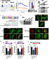
Figure 1
Figure 1
Phosphorylated PBF represses RAIU in breast cancer cells. (A) Left – schematic of the interaction between NIS and PBF proteins tagged at the C-terminus with the NanoLuc luciferase subunits LgBiT and SmBiT, respectively. In close proximity, LgBiT and SmBiT form a functional enzyme that uses the substrate furimazine to produce a high-intensity, luminescent signal. Created using Alphafold (59, 60) and BioRender. Right – NanoBiT analysis of NIS:PBF interaction in MCF-7 cells, with bar graph showing results at 20 min post-addition of Nano-Glo live cell assay substrate. (B) Co-immunoprecipitation assay showing PBF-HA and NIS-MYC interaction in MCF-7 breast cancer cells. (C) Schematic of PBF highlighting the overlapping phosphorylation site and endocytosis motif at the C-terminus, and the PBF-Y174A and E170_N172delinsAAA (PBF-EEN MUT) mutants. (D) Western blot showing the phosphorylation status of PBF-Y174A and PBF-EEN MUT compared with PBF-WT in MCF-7 cells. (E) Immunofluorescent images showing PBF-WT, PBF-Y174A and PBF-EEN MUT localisation in MCF-7 cells. (F) Immunofluorescent images showing subcellular localisation following the co-transfection of NIS-MYC (green) with VO, PBF-WT, PBF-Y174A or PBF-EEN MUT (red) in MCF-7 cells. Co-localisation is seen in yellow. (G) The effect of PBF and phosphomutants (Y174A/EEN-MUT) on RAIU in MCF-7 (i) and MDA-MB-231 (ii) cells transiently co-transfected with NIS-MYC. (H) The effect of PBF and phosphomutants (Y174A/EEN-MUT) on RAIU in MCF-7 cells treated with ATRA and dexamethasone. VO = vector only control. Bars = 10 μm. n = 3 for all experiments. Error bars = SEM. Significance shown compared with VO unless otherwise shown. ns = not significant (P > 0.05), * = P < 0.05, ** = P < 0.01, *** = P < 0.001.
Figure 2
Figure 2
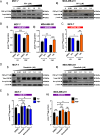
Src inhibition inhibits PBF phosphorylation and restores RAIU. (A) MCF-7 (i) and MDA-MB-231 (ii) cells were treated with varying doses of PP1 (0–2 μM) for 24 h before PBF-pY174 and total PBF expression levels were determined by Western blot. (B) Effect of 2 μM PP1 treatment on PBF-mediated RAIU repression in MCF-7 (i) and MDA-MB-231 (ii) cells transiently co-transfected with NIS-MYC. (C) Effect of 2 μM PP1 treatment on PBF-mediated RAIU repression in MCF-7 cells treated with ATRA and dexamethasone. (D) PBF-pY174 and total PBF expression levels following dasatinib dose response (0–2 μM) treatment for 24 h in MCF-7 (i) and MDA-MB-231 (ii) cells. (E) Effect of 1 nM dasatinib or 10 nM saracatinib treatment on PBF-repressed RAIU in MCF-7 (i) and MDA-MB-231 (ii) cells transiently co-transfected with NIS-MYC. n = 3 for all experiments. Error bars = SEM. * = P < 0.05, ** = P < 0.01, *** = P < 0.001.
Figure 3
Figure 3
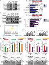
(A) Detection of PBF and PBF-pY174 levels following Src overexpression for 48 h in MCF-7 and MDA-MB-231 cells. (B) RAIU in ATRA/Dex-treated MCF-7 cells with stable expression of VO, PBF-WT, PBF-EEN-MUT or PBF-Y174A following transient transfection with Src or VO control. (C) Expression levels of PBF and PBF-pY174 in MCF-7 cells transiently transfected for 48 h with VO, Src-WT or the gatekeeper mutant Src-T341I and treated for 24 h with either DMSO or 10 nM dasatinib. (D) RAIU in ATRA/Dex-treated MCF-7 cells stably transfected with VO and PBF, transiently transfected with VO, Src-WT or Src-T341I for 48 h and treated with either DMSO or 10 nM dasatinib for 24 h. (E) Example of a heterozygous mutation induced by CRISPR-Cas9 targeting to PBF exon 1 in the MCF-7 PBF-KO #1 clonal cell line. (F) Confirmation of PBF deletion by Western blotting in MCF-7 and MDA-MB-231 PBF-KO cells generated using gRNA#1 (PBF-KO #1) and gRNA#2 (PBF-KO #2). (G) RAIU in parental MCF-7 cells and two PBF knockout cell lines (PBF-KO #1 and #2) following NIS, Src and NIS + Src overexpression. Confirmation of successful transfection shown below by Western blotting. (H and I) RAIU in parental MDA-MB-231 cells and PBF-KO cell lines (PBF-KO #1 and #2) following NIS, Src and NIS + Src overexpression. Confirmation of successful transfection shown below by Western blotting (J). n = 3 for all experiments. Error bars = SEM. Significance shown compared with VO unless otherwise shown. ns = not significant (P > 0.05), * = P < 0.05, ** = P < 0.01, *** = P < 0.001.
Figure 4
Figure 4
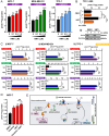
(A) RAIU in MCF-7 and MDA-MB-231 breast cancer cells and TPC-1 thyroid cancer cells transfected with NIS-MYC for 48 h and treated with varying doses of an NMT inhibitor (NMTi; 0–1 μM). The breast cancer cells were treated with NMTi for 4 h before RAIU and thyroid cancer cells were treated for 24 h. (B) RAIU in TPC-1 cells with stable NIS expression transfected with Src siRNA (or scrambled control) for 24 h before treatment with 1 μM NMTi (or DMSO vehicle control) for a further 24 h. (C) RAIU in parental MCF-7 (i), MDA-MB-231 (ii) and TPC-1 (iii) cells and two corresponding PBF knockout cell lines (PBF-KO #1 and #2) for each following VO or NIS transfection (48 h) and DMSO or 1 μM NMTi treatment (4 h – MCF-7/MDA-MB-231; 24 h – TPC-1). (D) TPC-1 cells were transfected with NIS-MYC for 48 h and then treated with DMSO, 1 nM dasatinib, 1 μM NMTi or a combination of the two drugs for 24 h before RAIU. (E) Schematic showing the NMT1/Src/PBF/NIS pathway. NMT1 myristoylates Src (1) and facilitates Src PM localisation and activation, resulting in PBF phosphorylation at Y174 (2). Phospho-PBF binds to NIS at the PM (3) leading to NIS internalisation and reduced RAIU (4). NMT1 inhibition (5) and Src inhibition (6) prevent PBF phosphorylation and NIS repression, while ATRA/Dex induces NIS expression and RAIU (7). n = 3 for all experiments. Error bars = SEM. Significance shown compared with VO and/or DMSO control unless otherwise shown. ns = not significant (P > 0.05), * = P < 0.05, ** = P < 0.01, *** = P < 0.001.
Figure 5
Figure 5
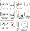
Elevated PBF and SRC expression in breast cancer. Box and whisker plots showing expression (log2) of PBF (A), SRC (B), NMT1 (C) and NIS (D) in various histotypes and molecular subtypes of the TGCA breast cancer (BRCA) RNA-Seq dataset versus normal (N; n = 112). BIDC = breast invasive ductal carcinoma (n = 812); BILC = breast invasive lobular carcinoma (n = 206); BIMMC = breast invasive mixed mucinous carcinoma (n = 16); BMDLC = breast mixed ductal and lobular carcinoma (n = 28); IBC = invasive breast carcinoma (n = 6); MBC = metaplastic breast cancer (n = 14); PDN = Paget’s disease of the nipple (n = 3); Luminal (ER+ and/or PR+; n = 565); HER2 = HER2-positive (n = 37); TNBC = triple negative breast cancer (n = 114). (E) Left – Box and whisker plots showing SRC and PBF expression in the BRCA cohort stratified by high versus low tumoural PBF/SRC expression in combination. Right – Correlation between those with high versus low tumoural PBF/SRC expression (n = 404/660). (F) Left – Proportion of molecular subtypes (%) within tumours with high versus low expression of both SRC and PBF. Right – Kaplan–Meier analysis of overall survival for BRCA patients with high versus low tumoural SRC and PBF expression. Number (n) of patients per sub-group. PL = log-rank test; PB = Breslow test. ns = not significant; ** = P < 0.01; *** = P < 0.001.

References

    1. Buffet C, Wassermann J, Hecht F, et al. Redifferentiation of radioiodine-refractory thyroid cancers. Endocr Relat Cancer 2020. 27 R113–R132. ( 10.1530/erc-19-0491) - DOI - PubMed
    1. Spitzweg C, Nelson PJ, Wagner E, et al. The sodium iodide symporter (NIS): novel applications for radionuclide imaging and treatment. Endocr Relat Cancer 2021. 28 T193–T213. ( 10.1530/erc-21-0177) - DOI - PubMed
    1. Tazebay UH, Wapnir IL, Levy O, et al. The mammary gland iodide transporter is expressed during lactation and in breast cancer. Nat Med 2000. 6 871–878. ( 10.1038/78630) - DOI - PubMed
    1. Wapnir IL, van de Rijn M, Nowels K, et al. Immunohistochemical profile of the sodium/iodide symporter in thyroid, breast, and other carcinomas using high density tissue microarrays and conventional sections. J Clin Endocrinol Metab 2003. 88 1880–1888. ( 10.1210/jc.2002-021544) - DOI - PubMed
    1. Knostman KA, McCubrey JA, Morrison CD, et al. PI3K activation is associated with intracellular sodium/iodide symporter protein expression in breast cancer. BMC Cancer 2007. 7 137. ( 10.1186/1471-2407-7-137) - DOI - PMC - PubMed

LinkOut - more resources