

Along-Tract Diffusion Alterations in the Dentato-Rubro-Thalamic Tract Correlate With Motor and Cognitive Decline in Huntington's Disease
- PMID: 40748199
- PMCID: PMC12315238
- DOI: 10.1002/hbm.70305
Along-Tract Diffusion Alterations in the Dentato-Rubro-Thalamic Tract Correlate With Motor and Cognitive Decline in Huntington's Disease
Abstract
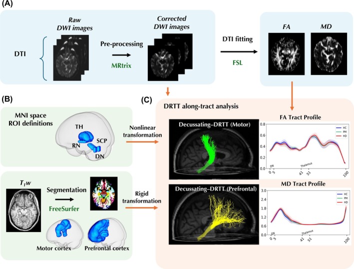
Huntington's disease (HD) is a progressive neurodegenerative disorder caused by cytosine-adenine-guanine repeat expansion in the huntingtin (HTT) gene, leading to widespread brain atrophy and white matter degeneration. Although cortico-striatal pathways have been extensively studied, the dentato-rubro-thalamic tract (DRTT), a key cerebellar efferent pathway integrating motor and cognitive functions, remains largely unexplored, despite increasing evidence of cerebellar involvement in these functions. By investigating microstructural alterations along the DRTT, we aim to elucidate its role in HD progression and its association with motor and cognitive impairments, providing insights into the potential contribution of the DRTT to disease severity and clinical outcomes. We retrospectively analyzed 1392 scans from 638 participants across three multinational HD cohorts (TRACK-HD/ON, PREDICT-HD, and IMAGE-HD) with standardized inclusion criteria, and applied the HD-ISS to categorize disease stages. Probabilistic tractography was performed on diffusion MRI data to reconstruct ipsilateral and decussating DRTT pathways ending in the motor or pre-frontal cortices, with fractional anisotropy (FA) and mean diffusivity (MD) values extracted along 100 nodes per tract. Along-tract analyses were conducted using linear mixed-effects models to assess group differences and correlations with motor and cognitive scores, while controlling for covariates. Significantly decreased FA and increased MD were observed in premanifest HD (PM, HD-ISS Stage 0 and 1) and manifest HD (HD, HD-ISS Stage 2 and 3) groups and over time compared to healthy controls (HC) across multiple regions along the DRTT, particularly in the dentate nucleus region and dentate nucleus-red nucleus projection. These microstructural changes were correlated to the greater motor and cognitive impairments. Conversely, the DRTT thalamo-cortical projection exhibited an opposite pattern, with higher FA in PM and HD than in HC. Both FA and MD were also positively correlated with motor score within this segment. Along-tract analysis revealed microstructural disruptions across DRTT in both premanifest and manifest HD individuals, suggesting that the DRTT plays a role in HD progression. Our findings also highlight the value of assessing regional changes along the tract. These segment-specific white matter alterations provide additional insights into HD pathology and may serve as biomarkers for motor and cognitive impairments in HD.
Keywords: Huntington's disease; cognitive impairment; dentato‐rubro‐thalamic‐tract; diffusion MRI; motor decline; tractography; white matter alterations.
© 2025 The Author(s). Human Brain Mapping published by Wiley Periodicals LLC.
Conflict of interest statement
The authors declare no conflicts of interest.
Figures


Similar articles
-
Mixed longitudinal and cross-sectional analyses of deep gray matter and white matter using diffusion weighted images in premanifest and manifest Huntington's disease.Neuroimage Clin. 2023;39:103493. doi: 10.1016/j.nicl.2023.103493. Epub 2023 Aug 9. Neuroimage Clin. 2023. PMID: 37582307 Free PMC article.
-
TRANSIENT ALTERATIONS IN THALAMO-CEREBELLAR FUNCTIONAL CONNECTIVITY IN PREMANIFEST HUNTINGTON'S DISEASE.medRxiv [Preprint]. 2025 Jan 15:2025.01.15.25320232. doi: 10.1101/2025.01.15.25320232. medRxiv. 2025. PMID: 39867412 Free PMC article. Preprint.
-
Upper Extremity Motor Performance During a Shirt Buttoning Task in Huntington's Disease.OTJR (Thorofare N J). 2025 Aug 16:15394492251355942. doi: 10.1177/15394492251355942. Online ahead of print. OTJR (Thorofare N J). 2025. PMID: 40817792
-
Physical exercise for people with Parkinson's disease: a systematic review and network meta-analysis.Cochrane Database Syst Rev. 2023 Jan 5;1(1):CD013856. doi: 10.1002/14651858.CD013856.pub2. Cochrane Database Syst Rev. 2023. Update in: Cochrane Database Syst Rev. 2024 Apr 08;4:CD013856. doi: 10.1002/14651858.CD013856.pub3. PMID: 36602886 Free PMC article. Updated.
-
Multi-domain interventions for the prevention of dementia and cognitive decline.Cochrane Database Syst Rev. 2021 Nov 8;11(11):CD013572. doi: 10.1002/14651858.CD013572.pub2. Cochrane Database Syst Rev. 2021. PMID: 34748207 Free PMC article.
References
-
- Bardile, C. F. , Garcia‐Miralles M., Caron N., et al. 2018. “A43 Intrinsic Mutant HTT‐Mediated Defects in Oligodendroglia Cells Contribute to Myelin Deficits and Behavioural Abnormalities in Huntington Disease.” Journal of Neurology, Neurosurgery, and Psychiatry 89, no. 1: A15–A16. 10.1136/jnnp-2018-EHDN.41. - DOI
MeSH terms
Grants and funding
LinkOut - more resources
Full Text Sources
Medical
Miscellaneous
