. 2025 Aug 12;97(31):16796-16804.
doi: 10.1021/acs.analchem.5c01702. Epub 2025 Aug 1.

Clear Native Gel Electrophoresis for the Purification of Fluorescently Labeled Membrane Proteins in Native Nanodiscs

Affiliations

Clear Native Gel Electrophoresis for the Purification of Fluorescently Labeled Membrane Proteins in Native Nanodiscs

Bence Ezsias et al. Anal Chem. .

Abstract

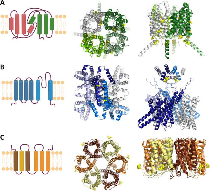
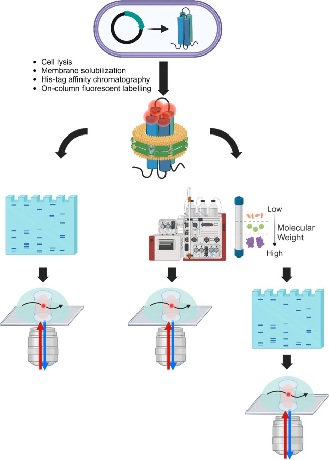
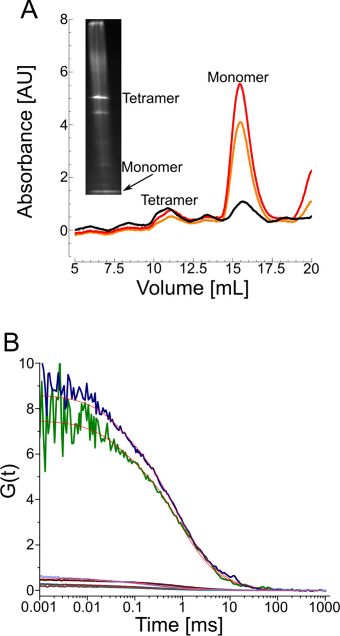
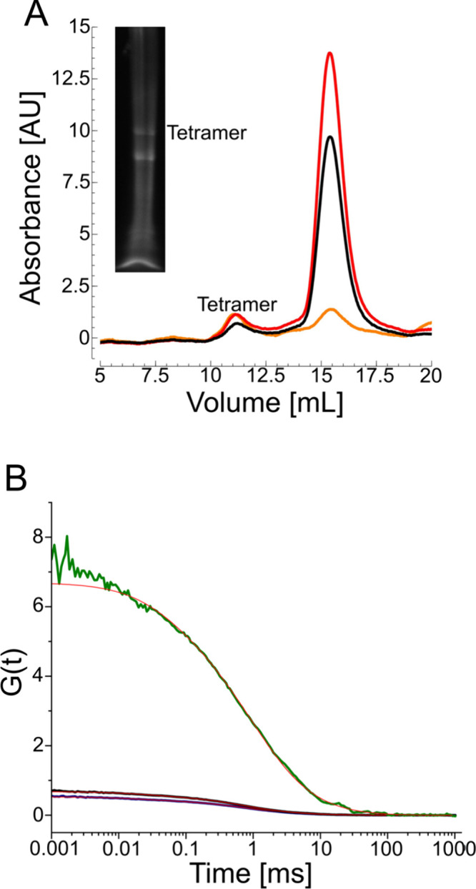
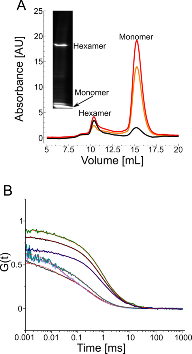
Native gel electrophoresis techniques, such as blue or clear native gel electrophoresis (BNE or CNE), are widely used to separate and characterize proteins. However, in high-resolution CNE, mild anionic or neutral detergents are often used at concentrations that are too low to prevent membrane-protein aggregation. Additionally, the identification of proteins is hampered by the lack of suitable molecular-weight markers such as those used in SDS-PAGE. Here, we introduce a novel approach that combines charged polymer-encapsulated nanodiscs and fluorescence correlation spectroscopy (FCS) to address both challenges. Membrane proteins are first extracted using Glyco-DIBMA, a negatively charged amphiphilic copolymer. This enables the spontaneous formation of nanodiscs harboring the fluorescently labeled target protein within a native-like lipid-bilayer environment, which is confirmed by FCS. The nanodiscs are then subjected to detergent-free CNE. As the number of protomers increases, the nanodiscs grow larger, resulting in increased migration distances in CNE due to higher charge densities. Crucially, the nanodiscs remain intact throughout the CNE, as demonstrated by FCS analysis of resolubilized bands excised from the gels. Moreover, the membrane proteins used in this study, a potassium channel (KvAP), a sodium channel (NavMs), a water channel (GlpF), and a urea channel (HpUreI), show only negligible aggregation, as evidenced by the fluorescent brightnesses and diffusion times of individual nanodiscs. In addition, the oligomeric states of membrane proteins can be deduced from the brightness per nanodisc. Since purified membrane proteins remain within a native-like lipid-bilayer environment and avoid detergent exposure, they are immediately suitable for downstream structural and functional studies.


Figures

1
1
Schematic representation of membrane proteins with different folds, shown in both monomeric and oligomeric forms. The oligomeric models were generated by using AlphaFold. This approach allows visualization of all labeling positions (yellow spheres), including those absent in the corresponding PDB structures. (A) Tetrameric glycerol uptake facilitator protein (GlpF) from . All six cysteine positions are shown, however not all can be labeled. (B) Tetrameric voltage-gated ion channels: KvAP from Aeropyrum pernix and NavMs from . Only the oligomeric model for KvAP is shown with a single cysteine at position 260. (C) Hexameric urea channel (HpUreI) from . L134C is the only available labeling position.
2
2
Schematic representation of the experimental workflow. Cell lysis and membrane solubilization by Glyco-DIBMA are followed by His-tag affinity chromatography and on-column labeling via the thiol reaction. The eluted protein is then either directly subjected to native PAGE or first to size-exclusion chromatography and then to native PAGE. The protein samples extracted from the gels and purified only by size-exclusion are analyzed by FCS.
3
3
Blue native PAGE (BNE) compared to size-exclusion chromatography (SEC) and fluorescence correlation spectroscopy (FCS) of the glycerol uptake facilitator, GlpF. (A) SEC and BNE fluorescent images show two major peaks, one for the tetramer (at 10–11 mL) and one for the monomer (at 15–16 mL). The black curve is the absorbance at 280 nm, the red curve at 650 nm, and the orange curve at 665 nm. (B) Autocorrelation functions of tetrameric (black) and monomeric (gray) GlpF measured after SEC; after extraction from the total protein fraction and separation by BNE (blue for tetramer and purple for monomer); or after extraction from SEC-purified fractions and separation by BNE (green for tetramer and cyan for monomer). Measured sample concentrations were 2.8 nM, 5.6 nM, 0.16 nM, 2.6 nM, 0.17 nM, and 7.6 nM, respectively. The estimated residence time and molecular brightness before BNE were 817 ± 85 μs and 33 ± 2 kHz for the tetramer and 360 ± 72.6 μs and 8.5 ± 0.15 kHz for the monomer. After BNE, these values were 854 ± 240 μs and 7.6 ± 0.9 kHz for the tetramer and 370 ± 48.2 μs and 3.3 ± 0.2 kHz for the monomer. The concentration of the tetramer extracted from the native gel was 0.16 nM.
4
4
High-resolution clear native PAGE (CNE) compared to size-exclusion chromatography (SEC) and fluorescence correlation spectroscopy (FCS) of the voltage-gated sodium ion channel NavMs. (A) SEC and high-resolution CNE fluorescent image of NavMs, labeled with Alexa Fluor 647 maleimide showing two major peaks and bands, one for a tetramer (at 10–11 mL) and one for smaller species (at 15–16 mL). The black curve is the absorbance at 280 nm, the red at 650 nm, and the orange at 665 nm. (B) Fitted autocorrelation functions of tetrameric NavMs measured after SEC (black); after extraction from the total protein fraction and separation by high-resolution CNE (blue); or after extraction from SEC-purified fractions and separation by high-resolution clear CNE (green). Measured sample concentrations were 1.75 nM, 3.1 nM, and 0.2 nM, respectively. The diffusion time and molecular brightness before CNE were 726 ± 37 μs and 23.4 ± 0.4 kHz, respectively. After CNE, these values were 740 ± 62 μs and 21.5 ± 0.9 kHz, respectively.
5
5
Detergent-free CNE of the urea channel HpUreI embedded in native nanodiscs compared to size-exclusion chromatography (SEC) and fluorescent correlation spectroscopy (FCS). (A) SEC and CNE fluorescent images of the total protein elution HpUreI; SEC shows two major peaks, one for a hexamer (at 10–11 mL) and one for a monomer (at 15–16 mL). The black curve is the absorbance at 280 nm, the red at 650 nm, and the orange at 665 nm. (B) Autocorrelation functions of hexameric (black) and monomeric (gray) HpUreI, measured after SEC; after extraction from the total protein fraction and separation by CNE (blue for the hexamer and purple for the monomer); or after extraction from SEC-purified fractions and separation by CNE (green for the hexamer and cyan for the monomer). Measured sample concentrations were 5.1 nM, 40 nM, 1.6 nM, 0.25 nM, 0.16 nM, and 0.5 nM, respectively. The diffusion time and molecular brightness before CNE were, respectively, 811 ± 58.7 μs and 36 ± 1.7 kHz for the tetramer and 320 ± 32.5 μs and 9 ± 0.5 kHz for the monomer. After CNE, these values were, respectively, 830 ± 84.2 μs and 37 ± 0.6 kHz for the tetramer and 270 ± 9.6 μs and 7 ± 0.2. kHz for the monomer.

Similar articles

  • Prescription of Controlled Substances: Benefits and Risks.
    Preuss CV, Kalava A, King KC. Preuss CV, et al. 2025 Jul 6. In: StatPearls [Internet]. Treasure Island (FL): StatPearls Publishing; 2025 Jan–. 2025 Jul 6. In: StatPearls [Internet]. Treasure Island (FL): StatPearls Publishing; 2025 Jan–. PMID: 30726003 Free Books & Documents.
  • Electrophoresis.
    Sonagra AD, Zubair M, Dholariya SJ. Sonagra AD, et al. 2025 Jul 14. In: StatPearls [Internet]. Treasure Island (FL): StatPearls Publishing; 2025 Jan–. 2025 Jul 14. In: StatPearls [Internet]. Treasure Island (FL): StatPearls Publishing; 2025 Jan–. PMID: 36251838 Free Books & Documents.
  • Home is where the lipids are: a comparison of MSP and DDDG nanodiscs for membrane protein research.
    Nakao K, Steinhauser A, Durand G, Soulié M, Rechberger GN, Züllig T, Keller S, Tych K. Nakao K, et al. Soft Matter. 2025 Aug 20;21(33):6596-6602. doi: 10.1039/d5sm00327j. Soft Matter. 2025. PMID: 40762966
  • Management of urinary stones by experts in stone disease (ESD 2025).
    Papatsoris A, Geavlete B, Radavoi GD, Alameedee M, Almusafer M, Ather MH, Budia A, Cumpanas AA, Kiremi MC, Dellis A, Elhowairis M, Galán-Llopis JA, Geavlete P, Guimerà Garcia J, Isern B, Jinga V, Lopez JM, Mainez JA, Mitsogiannis I, Mora Christian J, Moussa M, Multescu R, Oguz Acar Y, Petkova K, Piñero A, Popov E, Ramos Cebrian M, Rascu S, Siener R, Sountoulides P, Stamatelou K, Syed J, Trinchieri A. Papatsoris A, et al. Arch Ital Urol Androl. 2025 Jun 30;97(2):14085. doi: 10.4081/aiua.2025.14085. Epub 2025 Jun 30. Arch Ital Urol Androl. 2025. PMID: 40583613 Review.
  • Topical antiseptics for chronic suppurative otitis media.
    Head K, Chong LY, Bhutta MF, Daw J, Veselinović T, Morris PS, Vijayasekaran S, Schilder AG, Brennan-Jones CG. Head K, et al. Cochrane Database Syst Rev. 2025 Jun 9;6(6):CD013055. doi: 10.1002/14651858.CD013055.pub3. Cochrane Database Syst Rev. 2025. PMID: 40484403

References

    1. Laemmli U. K.. Cleavage of structural proteins during the assembly of the head of bacteriophage T4. Nature. 1970;227(5259):680–685. doi: 10.1038/227680a0. - DOI - PubMed
    1. Reinman S., Thornber J. P.. The electrophoretic isolation and partial characterization of three chlorophyll-protein complexes from blue-green algae. Biochim. Biophys. Acta. 1979;547(2):188–197. doi: 10.1016/0005-2728(79)90002-1. - DOI - PubMed
    1. Allen K. D., Staehelin L. A.. Resolution of 16 to 20 chlorophyll-protein complexes using a low ionic strength native green gel system. Anal. Biochem. 1991;194(1):214–222. doi: 10.1016/0003-2697(91)90170-X. - DOI - PubMed
    1. Tucker D. L., Sherman L. A.. Analysis of chlorophyll-protein complexes from the cyanobacterium Cyanothece sp. ATCC 51142 by non-denaturing gel electrophoresis. Biochim. Biophys. Acta. 2000;1468(1–2):150–160. doi: 10.1016/S0005-2736(00)00263-7. - DOI - PubMed
    1. Schägger H., von Jagow G.. Blue native electrophoresis for isolation of membrane protein complexes in enzymatically active form. Anal. Biochem. 1991;199(2):223–231. doi: 10.1016/0003-2697(91)90094-A. - DOI - PubMed

MeSH terms

LinkOut - more resources