. 2025 Aug 1;150(1):11.
doi: 10.1007/s00401-025-02918-y.

Myocardial sympathetic distal axon loss in subjects with Lewy pathology in three autopsy cohorts

Affiliations

Myocardial sympathetic distal axon loss in subjects with Lewy pathology in three autopsy cohorts

Ville Kivistö et al. Acta Neuropathol. .

Abstract

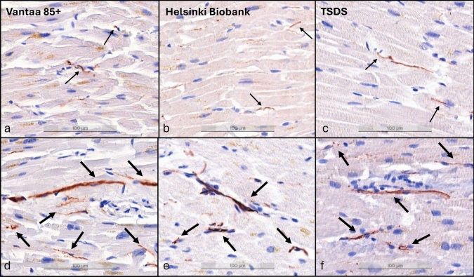
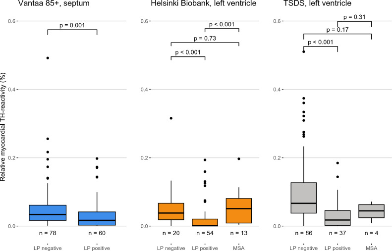
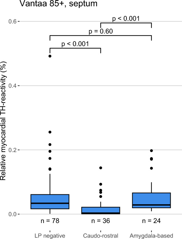
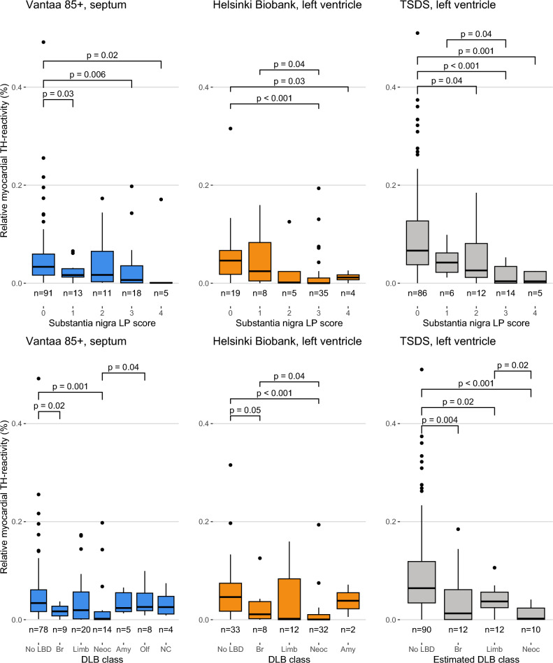
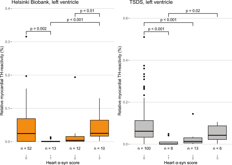
Cardiac manifestations are associated with Lewy body disease, but studies addressing the underlying histopathological mechanisms at the myocardial level are sparse. Here, we generated an artificial intelligence-based algorithm to quantify tyrosine hydroxylase (TH)-immunoreactive sympathetic distal axons at the myocardial level. This novel tool was applied to septal samples of the Vantaa 85 + study (n = 138), which is a population-based autopsy study representing all subjects aged 85 years or older living in the city of Vantaa (southern Finland) in 1991. In addition, the tool was applied to left ventricle samples of the Helsinki Biobank (n = 87) and the forensic Tampere Sudden Death Study (TSDS, n = 127). The level of myocardial TH reactivity was compared between subjects with and without Lewy pathology in the central nervous system in all datasets. In the Vantaa 85 + study, TH reactivity was also compared between subjects with caudo-rostral and amygdala-based subtypes, and potential confounding factors (age at death, sex, myocardial infarction, senile systemic amyloidosis, and diabetes medication) were controlled for using multiple linear regression models. Presence of Lewy pathology was strongly associated with loss of TH reactivity at the myocardial level in all three autopsy cohorts (Vantaa 85 + p = 0.001, Helsinki Biobank p < 0.001, TSDS p < 0.001)). In the Vantaa 85 + study, the caudo-rostral subtype (p < 0.001), but not the amygdala-based subtype (p = 0.60), was associated with myocardial denervation/dysfunction, and this association was independent of other known causes of sympathetic denervation/dysfunction. Caudo-rostral subtype and myocardial infarction were the strongest predictors of myocardial sympathetic denervation/dysfunction in the oldest-old population (Vantaa 85 +). In conclusion, our results show that Lewy pathology in the central nervous system, and particularly its caudo-rostral subtype, is strongly associated with loss of sympathetic distal axons at the myocardial level. We also provide evidence that the caudo-rostral subtype is one of the strongest predictors of myocardial sympathetic denervation/dysfunction in the oldest-old population.

Keywords: Image recognition tool; Lewy body disease; Myocardium; Neurodegeneration; Neuropathology; Post-mortem.


Conflict of interest statement

Declarations. Conflict of interest: P.J.T. holds a patent on C9orf72 in the diagnostics and treatment of ALS/FTD and has made paid consultations for Roche, Biogen, Merck, Lundbeck, Sanofi-Genzyme and Novartis unrelated to the subject of this study. H.P. has provided paid pathology consulting services for Aiforia Technologies Oyj in unrelated projects. All other authors report no competing interests. Ethical approval: All procedures performed in studies involving human participants were in accordance with the ethical standards of the institutional and/or national committee and with the 1964 Helsinki Declaration and its later amendments or comparable ethical standards. Consent to participate: In the Vantaa 85 + study, each participant and/or their relatives have given informed consent for the study and relatives have given written consent for autopsy. In accordance with the Finnish national ethical standards and the Act on the Medical Use of Human Organs, Tissues and Cells (101/2001), which allow using post-mortem samples collected as part of medical or forensic autopsies in research, informed consent was not obtained (or required) for the Helsinki Biobank and TSDS cohorts.

Figures

Fig. 1
Fig. 1
Example myocardial samples with decreased and preserved TH reactivity. a-c: subjects with decreased TH reactivity (thin arrows). d-f: subjects with preserved TH reactivity (thick arrows). Pictures taken at 200 × magnification. Scale bar shown at the bottom of each image. TH tyrosine hydroxylase
Fig. 2
Fig. 2
Relationship between relative myocardial TH reactivity and CNS Lewy pathology and MSA. Relative myocardial TH reactivity is defined as the area of TH-positive staining divided by analyzed tissue area, excluding the tissue and TH-positive staining in larger nerve fascicles (diameter > 20 µm). Box-and-whiskers plots with median, 1st to 3rd interquartile range, and minimum and maximum. Outliers shown as dots. P values for group differences calculated using Mann–Whitney U test. TH tyrosine hydroxylase, CNS central nervous system, LP positive CNS Lewy pathology-positive, LP negative CNS Lewy pathology-negative, MSA multiple system atrophy
Fig. 3
Fig. 3
Relationship between the LBD subtypes and relative myocardial TH reactivity in Vantaa 85 + subjects. Relative myocardial TH reactivity is defined as the area of TH-positive staining divided by analyzed tissue area, excluding the tissue and TH-positive staining in larger nerve fascicles (diameter > 20 µm). Box-and-whiskers plot with median, 1st to 3rd interquartile range, and minimum and maximum. Outliers shown as dots. P values calculated using Mann–Whitney U test. LBD Lewy body disease, TH tyrosine hydroxylase, CNS central nervous system, LP negative CNS Lewy pathology-negative
Fig. 4
Fig. 4
Relative myocardial TH reactivity compared to the semi-quantitative substantia nigra LP scores and DLB consortium classification. Relative myocardial TH reactivity is defined as the area of TH-positive staining divided by analyzed tissue area, excluding the tissue and TH-positive staining in larger nerve fascicles (diameter > 20 µm). Box-and-whiskers plots with median, 1st to 3rd interquartile range, and minimum and maximum. Outliers shown as dots. P values calculated using Mann–Whitney U test, only comparisons where P < 0.05 are shown. MSA cases are excluded in the substantia nigra LP analyses. Substantia nigra was missing for three non-MSA Helsinki Biobank cases. In TSDS, the estimated DLB consortium class is based on LP positivity in the substantia nigra, hippocampus and frontal cortex (No LBD = SN-, HC-, FC-; Br = SN + , HC-, FC-; Limb = SN + , HC + , FC-; Neoc = SN + , HC + , FC +). Cases with any of SN, HC or FC missing are excluded (n = 3). SN substantia nigra, HC hippocampus, FC frontal cortex, DLB dementia with Lewy bodies, LBD Lewy body disease, Br brainstem-predominant, Limb limbic, Neoc diffuse neocortical, Amy Amygdala-predominant, Olf olfactory bulb only, NC non-classifiable
Fig. 5
Fig. 5
Severity of α-syn pathology in the heart compared to relative myocardial TH reactivity. Relative myocardial TH reactivity is defined as the area of TH-positive staining divided by analyzed tissue area, excluding the tissue and TH-positive staining in larger nerve fascicles (diameter > 20 µm). Box-and-whiskers plots with median, 1st to 3rd interquartile range, and minimum and maximum. Outliers shown as dots. α-syn pathology in the heart was scored on a scale of 0–3 (0 = none; 1 = focal, sparse; 2 = local, moderate; 3 = widespread, severe). Data of the septal samples of the Vantaa 85 + study are not shown due to very low number of cases with cardiac α-syn pathology. P values calculated using Mann–Whitney U test, only comparisons where P < 0.05 are shown

Similar articles

References

    1. Amino T, Orimo S, Itoh Y, Takahashi A, Uchihara T, Mizusawa H (2006) Profound cardiac sympathetic denervation occurs in Parkinson disease. Brain Pathol 15:29. 10.1111/J.1750-3639.2005.TB00097.X - PMC - PubMed
    1. Andersen KB, Krishnamurthy A, Just MK, Van Den Berge N, Skjærbæk C, Horsager J et al (2025) Sympathetic and parasympathetic subtypes of body-first Lewy body disease observed in postmortem tissue from prediagnostic individuals. Nat Neurosci. 10.1038/S41593-025-01910-9 - PMC - PubMed
    1. Borghammer P (2021) The α-Synuclein origin and connectome model (SOC Model) of Parkinson’s disease: explaining motor asymmetry, non-motor phenotypes, and cognitive decline. J Parkinsons Dis 11:455. 10.3233/JPD-202481 - PMC - PubMed
    1. Borghammer P, Van Den Berge N (2019) Brain-first versus gut-first Parkinson’s disease: a hypothesis. J Parkinsons Dis 9:S281–S295. 10.3233/JPD-191721 - PMC - PubMed
    1. Borghammer P, Horsager J, Andersen K, Van Den Berge N, Raunio A, Murayama S et al (2021) Neuropathological evidence of body-first vs. brain-first Lewy body disease. Neurobiol Dis 161:105557. 10.1016/J.NBD.2021.105557 - PubMed

Substances