Review
. 2025 Aug 1:10.1152/physiol.00015.2025.
doi: 10.1152/physiol.00015.2025. Online ahead of print.

Brain Capillary Ion Channels: Physiology and Channelopathies

Affiliations
Review

Brain Capillary Ion Channels: Physiology and Channelopathies

Osama F Harraz et al. Physiology (Bethesda). .

Abstract

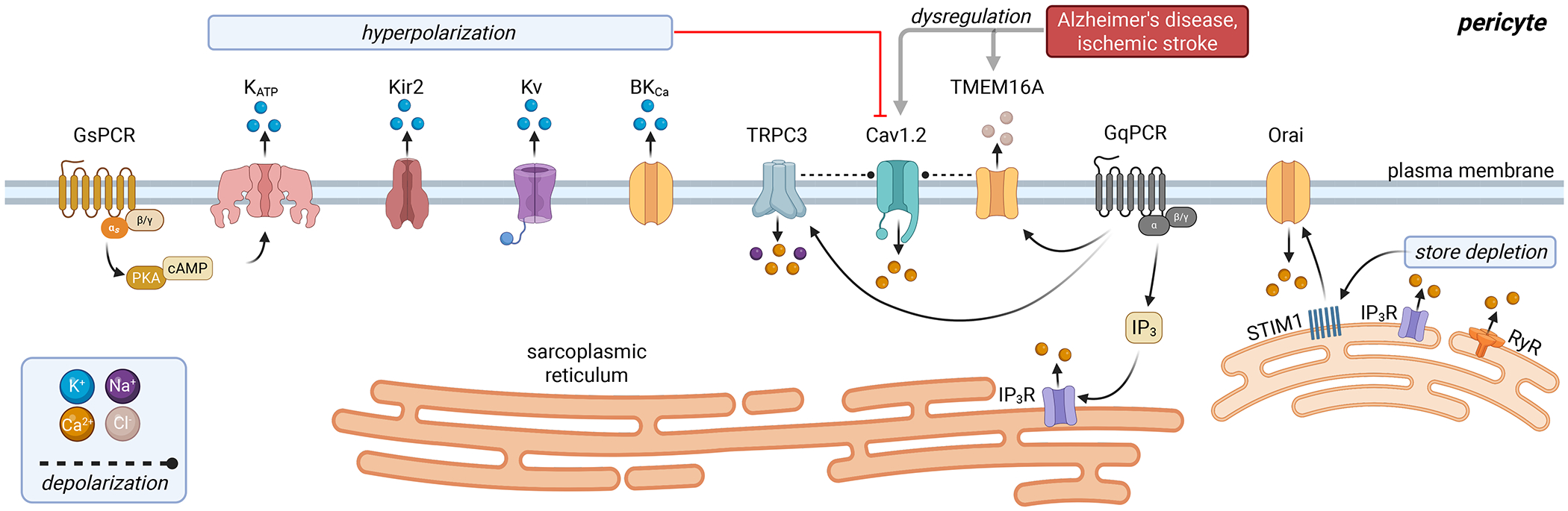
The brain relies on an intricate vascular network to deliver oxygen and nutrients through functional hyperemia, a process critical for matching blood flow to neuronal activity. This review explores the roles of ion channels in brain capillary endothelial cells and pericytes, focusing on their contributions to neurovascular coupling. Key endothelial ion channels, including Kir2.1, KATP, TRPV4, TRPA1, and Piezo1, regulate membrane potential and calcium dynamics, facilitating rapid electrical and chemical signaling that modulates blood flow. Pericytes, categorized as ensheathing and thin-strand, express ion channels such as KATP, voltage-gated calcium channels, TRPC, and TMEM16A, which govern contractility and orchestrate blood flow responses. Additionally, we discuss channelopathies in conditions like Alzheimer's disease, cerebral small vessel diseases, hypertension, and ischemic stroke, where ion channel dysfunction impairs brain blood flow regulation. Emerging evidence underscores the therapeutic potential of targeting capillary ion channels to restore neurovascular function in these disorders.

Keywords: brain capillaries; cerebral blood flow; endothelial cells; ion channels; pericytes.


Conflict of interest statement

DECLARATION OF INTERESTS

The authors declare no competing interests.

Figures

Figure 1.
Figure 1.. Simplified anatomy of the brain vasculature.
Pial arteries run on the brain surface, from which penetrating arterioles orthogonally dive into the brain parenchyma. Penetrating arterioles branch at regular intervals forming capillary networks that lie near all neurons (not shown). Capillary networks are primarily comprised of endothelial cells (ECs) throughout the network, and pericytes that are embedded in the capillary basement membrane. Pericyte characteristics and morphology change with the capillary branch order: proximal capillaries (~1st-4th order) are covered with ensheathing (transition zone) pericytes, and deeper capillaries are covered with thin-strand (distal) pericytes.
Figure 2.
Figure 2.. Capillary endothelial ion channels.
Kir2.1 channels are expressed in capillary ECs and are activated by neuronal derived extracellular K+. KATP channels are also expressed but are rather activated downstream of GsPCR stimulation. K+ efflux—through Kir2.1 and/or KATP channels—hyperpolarizes ECs, and the electrical signal is transmitted upstream through gap junctions to dilate arterioles and increase blood flow. The Ca2+-permeable TRPV4 channel is activated downstream to GqPCR stimulation. Endothelial Ca2+ events are also mediated by IP3-mediated Ca2+ release from the intracellular stores. Hyperpolarization, by Kir2.1 and KATP channels, influence endothelial Ca2+ dynamics by increasing the driving force of Ca2+ entry (electrocalcium coupling). TRPA1 channel is also present in capillary ECs, where neuronally mediated TRPA1 activation leads to Ca2+ influx to activate Pannexin-1 channel and then purinergic receptors to facilitate additional Ca2+ influx. The resulting Ca2+ wave propagates to upstream segments to hyperpolarize them and relax smooth muscle cells. Capillary ECs expresses the mechanosensor Piezo1. During functional hyperemia, Piezo1 activation could depolarize ECs, counteract endothelial hyperpolarization and therefore help repolarize ECs. Several pathologies are associated with Kir2.1 dysfunction in brain capillaries, which in turn is involved in the associated CBF defects.
Figure 3.
Figure 3.. Ion channels in brain capillary pericytes.
Several K+ channels have been reported in capillary pericytes, among which KATP is highly expressed. GsPCR activation in distal pericytes activates KATP channels and hyperpolarizes the cell. Pericyte hyperpolarization is transmitted to adjacent ECs to activate Kir2.1 channels; retrograde hyperpolarization subsequently dilates feeding arterioles. Pericytes also express Kir2, Kv and BKCa channels, but their exact roles in CBF regulation are not clear. Pericyte GqPCR activation is associated with increased cytosolic [Ca2+], not only through downstream IP3R activation but also via several other ion channels (Cav1.2, TMEM16A, TRPC3, Orai). VGCCs are the main Ca2+ influx pathway in ensheathing pericytes. TMEM16A and TRPC3 potentiate VGCC function, by depolarizing pericytes. TMEM16A is activated via IP3-mediated Ca2+ release from the sarcoplasmic reticulum stores, and TRPC3 is likely activated downstream of mechanosensitive GqPCR. The role of Cav1.2 in Ca2+ influx is minimal in distal thin-strand pericytes, a function that is likely mediated via store-operated Ca2+ entry, where STIM1/Orai signaling transduces when Ca2+ stores are depleted. Pericyte contraction leading to capillary constriction has been observed in AD and ischemic stroke. The excessive contraction involves TMEM16A upregulation, and excessive Ca2+ influx via VGCCs. Cav1.2 blockers decrease capillary stalling and enhance CBF in AD models, highlighting their involvement in AD pathogenesis.

Similar articles

References

    1. Iadecola C The Neurovascular Unit Coming of Age: A Journey through Neurovascular Coupling in Health and Disease. Neuron 96: 17–42, 2017. doi: 10.1016/j.neuron.2017.07.030. - DOI - PMC - PubMed
    1. Schaeffer S, Iadecola C. Revisiting the neurovascular unit. Nat Neurosci 24: 1198–1209, 2021. doi: 10.1038/S41593-021-00904-7. - DOI - PMC - PubMed
    1. Blinder P, Tsai PS, Kaufhold JP, Knutsen PM, Suhl H, Kleinfeld D. The cortical angiome: an interconnected vascular network with noncolumnar patterns of blood flow. Nat Neurosci 16: 889–897, 2013. doi: 10.1038/nn.3426. - DOI - PMC - PubMed
    1. Garcia DCG, Longden TA. Ion channels in capillary endothelium. Curr Top Membr 85: 261–300, 2020. doi: 10.1016/bs.ctm.2020.01.005. - DOI - PubMed
    1. Hariharan A, Weir N, Robertson C, He L, Betsholtz C, Longden TA. The Ion Channel and GPCR Toolkit of Brain Capillary Pericytes. Front Cell Neurosci 14, 2020. doi: 10.3389/FNCEL.2020.601324. - DOI - PMC - PubMed

LinkOut - more resources