

Protocol to derive postganglionic parasympathetic neurons using human pluripotent stem cells for electrophysiological and functional assessment
- PMID: 40748761
- PMCID: PMC12337169
- DOI: 10.1016/j.xpro.2025.104001
Protocol to derive postganglionic parasympathetic neurons using human pluripotent stem cells for electrophysiological and functional assessment
Abstract
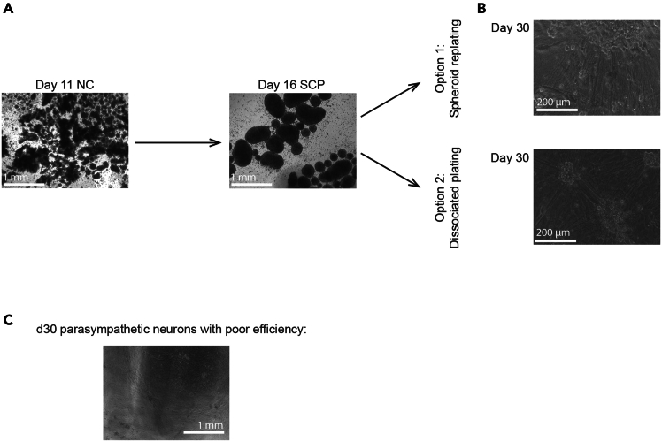
Accessing untransformed human primary parasympathetic neurons is extremely challenging due to their inaccessibility in patients. Therefore, a human pluripotent stem cell-based approach to generate pure parasympathetic neurons is valuable for studying their development, maturation, and pathology. Here, we present a protocol to derive parasympathetic neurons from human pluripotent stem cells. We describe steps for human pluripotent stem cell replating, neural crest replating, cryobanking, and parasympathetic neuron differentiation. We then detail procedures for functional assays via multielectrode array (MEA) analysis. For complete details on the use and execution of this protocol, please refer to Wu et al.1.
Keywords: Developmental biology; Neuroscience; Stem Cells.
Copyright © 2025 The Author(s). Published by Elsevier Inc. All rights reserved.
Conflict of interest statement
Declaration of interests A US patent application entitled “Composition and methods for making parasympathetic neurons” was filed under U.S.S.N. 18/503,100.
Figures


Similar articles
-
Protocol for the induction of human spinal motor neurons from human induced pluripotent stem cells for studying amyotrophic lateral sclerosis.STAR Protoc. 2025 Aug 5;6(3):104016. doi: 10.1016/j.xpro.2025.104016. Online ahead of print. STAR Protoc. 2025. PMID: 40773352 Free PMC article.
-
Protocol for generating postganglionic sympathetic neurons using human pluripotent stem cells for electrophysiological and functional assessments.STAR Protoc. 2024 Jun 21;5(2):102970. doi: 10.1016/j.xpro.2024.102970. Epub 2024 Mar 21. STAR Protoc. 2024. PMID: 38517897 Free PMC article.
-
How to differentiate induced pluripotent stem cells into sensory neurons for disease modelling: a functional assessment.Stem Cell Res Ther. 2024 Apr 5;15(1):99. doi: 10.1186/s13287-024-03696-2. Stem Cell Res Ther. 2024. PMID: 38581069 Free PMC article.
-
Interventions to reduce harm from continued tobacco use.Cochrane Database Syst Rev. 2016 Oct 13;10(10):CD005231. doi: 10.1002/14651858.CD005231.pub3. Cochrane Database Syst Rev. 2016. PMID: 27734465 Free PMC article.
-
Advancing Parkinson's disease treatment: cell replacement therapy with neurons derived from pluripotent stem cells.Stem Cells. 2024 Sep 10;42(9):781-790. doi: 10.1093/stmcls/sxae042. Stem Cells. 2024. PMID: 38902932 Review.
References
-
- Wu H.F., Saito-Diaz K., Huang C.W., McAlpine J.L., Seo D.E., Magruder D.S., Ishan M., Bergeron H.C., Delaney W.H., Santori F.R., et al. Parasympathetic neurons derived from human pluripotent stem cells model human diseases and development. Cell Stem Cell. 2024;31:734–753.e8. doi: 10.1016/j.stem.2024.03.011. - DOI - PMC - PubMed
-
- Goldsteen P.A., Sabogal Guaqueta A.M., Mulder P.P.M.F.A., Bos I.S.T., Eggens M., Van der Koog L., Soeiro J.T., Halayko A.J., Mathwig K., Kistemaker L.E.M., et al. Differentiation and on axon-guidance chip culture of human pluripotent stem cell-derived peripheral cholinergic neurons for airway neurobiology studies. Front. Pharmacol. 2022;13 doi: 10.3389/fphar.2022.991072. - DOI - PMC - PubMed
LinkOut - more resources
Full Text Sources
