. 2025 Aug 1;20(8):e0329626.
doi: 10.1371/journal.pone.0329626. eCollection 2025.

Identification of the Francisella novicida FTN_0096 as a factor involved in intracellular replication and host response

Affiliations

Identification of the Francisella novicida FTN_0096 as a factor involved in intracellular replication and host response

Dhandy Koesoemo Wardhana et al. PLoS One. .

Abstract

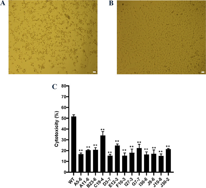
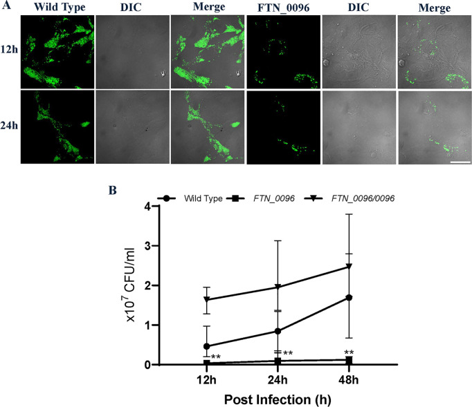
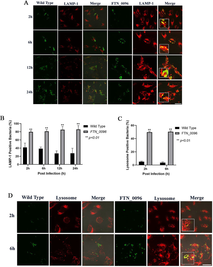
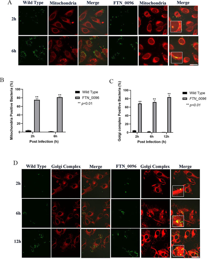
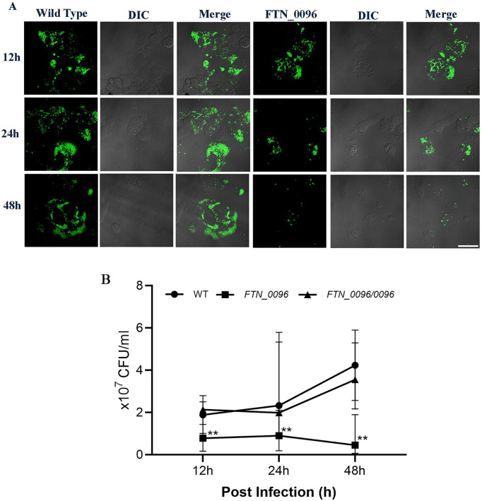
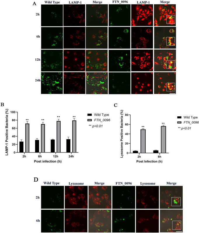
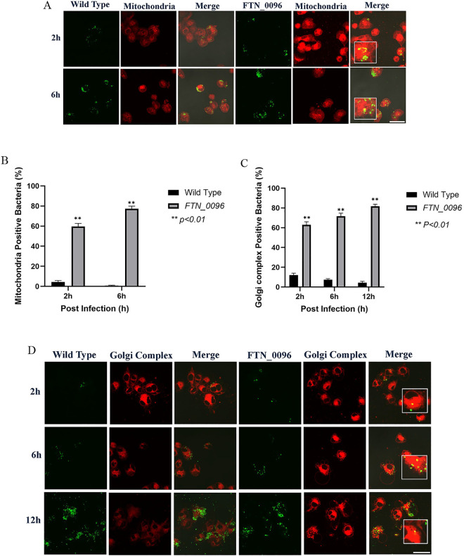
Francisella tularensis is the causative agent of the zoonotic disease tularemia. We investigated a pathogenic factor of F. tularensis subsp. novicida (F. novicida). Accordingly, we established a novel infection model using HeLa cells. F. novicida usually infects macrophage lineage cells and less frequently epithelial cells. We successfully infected HeLa cells expressing the Fc receptor (HeLa-FcγRII cells) using F. novicida supplemented with mouse serum containing F. novicida antibodies. A total of 2,232 transposon mutants of F. novicida were screened to determine the relatively fewer cytotoxic strains of the HeLa-FcγRII cells, and 13 strains were thus isolated. Sequencing analysis of transposon insertion sites identified 13 genes, including FTN_0096. We focused on FTN_0096. Although the F. novicida wild-type strain proliferated in HeLa-FcγRII and THP-1 cells, the number of intracellular FTN_0096 mutant decreased. FTN_0096 mutant cannot escape from phagolysosomes in the initial phases of infection. Moreover, FTN_0096 mutant was detected in the mitochondria and Golgi complex. These findings indicate the importance of FTN_0096 of F. novicida for intracellular replication in the cells.


Conflict of interest statement

The authors have declared that no competing interests exist.

Figures

Fig 1
Fig 1. Screening of the Transposon Mutant Library.
(A) HeLa–FcγRII cells were infected with F. novicida wild-type strain, preincubated with mouse serum containing F. novicida antibodies and incubated at 37°C for 24 h. Scale bar = 20 μm. (B) HeLa–FcγRII cells were infected with E12-3 transposon mutant of F. novicida, preincubated with mouse serum containing F. novicida antibodies, and incubated at 37°C for 24 h. Scale bar = 20 μm. (C) The transposon mutant library was screened using LDH cytotoxicity assay. HeLa–FcγRII cells were infected with the wild-type strain of F. novicida and transposon mutants of F. novicida preincubated with mouse serum containing F. novicida antibodies and incubated at 37°C for 24 h. LDH release was measured as an indicator of cytotoxicity. Data represent the mean and standard deviations of three identical experiments. Significant differences were evaluated in comparison to the wild-type strain using multiple comparison analyses as indicated by asterisks, **P <0.01.
Fig 2
Fig 2. Intracellular growth and cytotoxicity of the wild-type strain and FTN_0096 mutant in HeLa–FcγRII cells.
(A) HeLa–FcγRII cells were infected with F. novicida wild-type strain and FTN_0096 mutant, preincubated with mouse serum containing F. novicida antibodies at MOI = 1, and following treatment with gentamicin (50 μg/mL) for 1 h. The cells were fixed and examined at 12 and 24 h post-infection. Scale bar = 20 μm. (B) HeLa–FcγRII cells were infected with F. novicida wild-type, FTN_0096 mutant, and complementary strain (FTN_0096/0096), preincubated with mouse serum containing F. novicida antibodies at MOI = 1, and following treatment with 50 μg/mL of gentamicin for 1 h. At 12, 24, and 48 h post-infection, cells were lysed with 0.1% Triton X-100, and plating serial dilutions on BHIc agar. Data represent the mean and standard deviations of three identical experiments. Significant differences were evaluated in comparison to the wild-type strain using multiple comparison analyses as indicated by asterisks, **P <0.01.
Fig 3
Fig 3. Recognition of F. novicida strains by lysophagosomes and lysosomes in HeLa–FcγRII cells.
(A) HeLa–FcγRII cells were infected with F. novicida wild-type strain and FTN_0096 mutant preincubated with mouse serum containing F. novicida antibodies at MOI = 1, and following treatment with gentamicin (50 μg/mL) for 1 h. At 2 to 24 h post-infection, cells were treated with an anti-LAMP-1 antibody and visualized using a TRITC-conjugated anti-rat IgG. Scale bar = 20 μm. (B) The percentage of F. novicida wild-type strain and FTN_0096 mutant colocalized with LAMP-1. The data represent the mean and standard deviation from three identical experiments. Significant differences were evaluated in comparison to the wild-type strain using multiple comparison analyses as indicated by asterisks, **P < 0.01. (C) The percentage of F. novicida wild-type strain and FTN_0096 mutant colocalized with lysosomes. The data represent the mean and standard deviation from three identical experiments. Significant differences were evaluated in comparison to the wild-type strain using multiple comparison analyses as indicated by asterisks, **P < 0.01. (D) HeLa–FcγRII cells were infected with F. novicida wild-type strain and FTN_0096 mutant preincubated with mouse serum containing F. novicida antibodies at MOI = 1, and following treatment with gentamicin (50 μg/mL) for 1 h. At 2 to 6 h post-infection, cells were stained with LysoTracker™ Red DND-99 to visualize lysosomes. Scale bar = 20 μm.
Fig 4
Fig 4. Recognition of F. novicida strains by the mitochondria and Golgi complex in HeLa–FcγRII cells.
(A) HeLa–FcγRII cells were infected with F. novicida wild-type strain and FTN_0096 mutant preincubated with mouse serum containing F. novicida antibodies at MOI = 1, and following treatment with gentamicin (50 μg/mL) for 1 h. Cells were stained with Mitotracker® Deep Red FM at 2 and 6 h post-infection. Scale bar = 20 μm. (B) Percentage of F. novicida wild-type strain and FTN_0096 mutant that colocalized with the mitochondria. The data represent the mean and standard deviation from three identical experiments. Significant differences were evaluated in comparison to the wild-type strain using multiple comparison analyses as indicated by asterisks,**P < 0.01. (C) The percentage of F. novicida wild-type strain and FTN_0096 mutant that colocalized with the Golgi complex. The data represent the mean and standard deviation from three identical experiments. Significant differences were evaluated in comparison to the wild-type strain using multiple comparison analyses as indicated by asterisks, **P < 0.01. (D) HeLa–FcγRII cells were infected with F. novicida wild-type strain and FTN_0096 mutant preincubated with mouse serum containing F. novicida antibodies at MOI = 1, and treated with 50 μg/mL gentamicin. At 2, 6, and 12 h post-infection, cells were stained with BODIPY TR. Scale bar = 20 μm.
Fig 5
Fig 5. Intracellular growth of the wild-type strain and FTN_0096 mutant in THP-1 cells.
(A) THP-1 cells were infected with F. novicida wild-type strain and FTN_0096 mutant at MOI = 1, following treatment with gentamicin (50 μg/mL) for 1 h. Cells were fixed and examined at 12, 24, and 48 h post-infection. Scale bar = 20 μm. (B) THP-1 cells were infected with F. novicida wild-type, FTN_0096 mutant, and FTN_0096/0096 at MOI = 1, and following treatment with gentamicin (50 μg/mL) for 1 h. At 12, 24, and 48 h post-infection, cells were lysed with 0.1% Triton X-100 and plating serial dilutions on BHIc agar. The data represent the mean and standard deviation from three identical experiments. Significant differences were evaluated in comparison to the wild-type strain using multiple comparison analyses as indicated by asterisks, **P <0.01.
Fig 6
Fig 6. Recognition of F. novicida strains by lysophagosomes and lysosomes in THP-1 cells.
(A) THP-1 cells were infected with F. novicida wild-type strain and FTN_0096 mutant at MOI = 1, following treatment with gentamicin (50 μg/mL) for 1 h. Cells were treated with an anti-LAMP-1 antibody and stained with a TRITC-conjugated anti-rat IgG at 2–24 h after the infection. Scale bar = 20 μm. (B) The percentage of F. novicida wild-type strain and FTN_0096 mutant colocalized with LAMP-1. The data represent the mean and standard deviation from three identical experiments. Significant differences were evaluated in comparison to the wild-type strain using multiple comparison analyses as indicated by asterisks, **P < 0.01. (C) The percentage of F. novicida wild-type strain and FTN_0096 mutant colocalized with lysosomes. The data represent the mean and standard deviation from three identical experiments. Significant differences were evaluated in comparison to the wild-type strain using multiple comparison analyses as indicated by asterisks, **P < 0.01. (D) THP-1 cells were infected with F. novicida wild-type strain and FTN_0096 mutant MOI = 1, following treatment with gentamicin (50 μg/mL) for 1 h. Cells were treated with LysoTracker™ Red DND-99 from 2 to 6 h after the infection. Scale bar = 20 μm.
Fig 7
Fig 7. Recognition of F. novicida strains by the mitochondria and Golgi complex in THP-1 cells.
(A) THP-1 cells were infected with F. novicida wild-type strain and FTN_0096 mutant, MOI = 1, and following treatment with gentamicin (50 μg/mL) for 1 h. Cells were stained with the Mitotracker® Deep Red FM at 2 and 6 h post-infection. Scale bar = 20 μm. (B) The percentage of F. novicida wild-type strain and FTN_0096 mutant that colocalized with mitochondria. The data represent the mean and standard deviation from three identical experiments. Significant differences were evaluated in comparison to the wild-type strain using multiple comparison analyses as indicated by asterisks, **P < 0.01. (C) The percentage of F. novicida wild-type strain and FTN_0096 mutant that colocalized with the Golgi complex. The data represent the mean and standard deviation from three identical experiments. Significant differences were evaluated in comparison to the wild-type strain using multiple comparison analyses as indicated by asterisks, **P < 0.01. (D) THP-1 cells were infected with F. novicida wild-type strain and FTN_0096, MOI = 1, and following treatment with gentamicin (50 μg/mL) for 1 h. Cells were stained with BODIPY TR at 2, 6, and 12 h post-infection. Scale bar = 20 μm.

Similar articles

References

    1. Petersen JM, Mead PS, Jameson JL, Fauci AS, Kasper DL, Hauser SL. Tularemia. In: Jameson JL, Fauci AS, Kasper DL, Hauser SL, Longo DL, Loscalzo J, editors. Harrison’s principles of internal medicine. 20e ed. New York, NY: McGraw-Hill Education; 2018. p. 1196–9.
    1. Nelson C, Kugeler K, Petersen J, Mead P. Tularemia-United States, 2001—2010. MMWR Morb Mortal Wkly Rep. 2013;62:963–6. - PMC - PubMed
    1. Telford SR 3rd, Goethert HK. Ecology of Francisella tularensis. Annu Rev Entomol. 2020;65:351–72. doi: 10.1146/annurev-ento-011019-025134 - DOI - PMC - PubMed
    1. Freudenberger Catanzaro KC, Inzana TJ. The Francisella tularensis Polysaccharides: What Is the Real Capsule? Microbiol Mol Biol Rev. 2020;84(1):e00065-19. doi: 10.1128/MMBR.00065-19 - DOI - PMC - PubMed
    1. Yeni DK, Büyük F, Ashraf A, Shah MSUD. Tularemia: a re-emerging tick-borne infectious disease. Folia Microbiol (Praha). 2021;66(1):1–14. doi: 10.1007/s12223-020-00827-z - DOI - PMC - PubMed

Substances

Supplementary concepts

LinkOut - more resources