. 2025 Aug;11(31):eadv2902.
doi: 10.1126/sciadv.adv2902. Epub 2025 Aug 1.

Glia-derived noncanonical fatty acid binding protein modulates brain lipid storage and clearance

Affiliations

Glia-derived noncanonical fatty acid binding protein modulates brain lipid storage and clearance

Jun Yin et al. Sci Adv. 2025 Aug.

Abstract

Glia-derived secretory factors are essential for brain development, physiology, and homeostasis, with their dysfunction linked to a variety of neurological disorders. Through genetic and biochemical approaches, we identified odorant binding protein 44a (Obp44a), a noncanonical α-helical fatty acid binding protein (FABP) highly expressed in Drosophila central nervous system glia. Obp44a binds long-chain fatty acids and shuttles between glia and neurons, acting as a secretory lipid chaperone and scavenger to support lipid storage, efflux, and redox homeostasis. Notably, Obp44a is recruited to apoptotic cells and injured axons, especially when glial engulfment is impaired, demonstrating its role in lipid waste management and clearance of cellular debris during development and in pathological states. Our findings highlight FABPs' importance in regulating brain lipid dynamics and neuronal response to stress and injury. By visualizing FABP function in vivo, this study provides insights into how defective lipid regulation may contribute to neuronal stress and disease progression.


Figures
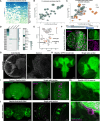
Fig. 1.
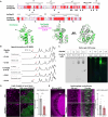
Fig. 1.. Obp44a is a highly abundant secretory protein produced by CNS glia in Drosophila.
(A) Heatmap showing the top 30 most highly expressed and enriched secreted proteins in L1 astrocytes. Obp44a ranks as the highest expressed secreted protein. Each column represents an individual astrocyte. (B) Expression and enrichment analysis of the top 30 secreted proteins in L1 astrocytes. (C) Comparative analysis of 13 secreted proteins expressed across L1, L3, and adult stages reveals that Obp44a consistently maintains high expression. (D) Left: Heatmap of the top 30 Obp family members in antenna, larval (L), and adult (A) brains. F, female; M, male. Unlike classical Obps, Obp44a is highly expressed in the brain rather than the antenna. Right: Tissue distribution heatmap shows Obp44a is prominently expressed in the brain and testis. RPKM, reads per kilobase per million mapped reads. (E) Single-cell atlas of the L1 larval brain highlights Obp44a expression in astrocytes, cortex glia, and ensheathing glia. UMAP-1, Uniform Manifold Approximation and Projection 1. (F) Top: Obp44a-Gal4 was generated using a 2.9-kb enhancer upstream of the transcriptional start site. Bottom: Obp44a-Gal4 drives CD8::GFP (membrane label) or redStinger (nuclear label) expression in a subset of glia in L3 larval brains. (G) Antibody staining shows that Obp44a protein is enriched in L3 larval neuropil. (H) An Obp44a::GFP knock-in line reveals in vivo localization of Obp44a in L3 larval brains. (Ha) Single-section images of astrocytes labeled with alrm > CD2::mCherry show low levels of Obp44a in astrocyte somas, consistent with its efficient secretion. (I) Left: Obp44a::GFP shows widespread and diffused distribution in the adult brain. Right: Single-section images reveal Obp44a’s localization in adult optic lobe chiasm (OLC) glia. (J) Blocking secretion via Obp44a-Gal4-driven Nsf2 RNAi knockdown causes Obp44a::GFP to accumulate in glial somas, confirming its glial production and secretion. Representative maximum intensity projections or single optic sections from confocal images are shown. L1, first instar larvae; L3, third instar larvae. Scale bars are as indicated.
Fig. 2.
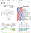
Fig. 2.. Obp44a is a FABP regulating lipid storage.
(A) Protein structure prediction and homology analysis reveal structural similarities between D. melanogaster Obp44a (DmObp44a) and A. aegypti Obp22 (AeObp22). Top: Protein sequence alignment excluding signal peptides highlights critical amino acids (black triangles) that form the hydrophobic pocket in DmObp44a, corresponding to the residues identified in AeObp22. Bottom: AlphaFold2-predicted 3D structures show structural similarities between DmObp44a (green) and AeObp22 (gray), both featuring six α helices and a hydrophobic pocket that accommodates fatty acid ligands, such as C20:4 arachidonic acid (magenta) shown in the model. (B) Fatty acid binding–induced conformational changes in Obp44a are detected by NMR. The Trp102 side-chain Hε1 proton (red arrow) is sensitive to ligand interactions. Distinctive shifts from the apo form are detected in the presence of palmitic acid (C16:0), stearic acid (C18:0), eicosenoic acid (C20:1), and 9-HODE (C18:2) but not in the case of docosanoic acid (C22:0). (C) Native PAGE binding assay demonstrates the interaction between Obp44a protein and C16 fatty acid. Left: Coomassie-stained gel shows purified Obp44a protein. Right: BODIPY FL-labeled C16 fatty acid (FA-C16) alone remains in the well without migration. Upon addition of 3.5 μM Obp44a to 10.5 μM FA-C16, a migrating complex is detected, and the colocalization (white star) indicates binding between Obp44a and FA-C16. (D and E) Obp44a mutants exhibit reduced numbers of lipid droplets in the L3 larval neuropil (D) and in the OLC region of adult brains (E), detected by Nile red staining (magenta). Astrocytes are labeled by alrm > CD8GFP (green) as landmarks. Statistical significance is assessed by unpaired t test with Welch’s correction. **P < 0.01 and ***P < 0.001. Error bars represent mean ± SEM; n = 10 in (D) and n = 14 and 19 in (E). WT, wild type.
Fig. 3.
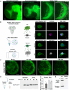
Fig. 3.. Obp44a maintains lipid homeostasis in the fly brain.
(A) Schematic diagram illustrating the sample preparation procedure for metabolomic profiling of L3 larval brains using HILIC-MS. Schematic image is created in BioRender. Yin, J. (2025) https://BioRender.com/sc2v2lu. (B) Principal components (PC) analysis of the L3 larval brain metabolome across all biological and replicates (n = 9) for wild type, Obp44a mutants, and blank controls. (C) Volcano plot highlighting changes in total detected metabolite between wild-type and Obp44a−/− L3 larval brains. Red and blue dots represent significantly increased or decreased metabolites, meeting the cutoff criteria of P < 0.05 and log2 fold change > 0.25. (D) Heatmap displays metabolites with significant changes in Obp44a mutant brains, including phosphatidylethanolamines (PE), diacylglycerol (DAG), phosphatidylinositol (PI), monoacylglycerol (MG), carnitines, and oxidated fatty acid 13-HODE. (E) Altered levels of all detected TAG and DAG species, most of which are reduced in Obp44a mutant brains. (F) Consistent reductions in multiple carnitine species were detected in Obp44a mutants. Red asterisks indicate P < 0.05. The color-coded bar (bottom) illustrates the average intensity of each metabolite within this category.
Fig. 4.
Fig. 4.. Obp44a deficiency leads to morphological and physiological defects.
(A) Obp44a mutants show disrupted astrocyte morphology (alrm > CD8GFP, in green) and nuclear arrangement (anti-repo, in gray) in the adult OLC (n = 28). The presence of large vacuoles and disorganized nuclei in mutants are marked by orange arrows and stars. (B) CRISPR-Cas9–mediated tissue-specific mutagenesis reduces Obp44a levels in glia. Left: Schematic of the mutagenesis. The image is created in BioRender. Yin, J. (2025) https://BioRender.com/sc2v2lu. Right: Western blot showing reduced Obp44a level in L3 larval brains following the knockdown. (C and D) Glia-specific Obp44a mutagenesis reduces light-elicited calcium responses in L3 larval LNvs, suggesting a role of Obp44a in supporting neuronal physiological response. (C) Left: Schematic diagram illustrating the setup for calcium imaging experiments. Light pulses detected by the Bolwig’s organ (BO) elicit calcium responses in LNvs, which are recorded at the axonal terminal (green dashed circle). Right: Representative images of Pdf > GCaMP7f recordings at 0 and 1 s after stimulation for control and glia-specific Obp44a knockdown brains. (D) Left: Traces showing average GCaMP responses. The shaded area represents SEM, and the dashed line indicates the 100-ms light pulse. Right: Quantification of peak amplitude of the change in GCaMP signals (ΔF/F), following light stimulations. Statistical analysis was performed using one-way analysis of variance (ANOVA) followed by Tukey post hoc test. **P < 0.01 and ***P < 0.001. Error bars represent mean ± SEM; n = 12, 23, and 23.
Fig. 5.
Fig. 5.. Obp44a is required for normal locomotion, sleep behavior, and resistance to oxidative stress in adult flies.
(A) Obp44a mutants exhibit significantly reduced climbing ability, indicated by a reduction in climbing index (top) and climbing height (bottom). Statistical significance was assessed using an unpaired t test with Welch’s correction for the climbing index and Welch’s ANOVA test with Dunnett’s multiple posttests for the climbing curve, **P < 0.01 and ***P < 0.001. Error bars represent mean ± SEM; n = 12 and 13 groups, 20 flies per group. (B) Obp44a mutants show altered locomotion patterns under light:dark (LD) and constant dark (DD) conditions. Representative actograms of average activity of 3- to 5-day-old adult flies are shown. (C) Activity curve and quantification demonstrate reduced activity levels in Obp44a mutants. ZT, Zeitgeber Time; CT, Circadian Time. (D) Quantifications show that, compared to wild-type controls, Obp44a mutants exhibit increased number of sleep episodes and decreased episode durations. Statistical significance was assessed using an unpaired t test with Welch’s correction, **P < 0.01 and ***P < 0.001; ns, not significant. Error bars represent mean ± SEM; n = 88, 94. (E) Glutathione redox (GSH/GSSG) biosensor (mito-roGFP2-Grx1) analysis reveals elevated redox potential in Obp44a mutant brains (10-day-old adults). Left: Representative raw and ratiometric images of optic lobes expressing mito-roGFP2-Grx1. Right: Quantification of 405/488 ratiometric fluorescent changes in the optic lobe region indicates increased redox potential in mutant brains. Statistical significance was assessed by unpaired t test with Welch’s correction, ***P < 0.001. Error bars represent mean ± SEM; n = 24, 30. (F) Survival curve of wild-type and Obp44a−/− adult flies subjected to 5% H2O2 treatment. Obp44a−/− flies exhibit significantly reduced survival probability after 24-hour treatment. Statistical significance was assessed by Welch’s ANOVA test with Dunnett’s multiple posttests, ***P < 0.001. Error bars represent mean ± SEM; n = 9 groups, 16 flies per group.
Fig. 6.
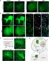
Fig. 6.. Obp44a traffics between neuron and glia and is secreted into the hemolymph.
(A) Expression of Obp44a::GFP transgene in astrocytes or neurons recapitulates its localization observed in the Obp44a::GFP knock-in line, demonstrating efficient secretion and dynamic trafficking across cell types. Right: Higher magnification, single optical section of the larval optic lobe shows that neuronally expressed Obp44a::GFP (elav-Gal4 > UAS-Obp44a::GFP) is found in the soma of repo-positive glia, demonstrating intercellular transfer from neurons to glia. (B) Obp44a traffics between glia and neuron. Left: Schematic of the experimental setup using acutely dissociated cells from L3 larval brains. Schematic image is created in BioRender. Yin, J. (2025) https://BioRender.com/sc2v2lu. Right: Obp44a::GFP produced in astrocytes (alrm-Gal4 > UAS-Obp44a::GFP) is observed in elav-positive neurons, while neuronal expressed Obp44a::GFP (elav-Gal4 > UAS-Obp44a::GFP) is detected in repo-positive glia, indicating Obp44a’s ability to traffic between glia and neuron. n = 74 (glia to neuron), n = 91 (neuron to glia). (C) Time-lapse live imaging of L3 larval brain explants reveals Obp44a::GFP mobilization from the neuropil toward surface glia. All 25 explants imaged exhibited similar phenotypes. (D) Obp44a::GFP is secreted from the brain into surrounding medium. Western blot of L3 larval brain explants incubated in physiological saline shows Obp44a::GFP in both the supernatant (secreted fraction) and brain lysate. Quantification from four biological replicates is shown in the right. (E) Obp44a is present in circulating hemolymph. Representative Western blot of L3 larval hemolymph reveals both monomeric and dimeric forms of Obp44a (arrow) in wild type larvae, which are absent in Obp44a mutants. Asterisk indicates a nonspecific band. DAPI, 4′,6-diamidino-2-phenylindole.
Fig. 7.
Fig. 7.. Obp44a functions as a lipid chaperone and scavenger during development and injury response.
(A) Endogenous Obp44a accumulates in apoptotic-like cells when phagocytosis is impaired in larval or adult drpr mutant brains. (B) Obp44a is recruited to Caspase-3–positive apoptotic like cells in both L3 larval and adult brains upon glial drpr knockdown. Right: Higher magnification, single-section images from adult optic lobes. Magenta arrowheads indicate representative cells colabeled with Obp44a::GFP and caspase-3. Nuclei are marked with DAPI (blue). (C) Obp44a responds to injury-induced lipid stress following antennal axotomy. Top left: Schematic of unilateral axotomy in adult flies. Right and bottom: Representative confocal images of Obp44a::GFP-labeled brains at 6, 24, and 48 hours postinjury. At 6 hours, Obp44a::GFP distribution is comparable on both sides of the brain. By 24 hours, Obp44a::GFP accumulates on the injured side, particularly in the AL and AMMC. At 48 hours, Obp44a::GFP signal intensifies and spreads to adjacent regions. (D) Schematic of Obp44a’s function as a lipid chaperone and scavenger. Produced by cortex and neuropil glia, Obp44a is secreted into the neuropil, traffics through multiple brain cell types, including neurons and surface glia, and ultimately enters the hemolymph. By interacting with both native and oxidized fatty acids, Obp44a regulates lipid trafficking and metabolism, facilitating the clearance and efflux of oxidized lipids during development and neuroinflammation and contributing to brain homeostasis. FFA, free fatty acid; FA, fatty acid. Schematic images are created in BioRender. Yin, J. (2025) https://BioRender.com/sc2v2lu.

References

    1. Bozek K., Wei Y., Yan Z., Liu X., Xiong J., Sugimoto M., Tomita M., Paabo S., Sherwood C. C., Hof P. R., Ely J. J., Li Y., Steinhauser D., Willmitzer L., Giavalisco P., Khaitovich P., Organization and evolution of brain lipidome revealed by large-scale analysis of human, chimpanzee, macaque, and mouse tissues. Neuron 85, 695–702 (2015). - PubMed
    1. Fitzner D., Bader J. M., Penkert H., Bergner C. G., Su M., Weil M. T., Surma M. A., Mann M., Klose C., Simons M., Cell-type- and brain-region-resolved mouse brain lipidome. Cell Rep. 32, 108132 (2020). - PubMed
    1. Vaughen J. P., Theisen E., Rivas-Serna I. M., Berger A. B., Kalakuntla P., Anreiter I., Mazurak V. C., Rodriguez T. P., Mast J. D., Hartl T., Perlstein E. O., Reimer R. J., Clandinin M. T., Clandinin T. R., Glial control of sphingolipid levels sculpts diurnal remodeling in a circadian circuit. Neuron 110, 3186–3205.e7 (2022). - PMC - PubMed
    1. Goodman L. D., Bellen H. J., Recent insights into the role of glia and oxidative stress in Alzheimer’s disease gained from Drosophila. Curr. Opin. Neurobiol. 72, 32–38 (2022). - PMC - PubMed
    1. Brandebura A. N., Paumier A., Onur T. S., Allen N. J., Astrocyte contribution to dysfunction, risk and progression in neurodegenerative disorders. Nat. Rev. Neurosci. 24, 23–39 (2023). - PMC - PubMed

MeSH terms

LinkOut - more resources