. 2025 Aug;11(31):eadv9311.
doi: 10.1126/sciadv.adv9311. Epub 2025 Aug 1.

A pan-respiratory virus attachment inhibitor with high potency in human airway models and in vivo

Affiliations

A pan-respiratory virus attachment inhibitor with high potency in human airway models and in vivo

Gregory Mathez et al. Sci Adv. 2025 Aug.

Abstract

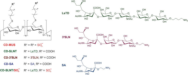
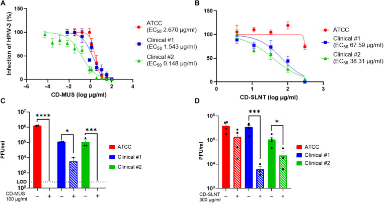
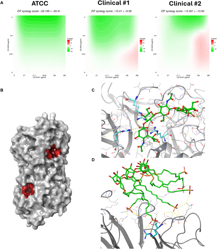
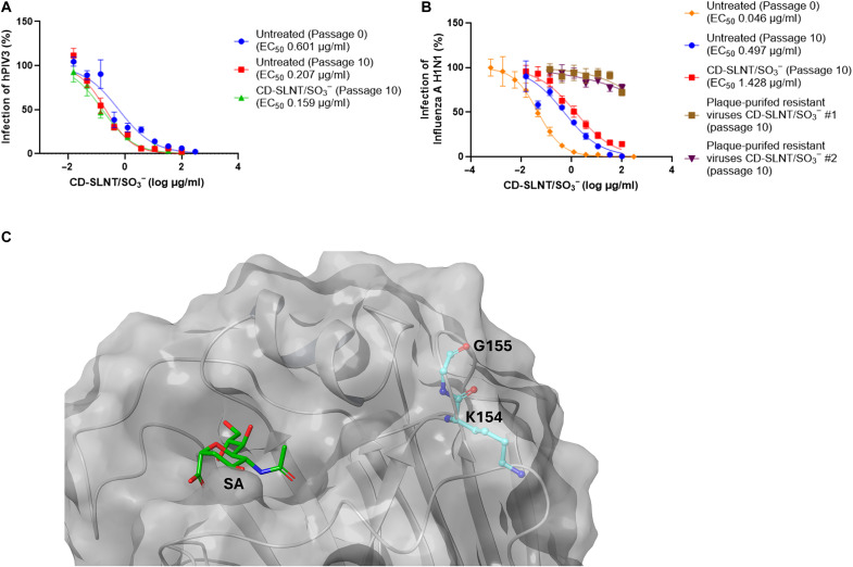
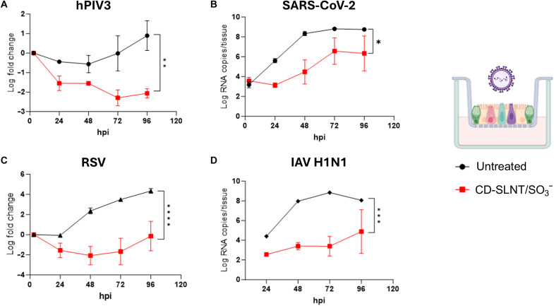
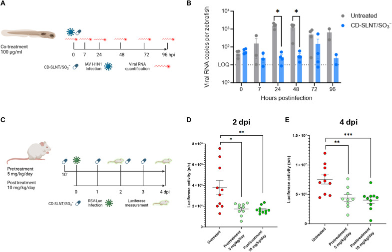
Respiratory viruses can cause severe infections, often leading to hospitalization or death, and pose a major pandemic threat. No broad-spectrum antiviral is currently available. However, most respiratory viruses use sialic acid or heparan sulfates as attachment receptors. Here, we report the identification of a pan-respiratory antiviral strategy based on mimicking both glycans. We synthesized a modified cyclodextrin that simultaneously mimics heparan sulfate and sialic acid. This compound demonstrated broad-spectrum antiviral activity against important human pathogens: parainfluenza virus 3, respiratory syncytial virus, influenza virus H1N1, and severe acute respiratory syndrome coronavirus 2 (SARS-CoV-2). In addition, the compound is active against avian strains of influenza virus, revealing its importance for pandemic preparedness. The compound retains broad-spectrum activity in ex vivo models of respiratory tissues and in vivo against respiratory syncytial virus and influenza virus, using prophylactic and therapeutic strategies. These findings contribute to the development of future treatments and preventive measures for respiratory viral infections.


Figures

Fig. 1.
Fig. 1.. Schematic representation of modified CDs.
Schemes of the different CD with the respective ligands. These drawings do not consider the possible configurations the substituents can adopt on the primary face of the CD but only the type of substituents.
Fig. 2.
Fig. 2.. Antiviral activity of CD-MUS and CD-SLNT against hPIV3.
Inhibition (A and B) and virucidal activity (C and D) of CDs. Inhibition was performed by incubating 1 hour at 37°C laboratory and clinical strains of hPIV3 with CD-MUS (A) or CD-SLNT (B) before infection on cells. Antiviral activity was assessed by plaque assay counted manually. Data represent means ± SEM of two [(A) ATCC], three [(A) clinical #1, (B)], and six [(A) clinical #2] independent experiments. Nonlinear regression with variable Hill slope and constraints for the bottom and top (0 and 100, respectively) were performed to compute EC50. (C and D) Virucidal experiments were done by incubating viruses for 1 hour with CD-MUS (100 μg/ml) (C) or 2 hours with CD-SLNT (300 μg/ml) (D) at 37°C. The virus-drug mix was then serially diluted. Viral titer was assessed by plaque assay counted manually. Data represent means ± SEM of two independent (C and D) experiments. Two-tailed t tests between untreated and treated conditions were performed. Limit of detection (LOD) *P < 0.0332, ***P < 0.0002, ****P < 0.0001.
Fig. 3.
Fig. 3.. Combination assays using CD-MUS and CD-SLNT.
(A) CD-MUS and CD-SLNT were mixed at different concentrations and incubated for 1 hour at 37°C with the laboratory or the two clinical strains of hPIV3 before infection of LLCMK2 cells. Inhibition percentages were calculated by plaque assay of two independent experiments and then evaluated with SynergyFinder 3.0 (34). The reference algorithms (highest single agent, Loewe, Bliss, and zero interaction potency) were applied. Green, white, and red areas indicate antagonistic, additive, and synergistic areas, respectively. (B) The two identical sialic acid–binding pockets (site I) are highlighted in red on the HN of hPIV3 (C). SLNT was docked on the boxed sialic acid–binding pocket [PDB 5B2D (44)]. (D) CD-MUS interactions with the HN protein were simulated by molecular dynamics and showed interaction with this pocket [PDB 4MZA (47)]. Ligands carbons are in green and key arginine residues are in cyan.
Fig. 4.
Fig. 4.. Resistance to CD-SLNT/SO3.
HPIV3 (A) and IAV H1N1 (B) viruses were passaged 10 times in the presence of an increasing concentration of CD-SLNT/SO3. At the final passage, the antiviral activity of CD-SLNT/SO3 against the untreated and treated viruses was assessed. Resistant plaque-purified IAV H1N1 viruses were obtained (B). Data represent means ± SEM of two [(B) plaque-purified resistant CD-SLNT/SO3 passage 10], three [(B) untreated passage 10, CD-SLNT/SO3 passage 10] or four (A) independent experiments. Each independent experiment with untreated or treated virus at passage 10 was carried out using the two independent virus replicates. Nonlinear regression with variable Hill slope and constraints for the bottom and top (0 and 100, respectively) were performed to compute EC50. (C) Mutations observed in resistant viruses present in the hemagglutinin protein [PDB 4JTV (59)] and sialic acid (SA) at the glycan-binding region are highlighted in cyan and green, respectively.
Fig. 5.
Fig. 5.. Ex vivo activity of CD-SLNT/SO3.
HPIV3 (A), SARS-CoV-2 (B), RSV (C), or IAV H1N1 (D) were incubated with CD-SLNT/SO3 (300 μg/ml) for 1 hour at 37°C. Human upper respiratory tract models [n = 2 (A), (C), and (D) or 6 (B) per group] were then infected at 33°C. The inoculum was removed after 3 hours. A daily apical wash was performed, and the level of viruses released was quantified by RT-qPCR. Data represent means ± SEM. Area under the curve followed by a two-tailed t test was performed. *P < 0.0332, **P < 0.0021, ***P < 0.0002, ****P < 0.0001. Cartoon created with BioRender.com, Cagno, V. (2025) https://BioRender.com/5w07cwy.
Fig. 6.
Fig. 6.. In vivo activity of CD-SLNT/SO3.
(A and B) Zebrafish larvae were infected with IAV H1N1. CD-SLNT/SO3 was coinjected at the same time at a concentration of 100 μg/ml. At 7 hpi and each day, 10 larvae are lysed for viral RNA quantification by RT-qPCR. Data represent means ± SEM of four independent experiments. (C to E) Mice (n = 10 per group) were infected with RSV. (C) Mice were either treated with CD-SLNT/SO3 (5 mg/kg) 10 min before infection (pretreatment) with a daily dose until 4 days postinfection (dpi) or with CD-SLNT/SO3 (10 mg/kg) starting 1 dpi (posttreatment). (D and E) RSV replication was assessed by luciferase activity at 2 (D) and 4 (E) dpi. Data represent means ± SEM of a single experiment. Two-tailed Mann-Whitney tests were performed to compare untreated and treated conditions. *P < 0.0332, **P < 0.0021, ***P < 0.0002. Schematic view created with BioRender.com, Cagno, V. (2025) https://BioRender.com/bllixw1.

Similar articles

References

    1. Leung N. H. L., Transmissibility and transmission of respiratory viruses. Nat. Rev. Microbiol. 19, 528–545 (2021). - PMC - PubMed
    1. Wang C. C., Prather K. A., Sznitman J., Jimenez J. L., Lakdawala S. S., Tufekci Z., Marr L. C., Airborne transmission of respiratory viruses. Science 373, eabd9149 (2021). - PMC - PubMed
    1. Zhou P., Yang X.-L., Wang X.-G., Hu B., Zhang L., Zhang W., Si H.-R., Zhu Y., Li B., Huang C.-L., Chen H.-D., Chen J., Luo Y., Guo H., Jiang R.-D., Liu M.-Q., Chen Y., Shen X.-R., Wang X., Zheng X.-S., Zhao K., Chen Q.-J., Deng F., Liu L.-L., Yan B., Zhan F.-X., Wang Y.-Y., Xiao G.-F., Shi Z.-L., A pneumonia outbreak associated with a new coronavirus of probable bat origin. Nature 579, 270–273 (2020). - PMC - PubMed
    1. Uyeki T. M., Milton S., Abdul Hamid C., Reinoso Webb C., Presley S. M., Shetty V., Rollo S. N., Martinez D. L., Rai S., Gonzales E. R., Kniss K. L., Jang Y., Frederick J. C., De La Cruz J. A., Liddell J., Di H., Kirby M. K., Barnes J. R., Davis C. T., Highly pathogenic avian influenza A(H5N1) virus infection in a dairy farm worker. N. Engl. J. Med. 390, 2028–2029 (2024). - PubMed
    1. Morse J., Coyle J., Mikesell L., Stoddard B., Eckel S., Weinberg M., Kuo J., Riner D., Margulieux K., Stricklen J., Dover M., Kniss K. L., Jang Y., Kirby M. K., Frederick J. C., Lacek K. A., Davis C. T., Uyeki T. M., Lyon-Callo S., Bagdasarian N., Influenza A(H5N1) virus infection in two dairy farm workers in Michigan. N. Engl. J. Med. 391, 963–964 (2024). - PubMed

LinkOut - more resources