. 2025 Aug 4;198(4):kiaf344.
doi: 10.1093/plphys/kiaf344.

Dynamic regulation of Arabidopsis β-AMYLASE1 by glutathione and thioredoxins affects starch in guard cells

Affiliations

Dynamic regulation of Arabidopsis β-AMYLASE1 by glutathione and thioredoxins affects starch in guard cells

Libero Gurrieri et al. Plant Physiol. .

Abstract

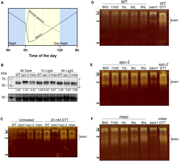
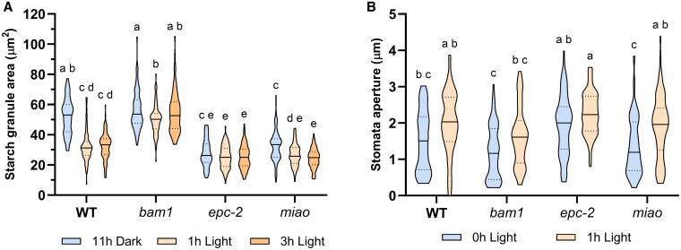
Guard cells control the opening and closure of stomatal pores in response to internal and external stimuli, ensuring gas exchange in plants. In Arabidopsis (Arabidopsis thaliana), β-AMYLASE1 (BAM1), assisted by α-AMYLASE3, begins degrading starch at dawn in guard cells to promote stomatal opening. Both enzymes are controlled by reversible disulfide bond formation, which decreases their activity. In the present study, we investigated the sensitivity of BAM1 to other redox-dependent post-translational modifications (PTM) both in vitro and in vivo. In vitro, H2O2 reversibly inactivates BAM1 and, in the presence of glutathione (GSH), induces S-glutathionylation of BAM1. Glutathionylated BAM1 is active and transiently protected from H2O2 inhibition. However, the glutathionylated state of BAM1 has limited stability and can be slowly resolved by a second cysteine with the formation of the intramolecular disulfide bond that inhibits BAM1 activity. Thioredoxin f can fully revert the inhibition by reducing the disulfide to a dithiol. In vivo, Arabidopsis mutants with lower plastidial GSH reductase activity, and consequently modified GSH homeostasis, showed higher BAM1 activity, lower starch levels in guard cells, and altered stomata aperture, indicating that GSH redox potential impacts stomatal physiology, possibly through BAM1. Moreover, plastidial BAM1 presents a prime example for the role of glutathionylation functioning as a transiently protective PTM, interfering with the formation of inhibitory disulfide bonds. This example illustrates how transitions between protein cysteinyl thiol PTMs can orchestrate dynamic responses involving several redox systems.


Conflict of interest statement

Conflict of interest statement. None declared.

Figures

Figure 1.
Figure 1.
BAM1 sensitivity to H2O2 and protective effect of GSH. A) Inactivation of BAM1 after 1 h incubation with 0.5 mm H2O2. The control sample was incubated for 60 min in the absence of H2O2. Rescue of BAM1 activity was recorded in response to an additional 30 min incubation with DTT. B) Inactivation kinetics of BAM1 incubated with 0.5 mm H2O2 with or without 2.5 mm GSH (reduced GSH). The control sample has been incubated in the absence of any reagent. All experiments were carried out in triplicate; error bars show standard deviation. Data were analyzed with Student's t-test and compared to the control sample; **, P < 0.01; ns, not significant.
Figure 2.
Figure 2.
Glutathionylation of BAM1 has no effect on catalysis. A) Upper panel: Western blot analysis with α-GSH antibodies on BAM1 samples untreated (CTR) and treated with 0.5 mm H2O2 and 2.5 mm GSH (reduced GSH). Samples were analyzed at 0 min and after 60 min of incubation. Lower panel: Coomassie staining of the same samples. B) Upper panel: Western blot analysis with α-GSH antibodies on BAM1 samples untreated (CTR) and treated with 1 mm GSSG (oxidized GSH). Samples were analyzed after 60 min of incubation. Lower panel: Coomassie staining of analogous samples. C) BAM1 activity was measured in untreated (Control) and treated sample (1 mm GSSG) after 1 h of incubation. Data were analyzed with Student's t-test and compared to the control sample; ns, not significant (P < 0.05). The experiment was carried out in triplicate; error bars show standard deviation. D) Upper panel: Western blot analysis with α-GSH antibodies on BAM1 pre-treated with 1 mm GSSG. The remaining free cysteine thiols were blocked with alkylating agents (20 mm IAM; and 20 mm NEM), the sample was diluted and glutathionylation has been removed with 20 mm DTT. All the reagents were removed by desalting and the protein treated with 0.5 mm H2O2 and 2.5 mm GSH. The signal observed in this latter sample indicates that the same cysteine can be a target of both glutathionylating treatments (GSSG or H2O2 and GSH). Lower panel: Ponceau staining of the membrane after protein transfer. E) Deglutathionylation of BAM1 assayed by Western blot analysis using α-GSH antibodies. Upper panel: BAM1 was treated with 1 mm GSSG (GSSG lane), desalted, and then incubated with 0.5 mm DTT, 2 mm GSH alone or in the presence of 1 µM GRX C5 or GRX S12. Lower panel: Ponceau staining of the membrane after protein transfer. A and E: The M lane images were acquired as colorimetric images by the same imaging system used to detect the chemiluminescence signal.
Figure 3.
Figure 3.
Spontaneous loss of GSH leads to BAM1 inhibition. A) Upper panel: BAM1 samples were incubated for 1 h with 0.5 mm H2O2 and 2.5 mm GSH or with an equal volume of buffer (CTR). After incubation both samples were desalted in 100 mm Tricine–NaOH pH 7.9 and at indicated time points, the enzyme activity were analyzed by Western blot analysis using α-GSH antibodies. Lower panel: Coomassie staining of the same samples. B) Upper panel: BAM1 samples were incubated for 1 h with 1 mm GSSG or with an equal volume of buffer (CTR). After incubation, both samples were desalted in 100 mm Tricine–NaOH pH 7.9 and at indicated time points, the enzyme activity were analyzed by Western blot analysis using α-GSH antibodies. Lower panel: Coomassie staining of the same samples. C) BAM1 activity measured on samples shown in A and B; the activity of every sample is expressed as percentage of the activity at 0 min after the desalting. D) Thiols were released from untreated (Control) and GSSG-treated BAM1 (as in B). Control and treated samples were desalted after 1 h and incubated for 90 min before measuring the released GSH in the flow-through of the ultrafiltered BAM1 samples. All experiments were carried out in triplicate; error bars show standard deviation. Data were analyzed with Student's t-test and compared to the untreated sample; **, P < 0.01; *, P < 0.05.
Figure 4.
Figure 4.
TRX f1 mediates rapid BAM1 reactivation. The reversibility of inactivation of BAM1 was assessed on inhibited BAM1 samples obtained after treatment with 1 mm GSSG for 1 h, desalted, and incubated for 90 min at 37 °C. Untreated sample followed the same procedure, except for the initial incubation that was with buffer. The recovery was tested incubating for 1 h with 1 µM of TRX f1 and 0.5 mm DTT, GRX C5, or GRX S12 and 2 mm GSH. The experiment was carried out in triplicate; error bars show standard deviation. Data were analyzed with 1-way ANOVA and Tukey's test with P < 0.01, where distinct lowercase letters denote significant group differences.
Figure 5.
Figure 5.
Alteration in GSH regeneration affects BAM1 activity. A) schematic representation of stomata aperture and starch levels in Arabidopsis chloroplasts of guard cells throughout the day. Adapted from Horrer et al. (2016). Sample collection times (9 and 11 h dark; 1, 3 and 9 h light) are marked by dotted lines. B) BAM1 protein levels visualized by Western blot using α-BAM1 antibodies. Protein extracts (40 µg) from the indicated genotypes were prepared at different timepoints and loaded onto gels. BAM1 appears as 2 bands due to the presence of precursor (P) and mature BAM1 (M) in protein extracts (Feike et al. 2022). Ponceau staining of Rubisco large subunit is shown as loading control. The levels of total BAM1 protein (precursor + mature) were quantified with ImageJ, normalized on the wild-type value and shown as numbers at the bottom of the Western blot. C) In-gel amylolytic activity on native acrylamide gel with 0.1% amylopectin. Protein samples were collected at 1 h of light and incubated with extraction buffer (left side of the gel) or reduced DTT (right side of the gel) for 1 h before loading. Arrowheads indicate amylolytic activities (Bands 1, 2, 3, and 4) other than BAM1. D)–F) In-gel amylolytic activity on native acrylamide gel with 0.1% amylopectin from protein samples of the indicated genotype collected at the different time points. Part of the extract at 1 h of light was reduced with 20 mm DTT and 40 µg of proteins were loaded onto gels to show the maximal activity. Arrowheads indicate amylolytic activities (Bands 1, 2, 3, and 4) other than BAM1.
Figure 6.
Figure 6.
GR2 deficiency affects guard cell starch and stomata aperture. A) Starch area per guard cell in wild-type, bam1, epc-2, and miao Arabidopsis plants. B) Stomata aperture of wild-type, bam1, epc-2, and miao plants. Solid lines represent the median; dotted line represent the highest and the lowest quartiles. For starch granule area, 70 to 80 guard cells were analyzed at each time point, for stomata aperture, 60 to 90 pores were analyzed at each timepoint. Data were analyzed using 2-way ANOVA and Tukey's test with P < 0.01, where distinct lowercase letters denote significant group differences.
Figure 7.
Figure 7.
Model of BAM1 redox regulation. BAM1 reacts with H2O2, undergoing sulfenylation (–SOH). The sulfenylation is either removed by forming a disulfide bond with a cysteine from the active site, which inactivates BAM1, or by reacting with GSH, leading to glutathionylation (–SSG) and maintaining the enzyme's activity. Glutathionylation can be removed by the active site cysteine, leading to intramolecular disulfide bond and inhibition of activity. Glutathionylation can occur also by direct reaction with oxidized GSH (GSSG). GRXs keep target cysteine redox states and GSH redox state near thermodynamic equilibrium. Depending on the GSH redox potential (EGSH), GRXs can catalyze glutathionylation or deglutathionylation. The inhibitory intramolecular disulfide bond can be reduced by chloroplast TRX using the reducing power provided by photosynthesis.

Similar articles

Cited by

References

    1. Akter N, Sobahan MA, Uraji M, Ye W, Hossain MA, Mori IC, Nakamura Y, Murata Y. Effects of depletion of glutathione on abscisic acid and methyl jasmonate-induced stomatal closure in Arabidopsis thaliana. Biosci Biotechnol Biochem. 2012:76(11):2032–2037. 10.1271/bbb.120384 - DOI - PubMed
    1. Ballicora MA, Frueauf JB, Fu Y, Schü Rmann P, Preiss J. Activation of the potato tuber ADP-glucose pyrophosphorylase by thioredoxin. J Biol Chem. 2000:275(2):1315–1320. 10.1074/jbc.275.2.1315 - DOI - PubMed
    1. Bohle F, Rossi J, Tamanna SS, Jansohn H, Schlosser M, Reinhardt F, Brox A, Bethmann S, Kopriva S, Trentmann O, et al. Chloroplasts lacking class I glutaredoxins are functional but show a delayed recovery of protein cysteinyl redox state after oxidative challenge. Redox Biol. 2024:69:103015. 10.1016/j.redox.2023.103015 - DOI - PMC - PubMed
    1. Bradford MM. A rapid and sensitive method for the quantitation of microgram quantities of protein utilizing the principle of protein-dye binding. Anal Biochem. 1976:72(1–2):248–254. 10.1016/0003-2697(76)90527-3 - DOI - PubMed
    1. Cejudo FJ, Sandalio LM, Van Breusegem F. Understanding plant responses to stress conditions: redox-based strategies. J Exp Bot. 2021:72(16):5785–5788. 10.1093/jxb/erab324 - DOI - PMC - PubMed

MeSH terms

LinkOut - more resources