Case Reports
. 2025 Jul 30;30(21):104027.
doi: 10.1016/j.jaccas.2025.104027.

Unexpected Ventricular Fibrillation During a Cardiovascular Magnetic Resonance Examination Documented With Simultaneous Quantitative Myocardial Perfusion

Affiliations
Case Reports

Unexpected Ventricular Fibrillation During a Cardiovascular Magnetic Resonance Examination Documented With Simultaneous Quantitative Myocardial Perfusion

Guillem Pons-Lladó et al. JACC Case Rep. .

Abstract

Background: Unexpected life-threatening arrhythmias, even when occurring in hospital, are frequently ill documented, hindering a proper understanding of their mechanism.

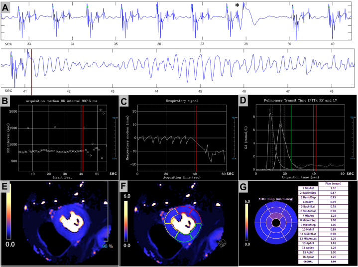
Case summary: We present a patient with coronary artery disease who developed an episode of ventricular fibrillation during a cardiovascular magnetic resonance examination. The arrhythmia appeared immediately after the injection of a contrast agent during a quantitative myocardial perfusion sequence at rest, with no stressor agent involved.

Discussion: To our knowledge, this is the first episode of ventricular fibrillation documented with simultaneous electrocardiographic, physiological recordings, and quantitative myocardial perfusion data. Such unique information allowed for a comprehensive analysis of the sequence of facts preceding the final arrhythmia and, in consequence, provided a well-reasoned conclusion on its mechanism.

Keywords: cardiac magnetic resonance; contrast agent; perfusion; ventricular fibrillation.


Conflict of interest statement

Funding Support and Author Disclosures Dr Vidorreta is an employee of Siemens Healthineers, Madrid, Spain. All other authors have reported that they have no relationships relevant to the contents of this paper to disclose.

Figures

None
Graphical abstract
Figure 1
Figure 1
Noncontrasted Computed Tomography Scan Showing Multiple Focal Calcifications on the Wall of Coronary Arteries LAD = left anterior descending; LCx = left circumflex; RCA = right coronary artery.
Figure 2
Figure 2
Invasive Angiography of the Left Coronary Artery and Percutaneous Intervention Coronary angiography showing (A) severe obstruction at the middle portion of the left anterior descending, at the level of the origin of a diagonal branch (arrow), which was successfully treated by percutaneous intervention (B).
Figure 3
Figure 3
Recording of the ECG Tracing Retrieved From the MR System Continuous electrocardiographic (ECG) tracing during the 65 seconds of the rest perfusion sequence, showing the presence of occasional, short-coupled, PVCs. One of them, at 41 seconds, was followed by a brief burst of polymorphic ventricular tachycardia degenerating into VF. Artifacts appearing at 50 seconds are due to the removal of the patient from the magnet followed by, at 55 seconds, regular defections of the ECG line caused by chest compression during CPR. Note the presence of spike signals grouped on the QRS-T complexes in regular sinus rhythm, which correspond to the set of radiofrequency excitations of the sequence. CPR = cardiopulmonary resuscitation; PVC = premature ventricular contraction; VF = ventricular fibrillation.
Visual Summary
Visual Summary
Information Retrieved From the System at the Time of Acquisition of the Quantitative Myocardial Perfusion Sequence (A) Selected electrocardiographic strips showing regular sinus rhythm at 74 beats/min with 1 short-coupled premature ventricular contraction (PVC) (asterisk), and initiation of ventricular fibrillation (VF) at 41 seconds after another PVC (red line). (B) Individual RR intervals of the electrocardiography during the whole sequence acquisition showing the regular rhythm interrupted by occasional post-extrasystolic pauses and its disruption, at 41 seconds from start, due to the onset of VF (red line). (C) Respiratory signal showing regular cycles at 15 per minute, and a respiratory standstill coincident with the onset of VF (red line). (D) Time curves of contrast passage through the right and left ventricular chambers with a green line signaling the approximate time of myocardial arrival of contrast, at 25 seconds from the start, and later, the time of initiation of VF (red line). (E) One of the 3 resultant quantitative myocardial perfusion mappings in short-axis orientation. (F) Automatic segmentation of the slice. (G) In-line automatic calculation of myocardial perfusion values (mL/min/g) at each of the 16 segments of the left ventricle.

References

    1. Bruder O., Wagner A., Lombardi M., et al. European cardiovascular magnetic resonance (EuroCMR) registry–multinational results from 57 centers in 15 countries. J Cardiovasc Magn Reson. 2013;15:9–18. - PMC - PubMed
    1. Uhlig J., Lücke C., Vliegenthart R., et al. Acute adverse events in cardiac MR imaging with gadolinium-based contrast agents: results from the European Society of Cardiovascular Radiology (ESCR) MRCT Registry in 72,839 patients. Eur Radiol. 2019;29:3686–3695. - PMC - PubMed
    1. Kellman P., Hansen M.S., Nielles-Vallespín S., et al. Myocardial perfusion cardiovascular magnetic resonance: optimized dual sequence and reconstruction for quantification. J Cardiovasc Magn Reson. 2017;19:43–56. - PMC - PubMed
    1. Power S., Talbot N., Kucharczyck W., Mandell D.M. Allergic-like reactions to the MR imaging contrast agent gadobutrol: a prospective study of 32 991 consecutive injections. Radiology. 2016;281:72–77. - PubMed
    1. Brown L.A.E., Gulsin G.S., Onciul S.C., et al. Sex- and age-specific normal values for automated quantitative pixel-wise myocardial perfusion cardiovascular magnetic resonance. Eur Heart J Cardiovasc Imaging. 2023;24:426–434. - PMC - PubMed

Publication types

LinkOut - more resources