. 2025 Aug;57(8):1727-1742.
doi: 10.1038/s12276-025-01510-4. Epub 2025 Aug 1.

Canonical Wnt signaling regulates Mbd3 protein stability during neurogenesis

Affiliations

Canonical Wnt signaling regulates Mbd3 protein stability during neurogenesis

Nhu Thi Quynh Mai et al. Exp Mol Med. 2025 Aug.

Abstract

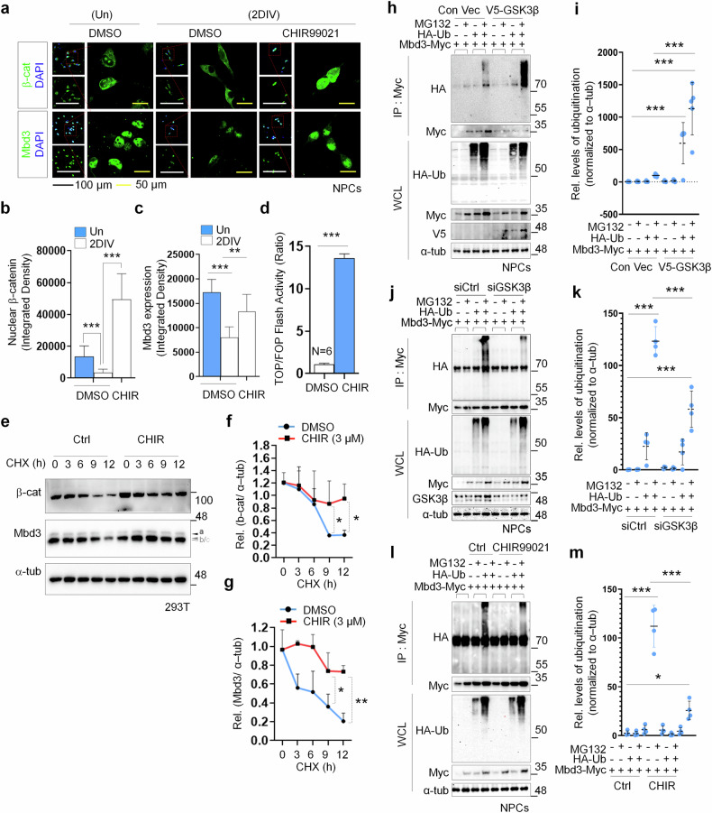
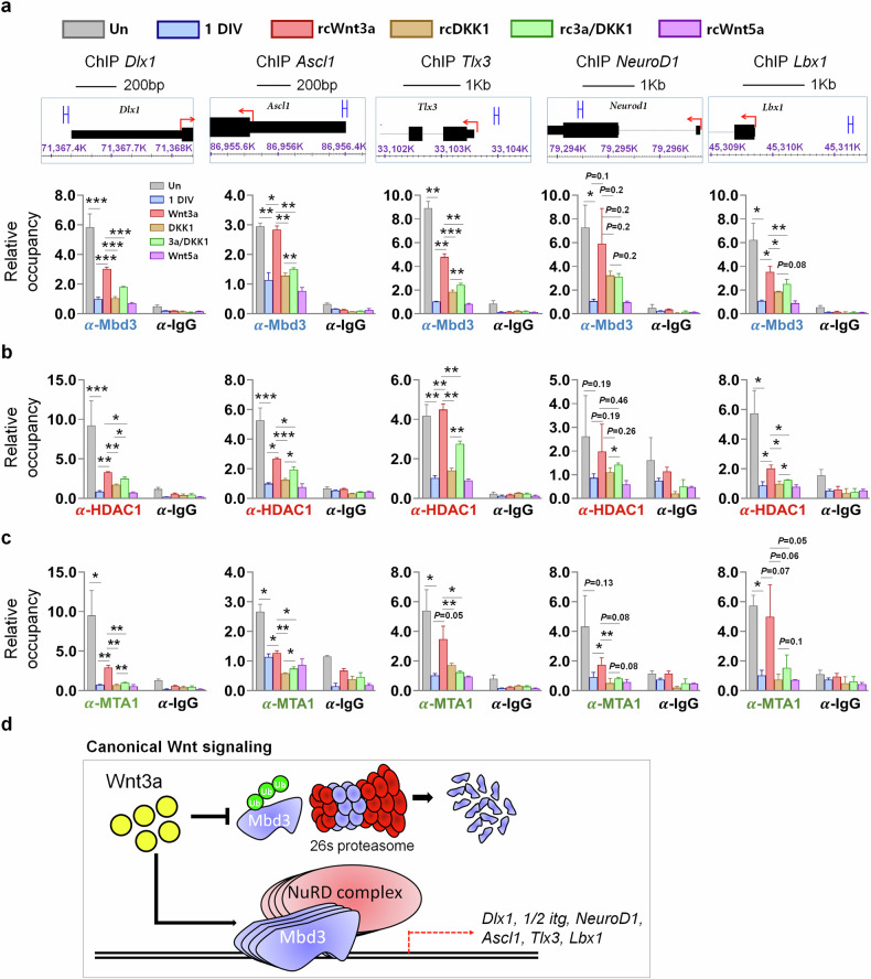
Acquisition of neural progenitor cell (NPC) homeostasis through balancing self-renewal and differentiation is essential for brain development and function. Among the mechanisms controlling these processes, canonical Wnt signaling and the Mbd3-NuRD complex, with prominent suppressive effects on neurogenesis, have been described as crucial parts of the core regulatory circuit. Here we explored Mbd3 as a downstream element of the canonical Wnt signalosomes. Specifically, dynamic modulation of Wnt signaling through activator (Wnt3a) and inhibitor (DKK1) resulted in parallel alterations in β-catenin and Mbd3 expression patterns. Also, overexpression and depletion of GSK3β respectively promoted and attenuated Mbd3 ubiquitination, highlighting that the canonical Wnt cascade promotes Mbd3 stability. Downstream of the Wnt-β-catenin pathway, Mbd3 represses transcription of neurogenesis-associated genes by triggering NuRD complex assembly, thereby promoting NPC stemness. This new Wnt-Mbd3 axis extends the current understanding of the canonical Wnt network in directing neuronal cell-fate determination in NPCs, suggesting this pathway as a potential target for driving neural stem cell reprogramming and neuronal lineage commitment.


Conflict of interest statement

Competing interests: The authors declare no competing interests.

Figures

Fig. 1
Fig. 1. Canonical Wnt signaling promotes Mbd3 stabilization.
a Immunostaining detecting nuclear localization of β-catenin (green) in undifferentiated (Un) and 2DIV differentiated NPCs in different states of Wnt signaling (N = 3). Scale bar, 50 µm. b Quantification of nuclear β-catenin intensity in undifferentiated and 2DIV differentiated cells in different states of Wnt signaling (N = 3). c A plot of the TOP/FOP Flash assay indicating the β-catenin level through luciferase activity in differentiated NPCs overexpressing Wnt activators (Wnt3a (canonical signaling), Wnt5a (noncanonical signaling)) or inhibitor (DKK1) (N = 6). d A plot of the TOP/FOP Flash assay indicating the β-catenin level through luciferase activity in differentiated NPCs under the supplements of the indicated rcWnt signaling stimuli (N = 6). e Immunostaining detecting nuclear localization of Mbd3 (green) in undifferentiated and 2DIV differentiated NPCs in different states of Wnt signaling. Scale bar, 50 µm. f Quantification of Mbd3 expression level in undifferentiated and 2DIV differentiated NPCs in different states of Wnt signaling (N = 3). g The expression of β-catenin and Mbd3 at the protein level (by immunoblot (IB), top) and mRNA level (by RT-PCR, bottom) in 2DIV differentiated NPCs treated with varying doses of Wnt3aCM (N = 3). Labels a–c denote Mbd3 isoforms. h Western blot showing β-catenin and Mbd3 expression level in HEK293T cells with dose-dependent supplement of Wnt3a protein (N = 3). Labels a–c denote Mbd3 isoforms. i Quantification of the relative expression of β-catenin and Mbd3 of h In all cases, data are presented as mean ± s.d.; one-way ANOVA was performed to calculate significance (*P < 0.05, **P < 0.01, ***P < 0.001).
Fig. 2
Fig. 2. Canonical Wnt signaling inhibits Mbd3 stabilization.
a Immunoblot (IB) analysis of β-catenin and Mbd3 in HEK293T cells treated time-dependently with CHX, followed by activation of canonical Wnt signaling through rcWnt3a supplementation. Labels a–c denote Mbd3 isoforms. b, c Corresponding quantification of the relative expression level of β-catenin (b) and Mbd3 (c) in the indicated conditions. d IP of HA-tagged ubiquitin (HA-Ub) in NPCs expressing full-length Myc-tagged Mbd3 (Myc-Mbd3) in LCM and Wnt3aCM (N = 3). Levels of each protein in the whole-cell lysate (WCL) are shown with western blot. IP was demonstrated using an anti-Myc antibody. e The relative level of Mbd3 ubiquitination in LCM and Wnt3aCM. f Myc-Mbd3 and HA-Ub expression vectors were transfected to NPCs and subjected to differentiation in Wnt3a or DKK1 treatment with or without MG132 (1 day later), then co-IP with anti-Myc antibody (N > 3). g The relative level of Mbd3 ubiquitination in Wnt3a and DKK1 conditions. h IP of HA-Ub in NPCs carrying control vector (FUIGW) or Wnt5a-overexpressing vector (FUIGW-Wnt5a) (N = 3). i The relative level of Mbd3 ubiquitination in negative control and FUIGW-Wnt5a conditions. In all cases, data are presented as mean ± s.d.; one-way ANOVA was performed to calculate significance (*P < 0.05, **P < 0.01, ***P < 0.001).
Fig. 3
Fig. 3. GSK3β inhibition promotes Mbd3 protein stability.
a Immunostaining detecting β-catenin and Mbd3 enrichment in undifferentiated NPCs and 2DIV differentiated NPCs treated with GSK3β inhibitor (CHIR). Nuclear staining is shown by DAPI (blue). Scale bars, 100 µm (black) and 50 µm (yellow). b, c Quantification of protein levels of β-catenin (b) and Mbd3 (c) in undifferentiated and 2DIV differentiated NPCs. d A plot of TOP/FOP Flash assay indicating β-catenin levels through luciferase activity in CHIR-treated differentiated NPCs. e Time-dependent levels of β-catenin and Mbd3 protein in HEK293T cells following CHX and with or without CHIR treatment (N = 3). Labels a–c denote Mbd3 isoforms. f, g Corresponding quantification of protein levels of β-catenin (f) and Mbd3 (g). h IP of HA-Ub in the NPCs expressing full-length Myc-Mbd3 with control or V5 tagged GSK3β (V5-GSK3β) expression vector (N = 3). IP was demonstrated using an anti-Myc antibody. Overexpression of GSK3β was shown through anti-V5 antibody. i Corresponding relative level of Mbd3 ubiquitination in GSK3β overexpression. j Myc-Mbd3 and HA-Ub expression vectors were transfected to NPCs carrying control siRNA or GSK3β siRNA and subjected to co-IP with anti-Myc antibody (N > 3). k Corresponding relative level of Mbd3 ubiquitination in GSK3β silence. l IP of HA-Ub in the NPCs expressing full-length Myc-tagged Mbd3 (Myc-Mbd3) with or without CHIR treatment (N = 3). m Corresponding relative level of Mbd3 ubiquitination in GSK3β overexpression induced by CHIR. In all cases, data are presented as mean ± s.d. Statistical significance was determined using an unpaired two-tailed Student’s t-test for bd (*P < 0.05, **P < 0.01, ***P < 0.001) and one-way ANOVA for f, g, i, k and m (*P < 0.05, **P < 0.01, ***P < 0.001).
Fig. 4
Fig. 4. Activation of canonical Wnt signaling maintains stemness of NPCs.
a Immunostaining to detect the expression of CD133 stemness biomarkers (red) in undifferentiated and 2DIV differentiated NPCs under different stimuli on Wnt signaling. Scale bars, 100 µm (black) and 50 µm (yellow). b Corresponding quantification of CD133 expression level of undifferentiated and 2DIV differentiated NPCs in each indicated condition. c Immunostaining detecting the expression of CD133 (red) in undifferentiated NPCs and 2DIV differentiated NPCs with CHIR treatment. Scale bars, 100 µm (black) and 50 µm (yellow). d Corresponding quantification of CD133 expression level in undifferentiated and 2DIV differentiated NPCs with CHIR supplement. e Immunostaining detecting the expression of Nestin neural stem cell marker (red) and Ki67 proliferative marker (green) in undifferentiated and 2DIV differentiated NPCs under different stimuli on Wnt signaling. Scale bars, 100 µm (black) and 50 µm (yellow). f, g Corresponding quantification of Nestin (f) and Ki67 (g) expression level of undifferentiated and 2DIV differentiated NPCs in each indicated condition. In all cases, data are presented as mean ± s.d. Statistical significance was determined using an unpaired two-tailed Student’s t-test for d (*P < 0.05, **P < 0.01, ***P < 0.001) and one-way ANOVA for b, f and g (*P < 0.05, **P < 0.01, ***P < 0.001).
Fig. 5
Fig. 5. Canonical Wnt signaling cascade activation abrogates neuronal differentiation of NPCs.
a Immunostaining to detect the expression of Tuj1 (red) and MAP2 (green) biomarkers for early and mature neurons in undifferentiated and 2DIV differentiated NPCs under different stimuli on Wnt signaling. Scale bars, 100 µm (black) and 50 µm (yellow). b, c Corresponding quantification of Tuj1-expressing (b) and MAP2-expressing (c) cells of undifferentiated and differentiated NPCs under each indicated condition. d Immunostaining detecting the expression of MAP2 (green) in undifferentiated NPCs and 2DIV differentiated NPCs treated with CHIR. Scale bar, 50 µm (black). e Corresponding quantification of MAP2 in undifferentiated and differentiated NPCs with CHIR supplement. In all cases, data are presented as mean ± s.d.; one-way ANOVA was performed to calculate the significance (*P < 0.05, **P < 0.01, ***P < 0.001).
Fig. 6
Fig. 6. Wnt signaling states regulate transcription of neuronal genes through the recruitment of Mbd3–NuRD complex components during differentiation of NPCs.
ac ChIP–qPCR analysis of Mbd3 (a), HDAC1 (b) and MTA1 (c) occupancy at Mbd3-binding loci in undifferentiated and differentiated cells in different stimuli for the Wnt signaling cascade (N = 3). Data are presented as mean ± s.d.; one-way ANOVA was performed to calculate the significance (*P < 0.05, **P < 0.01, ***P < 0.001). d A model of the regulatory role of canonical Wnt signaling on cell-fate determination during neurogenesis, in which Mbd3–NuRD functions downstream to suppress the transcription of neural differentiation genes, thereby maintaining the stem cell pool.
Fig. 7
Fig. 7. Mbd3 stabilization by GSK3β inhibitor (CHIR) treatment promotes recruitment of Mbd3–NuRD transcription-repressive complex components on neurogenesis-asociated gene loci.
ac, ChIP–qPCR analysis of Mbd3 (a), HDAC1 (b) and MTA1 (c) occupancy at Mbd3-binding loci in undifferentiated and differentiated cells with or without CHIR treatment (N = 3). Data are presented as mean ± s.d.; one-way ANOVA was performed to calculate the significance (*P < 0.05, **P < 0.01, ***P < 0.001).
Fig. 8
Fig. 8. Enrichment of the Mbd3–NuRD complex on neurogenesis-associated genes is facilitated by Mbd3–HDAC1 interaction.
a, b Reciprocal ChIP–qPCR targeting Mbd3 (a) and HDAC1 (b) in Mbd3-knockdown cells, with or without Wnt3a supplementation (N = 3). c, d Reciprocal ChIP–qPCR targeting Mbd3 (c) and HDAC1 (d) in HDAC1-knockdown cells, with or without Wnt3a supplementation (N = 3). Data are presented as mean ± s.d.; one-way ANOVA was performed to calculate the significance (*P < 0.05, **P < 0.01, ***P < 0.001).

References

    1. Temple, S. The development of neural stem cells. Nature414, 112–117 (2001). - PubMed
    1. Götz, M. & Huttner, W. B. The cell biology of neurogenesis. Nat. Rev. Mol. Cell Biol.6, 777–788 (2005). - PubMed
    1. Varela-Nallar, L. & Inestrosa, N. C. Wnt signaling in the regulation of adult hippocampal neurogenesis. Front. Cell Neurosci.7, 100 (2013). - PMC - PubMed
    1. Stephano, F. et al. Impaired Wnt signaling in dopamine containing neurons is associated with pathogenesis in a rotenone triggered Drosophila Parkinson’s disease model. Sci. Rep.8, 2372 (2018). - PMC - PubMed
    1. Buechler, J. & Salinas, P. C. Deficient Wnt signaling and synaptic vulnerability in Alzheimer’s disease: emerging roles for the LRP6 receptor. Front. Synaptic Neurosci.10, 38 (2018). - PMC - PubMed

LinkOut - more resources