. 2025 Aug 1;10(1):244.
doi: 10.1038/s41392-025-02321-9.

Targeting AKR1B1 inhibits metabolic reprogramming to reverse systemic therapy resistance in hepatocellular carcinoma

Affiliations

Targeting AKR1B1 inhibits metabolic reprogramming to reverse systemic therapy resistance in hepatocellular carcinoma

Qi Wang et al. Signal Transduct Target Ther. .

Abstract

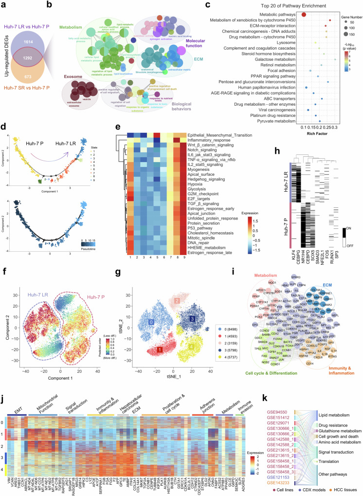
Hepatocellular carcinoma (HCC) is a leading cause of cancer-related mortality, and resistance to systemic therapies remains a significant clinical challenge. This study investigated the mechanisms by which metabolic reprogramming contributes to systemic treatment resistance in HCC. We established HCC cell lines with multidrug resistance characteristics and observed enhanced metabolic activity in these cells. Integrated multiomics analyses revealed hyperactive glucose‒lipid and glutathione metabolic pathways that play critical roles in supporting tumor cell proliferation and survival. We constructed a metabolic reprogramming atlas for HCC-resistant cells and identified aldo-keto reductase (Aldo-keto reductase family 1 Member B1, AKR1B1) as a key regulator of this reprogramming, which sustains drug resistance by regulating energy metabolism and enhancing stress tolerance. Importantly, AKR1B1 expression levels are closely associated with drug resistance and poor prognosis in HCC patients. The secretory nature of AKR1B1 not only underscores its predictive value but also facilitates the intercellular transmission of drug resistance. In terms of overcoming resistance, the AKR1B1 inhibitor epalrestat significantly mitigated drug resistance when it was used in combination with standard therapies. These findings underscore the importance of metabolic reprogramming in the development of HCC resistance. AKR1B1, a key enzyme that regulates metabolic reprogramming, has been identified as a potential biomarker and therapeutic target, providing new insights into overcoming resistance in HCC treatment.


Conflict of interest statement

Competing interests: The authors declare no competing interests.

Figures

Fig. 1
Fig. 1
Multidrug resistance and associated metabolic adaptations in drug-resistant cells. a Schematic representation of the drug-resistant cell model development process alongside lenvatinib and sorafenib IC50 values. The drug concentrations used for induction ranged from 1–20 μM for the Huh-7 LR cells and from 1–5 μM for the Huh-7 SR cells. Schematic figures were generated with BioRender (https://app.biorender.com/). b Drug sensitivity testing for targeted therapies and chemotherapeutic agents in drug-resistant and parental cells, presented as IC50 values. c Drug sensitivity evaluation in CDX models derived from drug-resistant and parental cells (n = 5/group). CDX model drug application concentrations: solvent: 5‰ carboxymethyl cellulose sodium; lenvatinib: 5 mg/kg/d. d Tumor growth kinetics in CDX models derived from drug-resistant and parental cells. Testing method: Unpaired Student’s t-test. e Multicolor IF staining of metabolic enzymes in tumor tissues from CDX models of drug-resistant and parental cells. GLUT1 (yellow), FABPs (purple), CD31 (red), FASN (blue), CD36 (green), and DAPI (gray). Scale bar = 100 μm. f Quantification of multicolor IF staining intensity in tumor tissues from CDX models. Testing method: Unpaired Student’s t-test. g Multicolor IF staining of metabolic enzymes in tumor tissues from HCC patients treated with or without systemic therapy. GLUT1 (yellow), FABPs (purple), CD31 (red), FASN (blue), CD36 (green), and DAPI (gray). Scale bar = 100 μm. h Quantification of multicolor IF staining intensity in patient-derived tumor tissues. Testing method: Unpaired Student’s t-test. *p < 0.05, **p < 0.01, ***p < 0.001, ****p < 0.0001
Fig. 2
Fig. 2
Transcriptomic analysis highlights stemness characteristics and metabolic reprogramming in drug-resistant cells. a Venn diagram depicting the upregulated genes shared between Huh-7 LR and Huh-7 SR (FC > 1.5, p < 0.05) based on bulk RNA sequencing. b PPI network analysis using ClueGo for commonly upregulated genes in the Huh-7 LR and Huh-7 SR. c KEGG analysis identified the top 20 enriched pathways of commonly upregulated genes in Huh-7 LR and Huh-7 SR cells. d Pseudotime trajectory analysis via Monocle2 and cell state clustering derived from single-cell sequencing of drug-resistant and parental cells. e Heatmap generated through GSVA via the MSigDB hallmark dataset for the 9 states derived from pseudotime analysis. f Stemness assessment of drug-resistant and parental cells from single-cell sequencing data conducted via CytoTRACE. g Clustering visualization through t-SNE dimensionality reduction from single-cell sequencing data. h Transcription factor analysis derived from single-cell sequencing of drug-resistant and parental cells. i PPI network analysis of downstream target gene clusters regulated by the transcription factors KLF4 and CEBPG. j GSEA was performed on clustering data from single-cell sequencing of drug-resistant and parental cells. k Proportional KEGG pathway enrichment analysis of upregulated genes across drug resistance-related datasets from multiple GEO database sources
Fig. 3
Fig. 3
Metabolic adaptations in drug-resistant HCC cells. a Nontargeted metabolomics profiling of parental and Huh-7 LR cells. b FFA profiling in parental and Huh-7 LR cells. c Enrichment analysis of upregulated metabolites (FC > 1.2, p < 0.05) in Huh-7 LR cells compared with parental cells. d Seahorse extracellular flux analysis of glycolytic activity (ECAR) in parental and drug-resistant cells. Testing method: Unpaired Student’s t-test. e Visualization of intracellular lipid droplet accumulation during drug resistance development. Lipid droplets (green), the cell membrane (red), and the nucleus (blue). Scale bar = 25 μm. IC50 values: Huh-7 LR_lenvatinib (S0: 13.02 μM; S1: 19.15 μM; S2: 36.40 μM; S3: 50.61 μM); Huh-7 SR_sorafenib (S0: 6.77 μM; S1: 9.46 μM; S2: 13.53 μM; S3: 19.46 μM). f Correlation analysis between the intracellular lipid droplet content and drug IC50 in drug-resistant cells. Testing method: Pearson’s correlation coefficient test. g Integrated metabolomic (four replicates per cell type, averaged in pairs), transcriptomic (two replicates per cell type), and supplementary kit-based assay profiling to map metabolic adaptations in drug-resistant cells. h High-resolution 13C-glucose metabolic flux analysis of glycolysis, the TCA cycle, and glutathione metabolism pathways. The “M+number” indicates the number of additional ¹³C atoms in the metabolite molecule. *p < 0.05, **p < 0.01, ***p < 0.001, ****p < 0.0001
Fig. 4
Fig. 4
AKR1B1 overexpression in drug-resistant cells modulates drug sensitivity. a Integrated transcriptomic (upregulated genes, FC > 2, p < 0.05) and metabolomic profiling identified the key enzymes implicated in HCC drug resistance. b IF validation of AKR1B1 expression in parental and drug-resistant cells. AKR1B1 (red) and DAPI (blue). Scale bar = 25 μm. c WB analysis of AKR1B1 expression in cells during the development of drug resistance. d Correlation analysis of AKR1B1 expression levels with drug resistance in drug-resistant cells. Testing method: Pearson’s correlation coefficient test. e IHC staining of AKR1B1 in tumor tissues from HCC patients treated with or without systemic therapy. The results of the IHC quantification of AKR1B1 are presented in the right panel. Scale bar = 100 μm. Testing method: Unpaired Student’s t-test. f Quantification of serum AKR1B1 levels in HCC patients treated with or without systemic therapy. Testing method: Unpaired Student’s t-test. g Heatmap illustrating drug sensitivity in Huh-7 LR cells following AKR1B1 knockdown. h Drug sensitivity evaluation in CDX models derived from drug-resistant cells following AKR1B1 knockdown (n = 5/group). CDX model drug application concentrations: solvent: 5‰ carboxymethyl cellulose sodium; lenvatinib: 5 mg/kg/d. i Tumor growth kinetics in CDX models generated from drug-resistant cells following AKR1B1 knockdown. Testing method: Unpaired Student’s t-test. j Tumor weight measurements in CDX models generated from drug-resistant cells following AKR1B1 knockdown. Testing method: Unpaired Student’s t-test. k Visualization of intracellular lipid droplet levels in drug-resistant cells following AKR1B1 knockdown. Lipid droplets (green), the cell membrane (red), and the nucleus (blue). Scale bar = 25 μm. Testing method: Unpaired Student’s t-test. l WB analysis of key glutathione-regulating enzymes in drug-resistant cells following AKR1B1 knockdown via statistical analysis. Testing method: Unpaired Student’s t-test. m Quantification of intracellular GSH levels in drug-resistant cells following AKR1B1 knockdown. GSH (teal green), the cell membrane (red), and the nucleus (blue). Scale bar = 25 μm. Testing method: Unpaired Student’s t-test. n Quantification of ROS levels in drug-resistant cells following AKR1B1 knockdown. ROS (red), nuclei (blue). Lenvatinib: 50.61 μM (IC50), 12 h; sorafenib: 16.70 μM (IC50), 12 h. Scale bar = 25 μm. Testing method: Unpaired Student’s t-test. *p < 0.05, **p < 0.01, ***p < 0.001, ****p < 0.0001
Fig. 5
Fig. 5
Associations of AKR1B1 expression with HCC patient prognosis and primary drug resistance. a The HCC cohort was divided into three clinical therapy subcohorts: no chemotherapy (NC), partial response (PR), and disease progression (DP). Clinical parameters are indicated in the heatmap. MVI: microvascular invasion; T stage: tumor stage; TACE: transcatheter arterial chemoembolization; HAIC: hepatic arterial infusion chemotherapy. b Comparison of AKR1B1 expression levels in tumor tissues among HCC patients in the NC, PR, and DP subcohorts. c OS rate of patients in the TCGA HCC iCluster_3 cohort in relation to AKR1B1 expression levels. Testing methods: Comparison of survival differences between the two groups: log-rank test; calculation of the hazard ratio (HR): Cox regression model. d Heatmap illustrating drug sensitivity in parental cells following AKR1B1 overexpression. e Schematic representation of CM extraction from drug-resistant cells and its application to parental cells. Schematic figures were generated with BioRender (https://app.biorender.com/). f Live/dead fluorescent probe analysis of drug resistance transfer and cell mortality in parental 3D microtissues cultured with conditioned medium from drug-resistant cells. Scale bar = 50 μm. Testing method: Unpaired Student’s t-test. g Cell tracing and IF analysis of AKR1B1 transfer from drug-resistant cells to parental cells. Cell tracing (green): drug-resistant cells; AKR1B1 (red); nuclei (blue). Scale bar = 20 μm. h WB analysis of AKR1B1 expression in various HCC cell lines and drug-resistant cells via statistical analysis. i Drug sensitivity evaluation was performed on the basis of IC50 values to assess drug resistance transfer between multiple HCC cell lines via CM. Testing method: Unpaired Student’s t-test. *p < 0.05, **p < 0.01, ***p < 0.001, ****p < 0.0001
Fig. 6
Fig. 6
A combination therapy strategy mitigates drug resistance in HCC. a Evaluation of the activity of drug-resistant cells treated with epalrestat (IC30 = 27.50 μM) in combination with lenvatinib (IC30 = 43.32 μM) or sorafenib (IC30 = 8.89 μM). Testing method: Unpaired Student’s t-test. b CI analysis for the coadministration of epalrestat with lenvatinib or sorafenib. c Drug sensitivity evaluation in CDX models derived from drug-resistant cells following coadministration of epalrestat and lenvatinib (n = 5/group). CDX model drug application concentrations: solvent: 5‰ carboxymethyl cellulose sodium; epalrestat: 50 mg/kg/d; lenvatinib: 5 mg/kg/d; combined regimen: epalrestat 50 mg/kg/d + lenvatinib 5 mg/kg/d. d Tumor weight measurements in CDX models generated from drug-resistant cells following coadministration of epalrestat and lenvatinib. Testing method: Unpaired Student’s t-test. e Tumor growth kinetics in CDX models generated from drug-resistant cells following coadministration of epalrestat and lenvatinib. Testing method: Unpaired Student’s t-test. f Ki-67 IHC staining of tumor tissues from nude mice treated with the combination therapy. Scale bar = 100 μm. g IF analysis of AKR1B1-expressing HCC PDOs. DAPI (gray), AFP (yellow), AKR1B1 (red), CK19 (orange), and Ki-67 (green) are shown. Scale bar = 10 μm/20 μm. h Brightfield microscopy image of the impact of coadministration of epalrestat and lenvatinib on HCC PDOs at 0 h and 48 h. Scale bar = 100 μm. Organoid drug application concentrations: Epalrestat: 53.96 μM; lenvatinib: 50.61 μM; combined regimen: Epalrestat 53.96 μM + lenvatinib 50.61 μM. i Resazurin assay to evaluate the viability of HCC PDOs after 48 h of coadministration of epalrestat and lenvatinib. Testing method: Unpaired Student’s t-test. j Live/dead probe staining was used to assess the survival rate of HCC PDOs following 48 h of coadministration of epalrestat and lenvatinib. Scale bar = 500 μm. k Schematic representation of metabolic reprogramming in drug-resistant cells and the role of AKR1B1 in HCC drug resistance. *p < 0.05, **p < 0.01, ***p < 0.001, ****p < 0.0001

References

    1. Bray, F. et al. Global cancer statistics 2022: GLOBOCAN estimates of incidence and mortality worldwide for 36 cancers in 185 countries. CA Cancer J. Clin.74, 229–263 (2024). - PubMed
    1. Kim, E. & Viatour, P. Hepatocellular carcinoma: old friends and new tricks. Exp. Mol. Med.52, 1898–1907 (2020). - PMC - PubMed
    1. Brouillet, A. & Lafdil, F. Risk factors for primary liver cancer initiation associated with tumor initiating cell emergence: novel targets for promising preventive therapies. eGastroenterology1, e100010 (2023). - PMC - PubMed
    1. Tan, K. et al. Evaluating Tislelizumab, Lenvatinib, and FOLFOX4-HAIC as a conversion therapy for unresectable hepatocellular carcinoma. iLIVER2, 163–169 (2023). - PMC - PubMed
    1. Li, P. et al. Alteration of chromatin high-order conformation associated with oxaliplatin resistance acquisition in colorectal cancer cells. Exploration3, 20220136 (2023). - PMC - PubMed

MeSH terms