. 2025 Aug 1;23(1):360.
doi: 10.1186/s12964-025-02312-x.

Synergic action of MicroRNAs and Wnts delivered by motor neuron EVs in promoting AChR clustering

Affiliations

Synergic action of MicroRNAs and Wnts delivered by motor neuron EVs in promoting AChR clustering

Rachele Agostini et al. Cell Commun Signal. .

Abstract

Background: The neuromuscular junction (NMJ) establishment occurs through complex communication events between motor neurons and muscle fibers; however, the molecular mechanisms leading to NMJ formation have yet to be fully elucidated. Little is known about the significance of extracellular vesicles (EVs) in mediating the interaction between motor neurons and muscle fiber in the NMJ establishment; this study investigates the role of motor neuron-derived EVs during the earliest stages of NMJ formation.

Methods: NSC-34 cells have been used as a model of motor neurons; EVs have been isolated during neurite development using a serial ultracentrifugation protocol specifically adjusted to isolate large and small EVs. Isolated EVs were quantified through Nanoparticles Tracking Assay and characterized by Western Blot and TEM analyses. The microRNA (miRNA) cargo of EV subpopulations was identified by small-RNA sequencing and the predicted miRNA downstream targets were investigated.

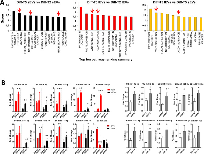
Results: NGS analysis of small RNAs carried by NSC-34-derived EVs identified a total of 245 EV specific miRNAs, most of which are up-regulated in NSC-34 cells and EVs during neurite stretching. Target prediction analysis evidenced how these miRNAs synergically target the Wnt signaling pathway. Moreover, we found that NSC-34-derived EVs carry Wnt proteins, including Wnt11, Wnt4 and Wnt3a. Since several studies suggested a role for the Wnt-associated signaling network in NMJ formation, we investigated the potential role of NSC-34 EVs in NMJ development and demonstrated that EV administration to myotubes increases acetylcholine receptor (AChR) cluster formation, as revealed by immunofluorescence staining with α-bungarotoxin. Moreover, myotube treatment with NSC-34-derived EVs led to GSK3β and JNK phosphorylation, followed by β-catenin nuclear translocation, suggesting that neuron-derived EVs can induce AChR clustering through Wnt pathway activation.

Conclusion: These data demonstrate that EVs released from differentiated motor neurons carry multimodal signals, miRNAs, and Wnts, which can stimulate AChR clustering in myotubes, a fundamental preparatory stage for NMJ formation. These new data highlight that EVs may play a role in the NMJ establishment and function under physiological and pathological conditions, particularly neurodegenerative diseases.

Keywords: Acetylcholine receptor; Agrin; Extracellular vesicles; Neuromuscular junctions; Wnt signalling; β-catenin.


Conflict of interest statement

Declarations. Ethical approval and consent to participate: All animal care and handling procedures were approved by the Institutional Animal Care and Use Committee of Istituto Italiano di Tecnologia, Genova, Italy. Consent for publication: All authors read and approved the final manuscript. Competing interests: The authors declare no competing interests.

Figures

Fig. 1
Fig. 1
Quantification and characterization of the EVs released during NSC-34 differentiation. Light microscopy images of the NSC-34 cells before (A; size bar: 200 μm) and after (B; size bar: 100 μm) inducing neuron process outgrowth. In particular, picture B clearly shows neurite development and branching obtained after 4 days of differentiation. Cell viability assayed after 4 days of differentiation is shown in Additional file 10. C) Time course of gene expression analysis of the key differentiation markers MAP2 (microtubule-associated protein), GAP-43 (growth associated protein 43) and ChAT (choline acetyltransferase), and (D) MotomiRs miR-9-3p and miR-124-3p in differentiating NSC-34 cells (n = 3, * p < 0.05, ** p < 0,01; one-way ANOVA test followed by Dunnett’s post-hoc test). The process of differentiation was carried out on NSC-34 cells for two days (Diff-T2), and four days (Diff-T4), and at each of these time points, the collection and characterisation of EVs was undertaken. E) Schematic representation of the serial ultracentrifugation protocol used to harvest EVs from conditioned medium, and separate large from small EVs (lEVs and sEVs, respectively); the NTA distribution plots of vesicle hydrodynamic diameter revealed the difference in size between lEVs and sEVs. F) The size distribution of lEVs and sEVs was also confirmed using transmission electron microscopy (in the panels b, c, d, e and f the size bar corresponds to 100 nm, while in the panels a and g the bar corresponds to 200 nm). G) Western blots analyses show the presence of the well-established EV markers: Alix, Hsp70 and Tsg101, and the negative marker for sEVs, Calnexin (5 µg of total proteins were loaded per lane). Molecular weight markers (kDa) are indicated. Original, uncropped immunoblots are reported in Additional file 7- Fig. S1
Fig. 2
Fig. 2
NGS analysis of miRNAs loaded into EVs and released during the NSC-34 differentiation process. A) Descriptive analyses of miRNAs detected in small and large EV samples (N = 12) at two differentiation time points (Diff-T2 and -T5) using small RNA seq. In the Venn diagrams, each area reports the number (%) of miRNAs detected in the corresponding group in at least two samples (N = 3 per group). The upper part of the panel A shows the comparison of the miRNAs detected in lEVs and sEVs regardless the differentiation time, while in the lower part lEVs and sEVs have been analysed separately, and the differences between the number of miRNAs detected at Diff-T2 and Diff-T5 have been reported. B) Unsupervised PCA analysis revealing the similarity between groups. The progressive separation of EV samples collected at Diff-T5 can be appreciated. C) and D) Volcano plot evidencing the number of miRNAs differentially abundant (Moderated t-test < 0.05, fold change > 1.5) in small and large EVs, respectively, when comparing Diff-T5 vs. Diff-T2. E) and F) Heatmap representation of the differentially abundant miRNAs in three EV replicate samples during neuronal differentiation. Red color indicates greater abundance, green color indicates lower abundance. The Panel G reports the list of miRNAs differentially abundant in large and small EVs at Diff-T5 differentiation and their relative expression. The X-axis represents the Log2 fold change in miRNA counts between Diff-T5-lEVs vs. Diff-T5-sEVs. Green bars are miRNAs more abundant in large EVs at Diff-T5, red bars are miRNAs more abundant in sEVs at Diff-T5
Fig. 3
Fig. 3
Dissecting the complex signals carried by neuron-derived EVs during NSC-34 differentiation. A) miRNAs significantly modulated in lEVs and sEVs collected from late stage of the NSC-34 neuron process outgrowth compared to early stage development using DIANA-mirPath v.3 (https://dianalab.e-ce.uth.gr/html/mirpathv3/). DIANA-mirPath is a miRNA pathway analysis web-server, providing accurate statistics, to predicted miRNA targets (in CDS or 3’-UTR regions) provided by the DIANA-microT-CDS algorithm or even experimentally validated miRNA interactions derived from DIANA-TarBase. In the three panels top ten pathway rankings were reported. B) Since, the prediction analysis of the significantly modulated EV-miRNAs, obtained by NGS, suggested that Axon guidance, neurotrophin, Wnt and TGF-𝛽 signalling pathways could be the targets of the miRNAs loaded into NSC-34-derived EVs, qRT-PCR analyses of selected key miRNAs (miR-9-3p, -16-5p, -335-5p, -669a-3p, -344b-3p, -218-1-3p, -124-3p, -709, -34c-3p, -185-5p) were performed both in cell bodies and EVs with the aim to confirm miRNA expression. MiRNA expression analysis performed in lEVs and sEVs at early (Diff-T2) and late (Diff-T5) is reported on the left part of the panel B (n = 3, * p < 0.05, ** p < 0,01; Diff-T5 vs Diff-T2 were compared by t-test for lEVs and sEVs separately); in the right part of the panel B, miRNA expression levels were quantified in cell bodies (n = 3, * p < 0.05, ** p < 0.01; t-test)
Fig. 4
Fig. 4
Expression analysis of Wnts during NSC-34 differentiation. (A) Gene expression analysis of Wnt isoforms in NSC-34 whole cell during neuron process outgrowth (Diff-T0: undifferentiated NSC-34 cells; Diff-T2: NSC-34 switched to neurobasal medium for 2 days; Diff-T5: NSC-34 switched to neurobasal medium for 5 days; n = 3, * p < 0.05, ** p < 0,01; one-way ANOVA test followed by Dunnett’s post-hoc test). (B) Western blot analysis of Agrin, Wnt3a, and Wnt11 in differentiating NSC-34 cells. In each lane 30 µg of total proteins were loaded and actin expression was used as loading control. (C) lEVs and sEVs were isolated from NSC-34 during differentiation as indicated in Fig. 1. Whole cell extracts of undifferentiated NSC-34 cells (CB Diff-T0) and extracellular vesicles (lEVs and sEVs) at indicated times were probed with Tsg101, CD81 and CD9, used as EV markers, and Calnexin as the negative control. The figure clearly shows that Wnt11 is present in both lEVs and sEVs whereas Wnt3a is expressed only in sEVs, with a decreasing trend during neurite process development. Five µg of total proteins were loaded in each lane. Molecular weight markers (kDa) are indicated. Original, uncropped immunoblots are reported in Additional File 8 - Fig. S2
Fig. 5
Fig. 5
Motor neuron-derived EVs stimulate AChR cluster formation in C2C12 plasma membrane. (A) C2C12 myotubes and differentiating NSC-34 cells were co-cultured and allowed to interact for 48 h without physical contact thanks to the presence of a membrane filter of 0.4 μm pores interposed between the two cell lines. (B) After 48 h co-culture, myotubes were harvested and mRNA extracted to evaluate the gene expression of selected targets; the obtained data show that the presence of NSC-34 cells (and likely their released signals) induced in C2C12 myotubes a decrease and an increase in AChR alpha and Rspo2 mRNAs, respectively (n = 3, * p < 0.05; t-test). (C) The α-Bungarotoxin confocal laser scanning microscopy analysis highlights a significant increase in AChR clusters in response to the EV administration. (D) The morphometric analysis of the AChR clusters shows that lEV and sEV treatments increased the number of AChR clusters per µm of fibre and reduces the cluster area; in detail the increase of the fluorescence intensity emitted from clusters in the treated samples, is more evident when the intensity was related to the cluster area (* p < 0.05, one-way ANOVA test followed by Dunnett’s post-hoc test). (E) At the molecular level, lEVs and sEVs induce the stimulation of Rspo2 in the EV-treated myotubes and a tendency to decrease of AChR alpha and Axin-1 mRNA expression (* p < 0.05, one-way ANOVA test followed by Dunnett’s post-hoc test). (F) An ex vivo model of primary motor neurons derived from spinal cord explants was used to corroborate the results obtained using the NSC-34 model. Total EVs were collected from motor neurons grown for 9–11 days after explantation and used to treat mature myotubes; the conditioned medium (CM) not centrifuged was used to compare bulk with vesicular signals. (G) In line with NSC34-derived EV effects, Rspo2 and Axin-1 mRNA levels are modulated by motor neuron EVs (* p < 0.05, one-way ANOVA test followed by Dunnett’s post-hoc test for Rspo2 and Bonferroni post-hoc test (Medium vs. EVs) for Axin-1)
Fig. 6
Fig. 6
NSC-34-derived EVs trigger β-catenin dependent and independent Wnt pathways for AChR clustering. (A) Target prediction of NSC-34-derived EV miRNAs and DIANAmirPath analysis of Wnt signalling (Kegg pathway), based on DIANA miRPath v.3.0 program. The miRNAs selected from the first two quartiles of Diff-T 2-T5 lEVs and sEVs have been used for the analysis. The miRNAs highlighted in red target the genes involved in the destruction complex of the 𝛽-catenin. (B) The α-Bungarotoxin confocal laser scanning microscopy analysis highlights a significant increase in AChR clusters in response to the Agrin and sEV treatments. (C) Expression analysis of some targets of the Wnt signalling pathway by qPCR analyses in C2C12 myotubes treated with Agrin or increasing quantities of NSC-34 sEVs (sEV concentrations were 5 × 108, 1 × 109, and 5 × 109 particles/mL indicated by symbols). * p < 0.05, one-way ANOVA test followed by Dunnett’s post-hoc test. (D) Western blotting analysis: the increased expression of GSK3 inactive form and JNK active form was measured as ratio of the phosphorylated isoform/not phosphorylated isoform. The fold change refers to the control condition (non-treated C2C12 myotubes). ** p < 0.01, *** p < 0.001, one-way ANOVA test followed by Dunnett’s post-hoc test. (E) Immunofluorescence staining of C2C12 myotubes treated with NSC-34-derived sEVs, representative pictures showing the increased nuclear localization of 𝛽-catenin (red) and DAPI (blue) following EV treatment. (F) Western blotting analysis of the 𝛽-catenin active form in C2C12 myotubes treated with NSC-34 sEVs ranging from 108 to 5 × 1010 particles/mL. Tubulin expression was used as loading control. Arbitrary units refer to the control condition. * p < 0.05; ** p < 0.01 *** p < 0.001, one-way ANOVA test followed by Dunnett’s post-hoc test. Original, uncropped immunoblots are reported in Additional File 9 - Fig. S3
Fig. 7
Fig. 7
Hek-WNT11 sEVs induce GSK3β phosphorylation. (A) NTA distribution plot of the hydrodynamic diameter size of Hek-WNT11 sEVs. (B) The Western blot analysis of the sEVs secreted by wild type and WNT11-HA tag-overexpressing HEK cells shows the presence of the well-established exosomal marker CD63, and the WNT11 or HA tag (10 µg of total proteins were loaded per lane). Molecular weight markers (kDa) are indicated. (C) Western blot analysis of GSK3α/β, JNK and β-catenin in C2C12 myotubes treated with increasing concentrations of Hek sEVs (lanes 2–4) or Hek-WNT11 sEVs (lanes 5–7) as indicated in the figure. Non-treated C2C12 myotubes represent the control (lane 1); Tubulin was used as the housekeeping protein. Molecular weight markers (kDa) are indicated. Original, uncropped immunoblots are reported in Additional File 11 - Fig. S4. (D) Expression of GSK3 inactive form e JNK active form was measured as ratio of the phosphorylated isoform/not phosphorylated isoform. The fold change refers to the control condition. *** p < 0.001, one-way ANOVA test followed by Dunnett’s post-hoc test

References

    1. Slater C. The structure of human neuromuscular junctions: some unanswered molecular questions. Int J Mol Sci. 2017;18:2183. 10.3390/ijms18102183. - PMC - PubMed
    1. Li L, Xiong W-C, Mei L. Neuromuscular junction formation, aging, and disorders. Annu Rev Physiol. 2018;80:159–88. 10.1146/annurev-physiol-022516-034255. - PubMed
    1. Kummer TT, Misgeld T, Sanes JR. Assembly of the postsynaptic membrane at the neuromuscular junction: paradigm lost. Curr Opin Neurobiol. 2006;16:74–82. 10.1016/j.conb.2005.12.003. - PubMed
    1. Anderson MJ, Cohen MW. Nerve-induced and spontaneous redistribution of acetylcholine receptors on cultured muscle cells. J Physiol. 1977;268:757–73. 10.1113/jphysiol.1977.sp011880. - PMC - PubMed
    1. Zong Y, Zhang B, Gu S, et al. Structural basis of agrin–LRP4–MuSK signaling. Genes Dev. 2012;26:247–58. 10.1101/gad.180885.111. - PMC - PubMed

LinkOut - more resources