. 2025 Dec;14(1):2538684.
doi: 10.1080/2162402X.2025.2538684. Epub 2025 Aug 2.

Systematic identification of lincRNA-derived immunogenic peptides in melanoma

Affiliations

Systematic identification of lincRNA-derived immunogenic peptides in melanoma

Emilie Dupré et al. Oncoimmunology. 2025 Dec.

Abstract

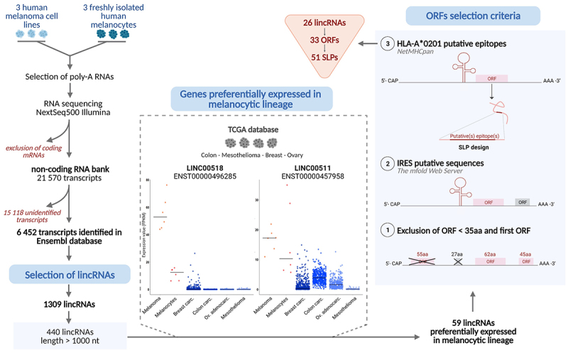
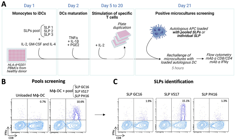
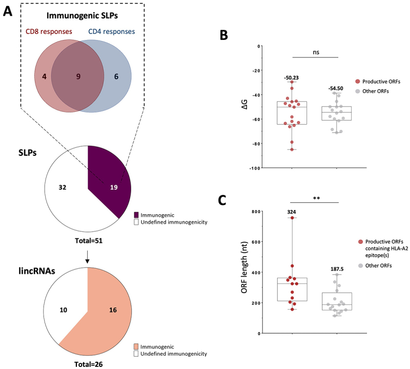
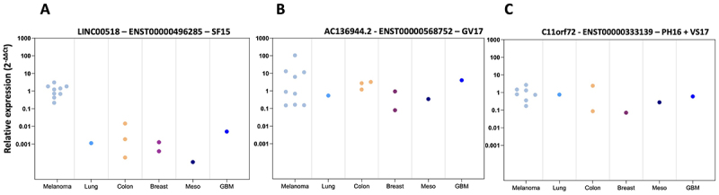
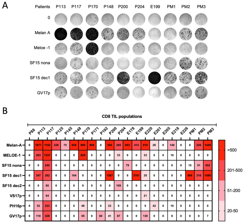
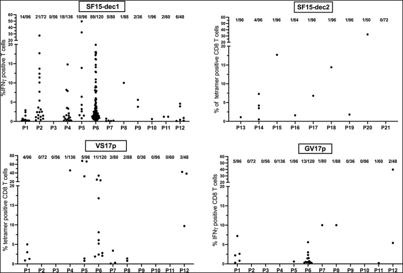
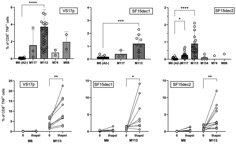
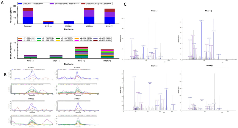
The search for reliable shared tumor-specific antigens (TSAs) to improve cancer immunotherapy is on-going. The so-called non-coding regions of the genome have recently been shown to give rise to immunogenic peptides, including the melanoma-specific antigen MELOE-1 which is translated from the long intergenic non-coding RNA (lincRNA) meloe in an IRES-dependent manner. Here, we present a strategy to systematically identify tumor-specific antigens produced by ORFs within lincRNAs with IRES-like upstream structures. We provide evidence suggesting that in the melanocytic lineage a significant proportion of the selected lincRNAs can produce immunogenic peptides. T cell repertoires against some of these peptides were found in peripheral blood mononuclear cells (PBMCs) from healthy donors and melanoma patients, and in tumor-infiltrating lymphocytes (TILs) from metastatic melanoma patients. Finally, CD8+ T cell lines from melanoma patients specific for three of the characterized HLA-A *0201 epitopes could recognize melanoma cell lines, which were enhanced by reticular stress. Thus, these peptides may represent a new class of shared TSAs in melanoma and are attractive candidates for evaluation as targets for immunotherapy in preclinical studies. In addition, our selection strategy has the potential to identify new lincRNA-derived antigens in other cancers.

Keywords: Cancer immunotherapy, T cell epitopes; long intergenic non-coding RNA; melanoma; tumor antigens.


Conflict of interest statement

No potential conflict of interest was reported by the author(s).

Figures

Figure 1.
Figure 1.
Pipeline of in silico selection of lincRNA, ORF and SLPs. After deep sequencing of poly-A RNA of 3 melanoma and 3 melanocytes cell lines, coding transcripts were excluded and lincRnas > 1,000nt were selected. 59 candidates were further selected for their high expression. ORF selection: i) exclusion of the first ORF and of ORFs < 35 aa, ii) presence of a putative IRES structure within the first 200 nt upstream of the ATG. Within these 33 ORFs, 51 putative HLA A * 0201 epitopes were identified and used to design SLPs.
Figure 2.
Figure 2.
Amplification of specific T cells and SLP identification. A. Schematic representation of in vitro specific T cell expansion. HLA-A *0201 PBMCs were seeded in 96 –well plates with pools of 2 to 3 SLPs in the presence of GM-CSF, IL4 and IL2. On D2, a DC maturation cocktail was added. Fresh IL-2 (50UI/ml) was replaced as needed until screening at D21. Microcultures were restimulated for 5 h with Mφ-DC pulsed with pools of SLPs and IFNγ secretion was assessed. B. Example of a positive microculture reacting against a pool of three SLP identified by IFNγ intracellular staining. C. Within positive pools, individual immunogenic SLP were identified by restimulation for 5 h with a SLP-pulsed HLA-A *0201 B cell line.
Figure 3.
Figure 3.
A high proportion of selected lincRnas code for immunogenic SLPs. A. 19 out of 51 SLPs tested were immunogenic, originating from 16 lincRnas. B. No difference in stability (γG) of the 200 nt secondary structures upstream of the selected ORFs between ORFs that produced CD8 and/or CD4 epitopes (red) and ORFs that did not (gray). C. ORFs producing HLA-A *0201 epitopes (red) are significantly longer than the non-producing ones (p < 0.01, Mann-Whitney test).
Figure 4.
Figure 4.
RT-qPCR of immunogenic transcripts in various cancer cell lines. Relative expressions of each transcript, normalized to RPLPO gene, (panel a for linc00518, panel B for transcript AC136944.2 and panel C for transcript C11orf72) were assessed in cancer cell-lines from various tissues (LUD, lung cancer; SW601, SW480, HCT116: colon; MCF7, MDA-MB-231: breast; Meso34/mesothelioma; U251 for glioblastoma (GBM)) compared to the mean expression within melanoma cell lines.
Figure 5.
Figure 5.
Detection of specific CD8+ T lymphocytes in TILs from HLA-A *0201 melanoma patients. A. Example of an IFNγ ELISpot assay performed on TILs stimulated with the indicated HLA A * 0201 epitopes. Unstimulated cells were used as negative controls. B. Heat map representing the number of spots/10 TIL (above background) for each epitope in 22 patients.
Figure 6.
Figure 6.
Detection of specific CD8+ T lymphocytes in PBMCs from HLA-A *0201 melanoma patients. PBMCs were stimulated with indicated HLA-A *0201 epitopes in 96 well plates and rechallenged at D21. Specific T cells were detected either by tetramer staining or by IFNγ production. The proportion of positive microcultures for each patient is indicated.
Figure 7.
Figure 7.
Expression of lincRNA-derived epitopes on melanoma cell lines. A. A panel of melanoma cell lines were co-cultured for 5 h with T cell lines specific for VS17p, SF15-dec1 or SF15-dec2 and melanoma recognition was assessed by intracellular TNFγ staining. The HLA-A *0201 negative melanoma cell line was used as control. ****p < 0.0001, Kruskal-Wallis test. B. M113 and M6 were treated with thapsigargin (200 nM) for 24 h before co-culture with the specific T cell populations and recognition was assessed as in A. **p < 0.01, Wilcoxon paired test. C. A representative experiment showing TNFγ production of the VS17p-specific T cell population after co-culture with melanoma cell-line M6 (negative control) or M113 melanoma cell-line, pre-treated or not with thapsigargin. Dot plots are shown after gating on CD8+ cells to exclude melanoma cells from the analysis.
Figure 8.
Figure 8.
Detection of the translation of ORF549-905 from LINC00518 in melanoma cells by mass spectrometry. The peptide THGPYVITGDYPR (SRM5) was detected in total cell extracts from cells in thapsigargin treated (+) or untreated (-) conditions. (A) distribution of peak areas for precursor and transition ions of SRM5 across the different conditions. (B) identification spectra of precursor and transition ions based on retention time during the PRM analysis targeting SRM5. (C) Representative MS/MS spectrum of SRM5 showing the fragment ions used for identification, with labeled transitions corresponding to those reported in the supplementary table 2.

Similar articles

References

    1. Ribas A, Wolchok JD.. Cancer immunotherapy using checkpoint blockade. Science. 2018;359(6382):1350–17. doi: 10.1126/science.aar4060. - DOI - PMC - PubMed
    1. Schumacher TN, Schreiber RD.. Neoantigens in cancer immunotherapy. Science. 2015;348(6230):69–74. doi: 10.1126/science.aaa4971. - DOI - PubMed
    1. Snyder A, Makarov V, Merghoub T, Yuan J, Zaretsky JM, Desrichard A, Walsh LA, Postow MA, Wong P, Ho TS, et al. Genetic basis for clinical response to CTLA-4 blockade in melanoma. N Engl J Med. 2014;371(23):2189–2199. doi: 10.1056/nejmoa1406498. - DOI - PMC - PubMed
    1. Rizvi NA, Hellmann MD, Snyder A, Kvistborg P, Makarov V, Havel JJ, Lee W, Yuan J, Wong P, Ho TS, et al. Mutational landscape determines sensitivity to PD-1 blockade in non–small cell lung cancer. Science. 2015;348(6230):124–128. doi: 10.1126/science.aaa1348. - DOI - PMC - PubMed
    1. Bassani-Sternberg M, Bräunlein E, Klar R, Engleitner T, Sinitcyn P, Audehm S, Straub M, Weber J, Slotta-Huspenina J, Specht K, et al. Direct identification of clinically relevant neoepitopes presented on native human melanoma tissue by mass spectrometry. Nat Commun. 2016;7(1):13404–13416. doi: 10.1038/ncomms13404. - DOI - PMC - PubMed

MeSH terms

LinkOut - more resources