

Genomic characterization and pre-clinical evaluation of a new polyvalent lytic Loughborough phage
- PMID: 40751804
- PMCID: PMC12317907
- DOI: 10.1007/s00253-025-13559-2
Genomic characterization and pre-clinical evaluation of a new polyvalent lytic Loughborough phage
Abstract
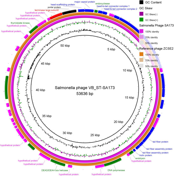
Carbapenem-resistant Acinetobacter baumannii (CRAB) has become a critical concern that necessitates the development of novel antimicrobial approaches. One of the most promising innovative approaches for combating CRAB infections is using effective and lytic bacteriophages (phages), known as phage therapy. Therefore, we recovered and characterized a polyvalent lytic Salmonella_phage_VB_ST-SA173, producing lytic activity against 6 CRAB clinical isolates and 3 multidrug-resistant (MDR) Salmonella serovars. Throughout pH 2-10, and thermal stability at up to 60 °C, the phage maintained its stability and lytic activity against the tested isolates. The presence of a tailed phage with a characteristic prolate head and a contractile tail was detected by the transmission electron microscope (TEM). According to the Oxford nanopore sequencing data, the genome of Salmonella_phage_VB_ST-SA173 was 53,636 bp in size, contained 45.9% G + C, and had 53 opening reading frames (ORFs). According to the TEM, ORFs, and BLASTn analysis findings, it was proved that the Salmonella_phage_VB_ST-SA173 belongs to the Loughboroughvirus genus. The efficacy of the phage-formulated Carbopol 940 hydrogel in wound healing was assessed preclinically in an infected burn wound animal model with a CRABa clinical isolate. The survival rate was enhanced in the phage-treated group compared to the untreated control groups. Histopathological analysis showed improved wound healing in the form of apparently healthy skin with apparently normal epidermal and dermis layers. In conclusion, depending on its in vitro and physicochemical traits, the phage-loaded hydrogel is expected to be a promising candidate for clinical trials against human CRAB-related skin infections. KEY POINTS: • A polyvalent Loughboroughvirus phage showed lytic activity against CRAB and Salmonella serovars. • The phage showed stability at a wide range of pH and temperature. • The phage hydrogel enhanced healing in the burn-wound animal model infected with CRABa.
Keywords: Acinetobacter baumannii; CRAB; Hydrogel; MDR Salmonella; Molecular analysisSalmonella_; Polyvalent phage; Thermal animal model.
© 2025. The Author(s).
Conflict of interest statement
Declarations. Ethical approval: The research ethical committee of Ain Shams University’s Faculty of Pharmacy in Egypt gave authorization to the study (Protocol approval number: ACUC-FP-ASU-RHDIRB2020110301-REC#206). The animals were handled according to the ARRIVE and Care and Use of Laboratory Animals requirements. ARRIVE Guidelines ( https://arriveguidelines.org ). Consent to participate: Not applicable. Consent for publication: Not applicable. Competing interests: The authors declare no competing interests.
Figures


References
-
- Abdelaziz AA, Abo Kamer AM, Nosair AM, Al-Madboly LA (2023) Exploring the potential efficacy of phage therapy for biocontrol of foodborne pathogenic extensively drug-resistant Escherichia coli in gastrointestinal tract of rat model. Life Sci 315:121362. 10.1016/j.lfs.2022.121362 - PubMed
-
- Abdelaziz NA, Elkhatib WF, Sherif MM, Abourehab MAS, Al-Rashood ST, Eldehna WM, Mostafa NM, Elleboudy NS (2022) In Silico docking, resistance modulation and biofilm gene expression in multidrug-resistant Acinetobacter baumannii via cinnamic and gallic acids. Antibiotics 11:870. 10.3390/antibiotics11070870 - PMC - PubMed
-
- Abo Kamer AM, Abdelaziz AA, Nosair AM, Al-Madboly LA (2022) Characterization of newly isolated bacteriophage to control multi-drug resistant Pseudomonas aeruginosa colonizing incision wounds in a rat model: in vitro and in vivo approach. Life Sci 310:121085. 10.1016/j.lfs.2022.121085 - PubMed
MeSH terms
LinkOut - more resources
Full Text Sources
