. 2025 Aug 2;15(1):28260.
doi: 10.1038/s41598-025-13102-3.

Tcf21 modulates fibroblast activation and promotes cardiac fibrosis after injury via Pdgfrb signaling

Affiliations

Tcf21 modulates fibroblast activation and promotes cardiac fibrosis after injury via Pdgfrb signaling

Yusuke Baba et al. Sci Rep. .

Abstract

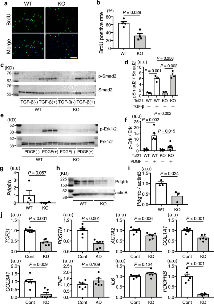
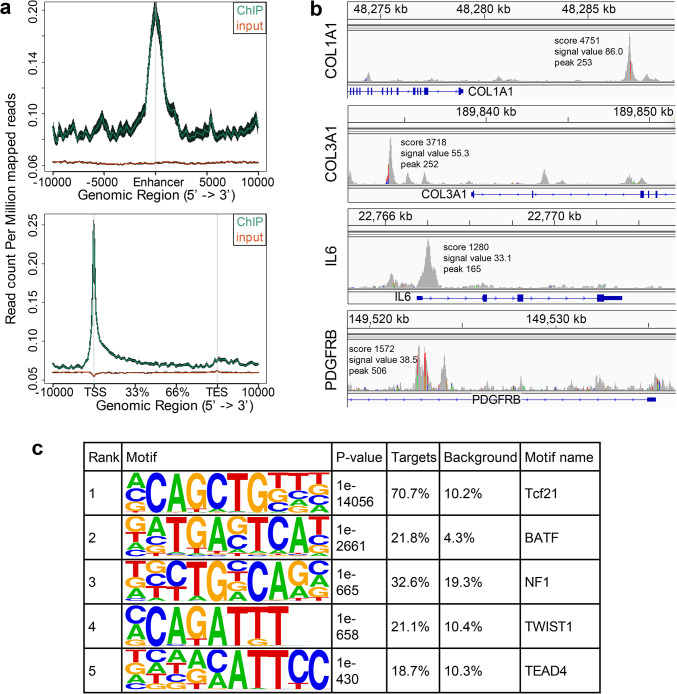
Transformation of cardiac fibroblasts into matrix-producing myofibroblasts plays an important role in tissue repair and fibrosis after myocardial injury. Tcf21 is a basic helix-loop-helix transcription factor that is essential for the development of cardiac fibroblasts and the epicardium. Myofibroblasts are derived from Tcf21 (+) residual fibroblasts; however, whether Tcf21 itself promotes fibrosis is still unknown. Since Tcf21 deficiency leads to perinatal lethality, cardiac fibroblasts were isolated from Tcf21-knockout mouse embryos, and mice that lack Tcf21 after birth were generated (inducible Tcf21 KO, iTcf21). In vitro analysis revealed that Tcf21 promoted cell proliferation and upregulated the expression of extracellular matrix genes. Notably, the Pdgfrb-Erk pathway was severely suppressed in Tcf21-knockout fibroblasts. Chromatin immunoprecipitation-sequencing assay demonstrated the direct binding of Tcf21 to COL1A1, COL3A1, IL6, and PDGFRB loci. Integrated analysis identified pathways involved in fibroblast activation, extracellular matrix production and cell proliferation. Moreover, iTcf21 mice were resistant to cardiac fibrosis induced by isoproterenol injection or metabolic overload with streptozotocin-induced diabetes and a high-fat diet. Our results demonstrate a critical role for Tcf21 in the transformation of cardiac fibroblasts into activated myofibroblasts. It directly binds to gene loci and works in a pro-fibrotic manner via Pdgfrb signaling.

Keywords: Cardiac fibroblasts; Cardiac fibrosis; Erk; Matrix production; Pdgfrb; Tcf21.


Conflict of interest statement

Declarations. Competing interests: The authors declare no competing interests.

Figures

Fig. 1
Fig. 1
Tcf21 knockout (KO) fibroblasts show attenuated proliferation capacity and reduced Pdgfrb signaling. (a,b) 5-bromo-2′-deoxyuridine (BrdU) uptake decreased in Tcf21 KO fibroblasts (biologically n = 4). Scale bar, 200 µm. (c) Western blots of Smad2 and phosphorylated Smad2 (p-Smad2), with TGF-β (10 ng/mL) stimulation for 15 min (biologically n = 3). (d) Quantitative analysis of p-Smad2/Smad2. (e) Western blots of Erk and phosphorylated Erk (p-Erk), with PDGF-BB (20 ng/mL) stimulation for 15 min (biologically n = 3). (f) Quantitative analysis of p-Erk/Erk. (g) qRT-PCR, (h,i) Western blot analysis of Pdgfrb (biologically n = 3). (j) qRT-PCR analysis using human cardiac fibroblasts with TCF21 knockdown by CRISPR-CAS9 showing decreased expression of TCF21, POSTN, ACTA2, COL1A1, COL3A1, and PDGFRB (biologically n = 3).
Fig. 2
Fig. 2
Tcf21 regulates extracellular matrix, proliferation, and inflammation-associated genes. (a,b) Results of gene expression cluster analysis obtained from DAVID 6.8 and Metascape showing altered expression of extracellular matrix and proliferation signaling genes in fibroblasts obtained from the heart of Tcf21 KO mice compared to those obtained from control mice (biologically n = 3). (c) Volcano plot shows that the expression of 440 genes was significantly altered by Tcf21. Upregulated genes in Tcf21 WT fibroblasts contain many extracellular matrix genes. Red dots represent significant genes upregulated in Tcf21 WT (P < 0.05). Blue dots represent significant genes downregulated in Tcf21 WT (P < 0.05). (d) Results of quantitative RT-PCR showing decreased expression of extracellular matrix and fibroblast activation genes in Tcf21 KO fibroblasts (biologically Control n = 15, iKO n = 24).
Fig. 3
Fig. 3
Tcf21 bound to specific chromosomal regions related to extracellular matrix and PDGFRB. (a) DNA pulled down by chromatin immunoprecipitation (ChIP) was distributed around the transcription start site (TSS) and enhancer region. (b) Strong Tcf21 binding peaks were observed around the TSS of COL1A1, COL3A1, and IL6. (c) Result of motif analysis obtained using HOMER2 with a size parameter 200. Tcf21 majorly bound to the E-box domain (CAGCTG), and other DNA sequences, such as AP-1, TEAD4, and TWIST1 domains, were also detected as the Tcf21 binding motifs.
Fig. 4
Fig. 4
Isoproterenol (ISO) injection leads to cardiac fibrosis and expansion of Tcf21-expressing cells. (a) Overview of the ISO injection schedule. (b) Cardiac fibrosis area of Masson trichrome-stained sections after ISO injection. Dunnett’s test was performed for post-hoc analysis (biologically n = 3). (c) Tcf21 expression before and after ISO injection assessed by qPCR. Dunnett’s test was performed for post-hoc analysis (biologically n = 3). (d) Tcf21-positive area calculated based on β-galactosidase staining (biologically n = 4). The detected area was normalized to the cardiac cross-sectional area. (e,f) Tcf21-positive cells accumulated in the fibrotic area after 3 or 7 days of ISO injection. Tcf21-positive cells were calculated in the Masson trichrome (MT) positive or negative area. Scale bar, 100 µm (biologically n = 3). (g) Immunostaining of a section of heart from Tcf21LacZ/+ transgenic mice. Tcf21 expression as assessed by β-galactosidase (green) and periostin (red) staining coexisted after 3 days of ISO injection. Scale bar, 100 µm. (h) Immunostaining of Ki67 (brown) and Tcf21 expression by β-galactosidase (blue). Ki67-positive cells accumulated in the Tcf21-positive area. Scale bar, 100 µm. (i) Proliferation capacity of Ki67-positive cells. Each sample was counted in a high-power microscopic field more than three times, and the average values were used for analysis. Unpaired two-tailed t-tests with Bonferroni correction were used for post-hoc analysis (biologically n = 4). (j) Fibroblast-associated gene expression increased after ISO injection. Dunnett’s test was performed for post-hoc analysis (biologically n = 3).
Fig. 5
Fig. 5
Lack of Tcf21 suppresses cardiac fibrosis and maintains cardiac function. (a) Overview of isoproterenol (ISO) injection schedule. (b) Cardiac fibrosis observed at each time point. Masson trichrome staining was perfoemed. Before ISO injection and 3, 7, and 28 days after the injection. Scale bar, 1 mm. Yellow arrowheads indicated fibrotic area. (c) Fibrosis area from Masson trichrome staining normalized with cardiac cross-sectional area (biologically n = 3 ~ 4). (d) Immunostaining for Ki67. Scale bar, 50 µm. Arrows indicate Ki67-positive cells. (e) Change in the number of Ki67-positive cells, 7 days after ISO injection (biologically n = 5). (f) M-mode echocardiographic view, one week after ISO injection. (g) Changes in left ventricular fractional shortening (FS) after ISO injection. Dunnett’s multiple comparison test was performed to compare baseline data (biologically Control n = 14, iKO n = 7). (h) Protocol for hyperglycemic and hyperlipidemic injury. (i) Masson trichrome-stained heart Sections 16 weeks after diabetes induction with HFD. Arrows show fibrosis. Scale bar, 50 µm. (j) Fibrosis area calculated from Masson trichrome staining normalized by the cardiac cross-sectional area (biologically n = 4 and 3). Student’s t-test was performed.

References

    1. Grossman, W., Jones, D. & McLaurin, L. P. Wall stress and patterns of hypertrophy in the human left ventricle. J. Clin. Invest.56(1), 56–64 (1975). - PMC - PubMed
    1. Depre, C. & Taegtmeyer, H. Metabolic aspects of programmed cell survival and cell death in the heart. Cardiovasc. Res.45(3), 538–548 (2000). - PubMed
    1. Talman, V. & Ruskoaho, H. Cardiac fibrosis in myocardial infarction-from repair and remodeling to regeneration. Cell Tissue Res.365(3), 563–581 (2016). - PMC - PubMed
    1. Kawaguchi, M. et al. Inflammasome activation of cardiac fibroblasts is essential for myocardial ischemia/reperfusion injury. Circulation123(6), 594–604 (2011). - PubMed
    1. Khalil, H. et al. Fibroblast-specific TGF-beta-Smad2/3 signaling underlies cardiac fibrosis. J. Clin. Invest.127(10), 3770–3783 (2017). - PMC - PubMed

MeSH terms

Substances

LinkOut - more resources