Review
. 2025 Aug 4;23(1):17.
doi: 10.1186/s12947-025-00354-2.

Mini 3D transesophageal probe: technical advances and clinical applications

Affiliations
Review

Mini 3D transesophageal probe: technical advances and clinical applications

Monica Barki et al. Cardiovasc Ultrasound. .

Abstract

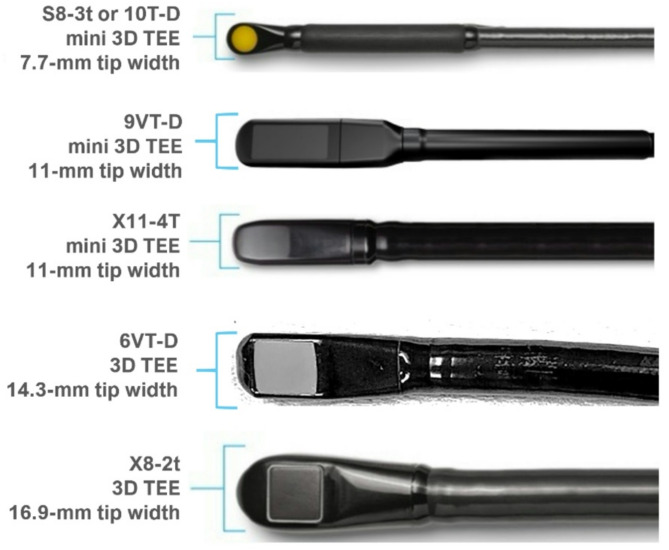
With the growing complexity of structural heart disease procedures, the need for advanced intraprocedural imaging has become increasingly critical. Transesophageal echocardiography remains the gold standard for procedural guidance but is associated with risks such as upper gastrointestinal tract injury and the need for general anesthesia for patient comfort and safety. Miniaturized three-dimensional transesophageal echocardiography (miniTEE) probes offer a promising solution by providing high-resolution imaging which could be performed under conscious sedation. Studies evaluating the miniTEE probe for safety, image quality, and ability to guide specific structural and non-structural heart disease procedures will be reviewed. The limitations and future developments will be discussed.

Keywords: Miniaturized; Three-dimensional transesophageal echocardiography.


Conflict of interest statement

Declarations. Ethics approval and consent to participate: Not applicable. Consent for publication: Not applicable. Competing interests: The authors declare no competing interests. Disclosures: Dr. Hahn reports speaker fees from Abbott Structural, Baylis Medical, Edwards Lifesciences and Philips Healthcare; she has institutional consulting contracts for which she receives no direct compensation with Abbott Structural, Anteris, Boston Scientific, Edwards Lifesciences, Medtronic, Novartis and Philips Healthcare; she is Chief Scientific Officer for the Echocardiography Core Laboratory at the Cardiovascular Research Foundation for multiple industry-sponsored valve trials, for which she receives no direct industry compensation.

Figures

Fig. 1
Fig. 1
Transesophageal echocardiography probes. The relative sizes of the transesophageal echocardiography (TEE) probe head are shown
Fig. 2
Fig. 2
Intraprocedural imaging with the miniTEE probe during left atrial appendage occlusion (LAAO). Panel A shows the biplane image of the LAA for sizing the device. Panel B shows the implantation of a Watchman FLX device with the final 3D imaging of the device (panel C)
Fig. 3
Fig. 3
Post-implant imaging with the miniTEE probe during transcatheter aortic valve implantation (TAVI). This case shows the use of the X11-4T phased array miniTEE probe during a transcatheter aortic valve implantation procedure under monitored anesthetic care. The patient had baseline renal dysfunction and in order to reduce contrast use, the probe was inserted immediately following the TAVI implantation to a mid-esophageal imaging level. Panel A shows biplane color Doppler showing a non-obstructed left main coronary artery. Panel B shows repositioning of the biplane image to the left ventricular outflow track and trivial paravalvular regurgitation at the site of a calcific nodule posteriorly. The probe was advanced to the deep transgastric (TG) level to image flow across the transcatheter valve. Pulsed wave and continuous wave Doppler (panel D) showed excellent valve hemodynamics
Fig. 4
Fig. 4
Imaging for mitral transcatheter edge-to-edge repair (M-TEER). In this case of an 83 year old patient with esophageal stricture, the X11-4T miniTEE probe was used to guide the M-TEER procedure under general anesthesia. Baseline imaging showed adequate mitral valve orifice area (MVOA, panel A) with severely increased effective regurgitant orifice area (EROA, panel B). Three-dimensional (3D) reconstruction could be used to guide introduction of the catheters and positioning of the device (panel C) with grasping of the mitral valve leaflets (panel D) performed using two-dimensional imaging for higher temporal/spatial resolution. After placement of the M-TEER devices, the final MVOA by 3D planimetry of the double orifice was 2.5 cm [2] with EROA of 0.07 cm [2] (panel D) consistent with very mild residual mitral regurgitation

Similar articles

References

    1. Agricola E, Ancona F, Bartel T, et al. Multimodality imaging for patient selection, procedural guidance, and follow-up of transcatheter interventions for structural heart disease: a consensus document of the EACVI task force on interventional cardiovascular imaging: part 1: access routes, transcatheter aortic valve implantation, and transcatheter mitral valve interventions. Eur Hear J Cardiovasc Imaging. 2023;24(9):E209–68. 10.1093/EHJCI/JEAD096. - PubMed
    1. Hahn RT, Kodali SK. State-of-the-art intra-procedural imaging for the mitral and tricuspid PASCAL repair system. Eur Heart J Cardiovasc Imaging. 2022;23(3):E94–110. 10.1093/EHJCI/JEAB040. - PMC - PubMed
    1. Hahn RT, Makkar R, Makar M, et al. EVOQUE tricuspid valve replacement system: State-of-the-Art screening and intraprocedural guidance. JACC Cardiovasc Interv. 2024;17(18):2093–112. 10.1016/J.JCIN.2024.07.034. - PubMed
    1. Hasnie AA, Parcha V, Hawi R, et al. Complications associated with transesophageal echocardiography in transcatheter structural cardiac interventions. J Am Soc Echocardiogr. 2023;36(4):381–90. 10.1016/j.echo.2022.12.023. - PMC - PubMed
    1. Gangwani MK, Aziz A, Dahiya DS, et al. Transesophageal echocardiography–associated Gastrointestinal injuries: systematic review and pooled rates of Gastrointestinal injuries. Bayl Univ Med Cent Proc. 2023;36(6):729–33. 10.1080/08998280.2023.2243381. - PMC - PubMed

MeSH terms

LinkOut - more resources