

Rheumatoid Nodule on the Hallux
- PMID: 40755468
- PMCID: PMC12310347
- DOI: 10.53045/jprs.2024-0005
Rheumatoid Nodule on the Hallux
Abstract
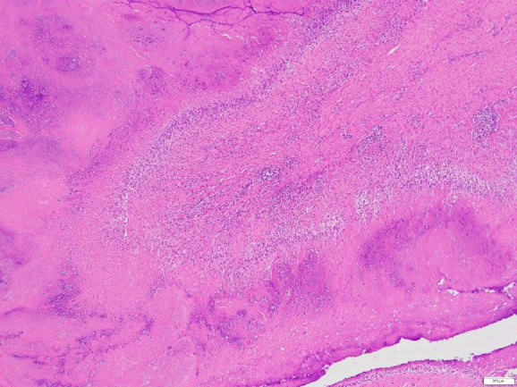
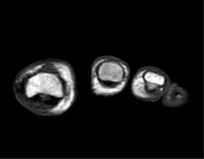
A subcutaneous rheumatoid nodule is a superficial soft tissue lesion that generally affects areas of frequent mechanical contact, such as the extensor aspect of the extremities, occiput, and sacrum. Rheumatoid nodules of the foot are rare, occurring in only 1% of cases. Herein, we present a case of a rheumatoid nodule on the plantar side of the left hallux in a 65-year-old woman with rheumatoid arthritis. The tumor was a 15 × 10 mm, dome-shaped, nontender, elastic hard mass in the plantar aspect of the left hallux. The discovery of rheumatoid nodules provides crucial information for the treatment and management of rheumatoid arthritis. Giant cell tumors may cause symptoms similar to those of rheumatoid nodules. Furthermore, difficulties are associated with differentiating rheumatoid nodules from giant cell tumors using imaging modalities. Therefore, the identification of these tumors requires careful observation.
Keywords: giant cell tumor; hallux; rheumatoid arthritis; rheumatoid nodule; soft tissue tumor.
© 2025 The Japan Society of Plastic and Reconstructive Surgery.
Conflict of interest statement
Conflicts of Interest: There are no conflicts of interest.
Figures


References
-
- Smolen JS, Aletaha D, Mcinnes IB. Rheumatoid arthritis. Lancet. 2016 Oct;388:2023-38. - PubMed
-
- Tastekin E, Btrtane M, Kilinc S, et al. From pathology to diagnosis: a symptom-free patient with a rheumatoid nodule in the foot. Turk J Rheumatol. 2012 Sep;27(3):195-9.
-
- Turesson C, Jacobsson LTH. Epidemiology of extra-articular manifestations in rheumatoid arthritis. Scand J Rheumatol. 2004;33(2):65-73. - PubMed