. 2025 Jul 25:2025:2208110.
doi: 10.1155/cdr/2208110. eCollection 2025.

Rivaroxaban Ameliorates Sunitinib-Induced Injury of Cardiomyocytes via Repressing MAPK Signaling Pathway

Affiliations

Rivaroxaban Ameliorates Sunitinib-Induced Injury of Cardiomyocytes via Repressing MAPK Signaling Pathway

Ying Qian et al. Cardiovasc Ther. .

Abstract

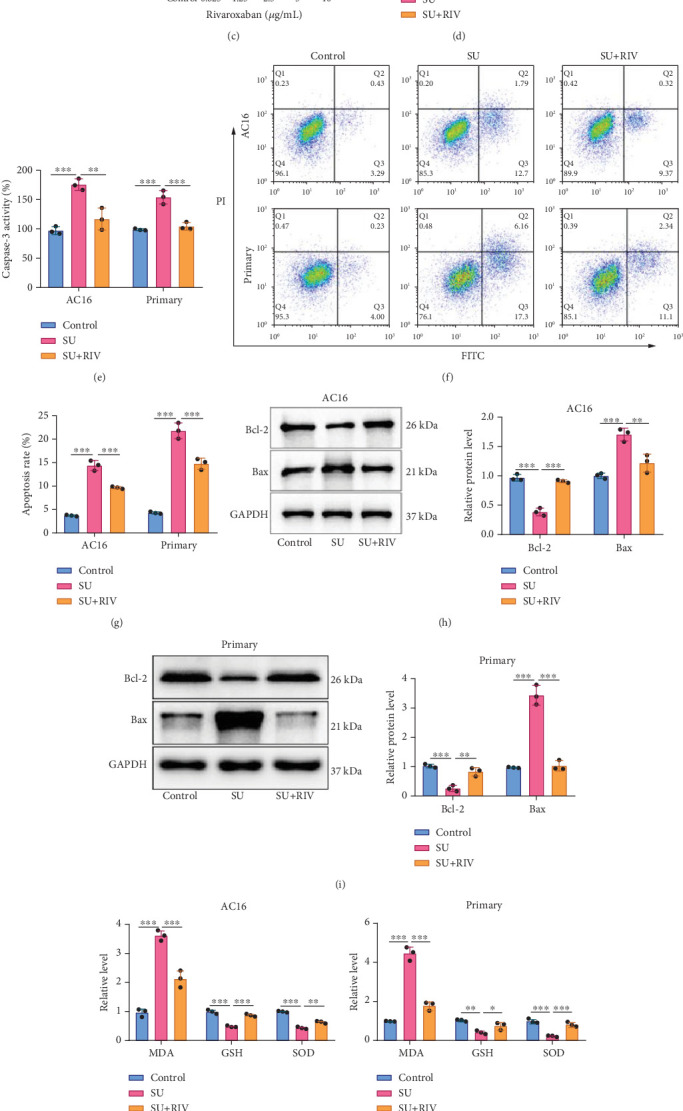
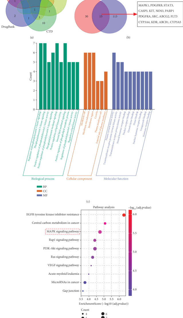
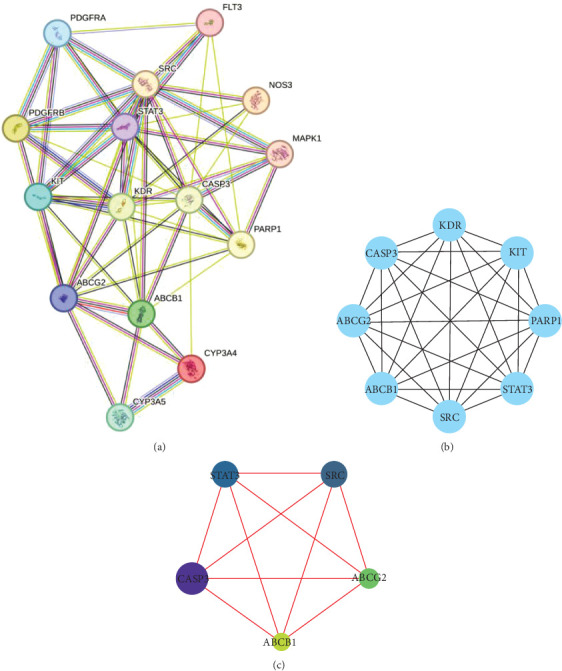
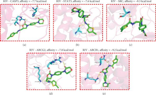
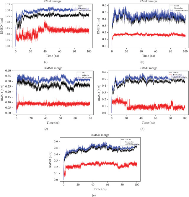
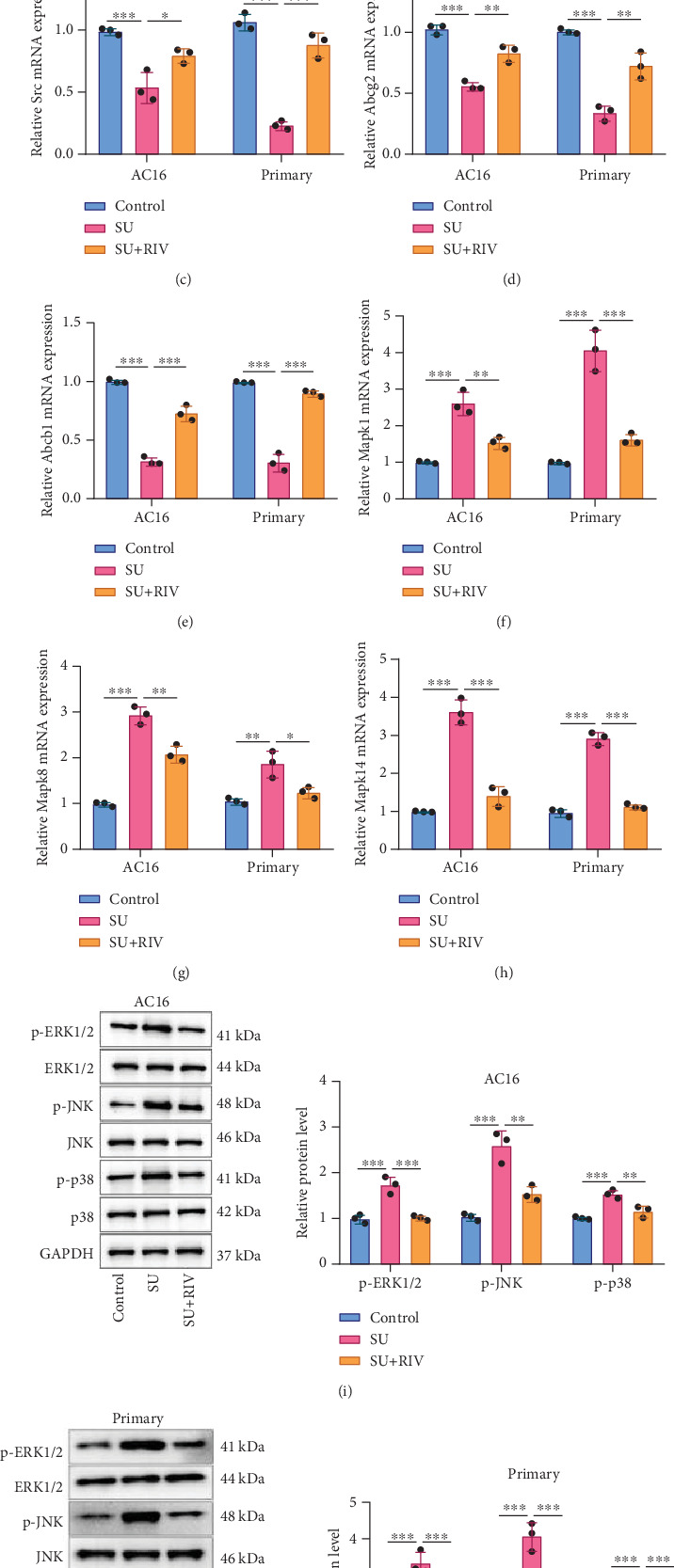
Background: Sunitinib (SU) is used to treat kidney cancer. However, it can also cause cardiotoxicity. This study is performed to investigate whether rivaroxaban (RIV) attenuates SU-induced cardiotoxicity (SIC). Methods and Materials: AC16 cells and primary cardiomyocytes of neonatal mouse were treated with different concentrations (2-10 μM) of SU for 24 h or with 6 μM SU and 10 μg/mL RIV for 24 h. The viability of cardiomyocytes was evaluated using the cell counting kit-8 (CCK-8) assay, and the apoptosis rate was evaluated using flow cytometry. The activity of caspase-3 was determined. The levels of malondialdehyde (MDA), glutathione (GSH), and superoxide dismutase (SOD) were also measured. The potential targets and downstream pathways of RIV in SIC treatment were investigated using network pharmacology, molecular docking, and molecular dynamics simulation. qPCR and western blotting were used to detect the regulatory effects of SU and RIV on mRNA and protein expression of MAPK pathway-related genes, respectively. Results: RIV treatment alleviated SU-induced cardiomyocyte injury by promoting viability and inhibiting apoptosis, oxidative stress, and the inflammatory response in AC16 cells and primary cardiomyocytes. Caspase 3 (CASP3), signal transducer and activator of transcription 3 (STAT3), SRC proto-oncogene, nonreceptor tyrosine kinase (SRC), ATP-binding cassette subfamily G member 2 (ABCG2), and ATP-binding cassette subfamily B member 1 (ABCB1) were candidate targets of RIV in SIC. The binding affinities between RIV and CASP3, STAT3, SRC, ABCG2, and ABCB1 were all less than -7.5 kcal/mol, indicating that RIV could bind stably to these targets. Bioinformatics analyses suggested that the mitogen-activated protein kinase (MAPK) pathway was involved in the mechanism by which RIV alleviated SIC. RIV treatment decreased the mRNA expression of CASP3 and increased the mRNA expression of STAT3, SRC, ABCG2, and ABCB1 in AC16 cells and primary cardiomyocytes. RIV also inhibited the SU-induced activation of the MAPK pathway. Conclusion: RIV exerts a protective effect against SU-induced cardiomyocyte injury by inhibiting the MAPK signaling pathway. RIV therapy may be a promising strategy to inhibit SU's cardiotoxicity in cancer patients.

Keywords: MAPK pathway; cardiomyocytes; rivaroxaban; sunitinib.


Conflict of interest statement

The authors declare no conflicts of interest.

Figures

Figure 1
Figure 1
SU induces injury of cardiomyocytes. (a) Chemical structure of SU. (b, c) AC16 cells and primary cardiomyocytes were treated with different concentrations (2, 4, 6, 8, and 10 μM) of SU for 24 h, and cell viability was detected by the CCK-8 method. (d) After treating AC16 cells and primary cardiomyocytes without or with 6 μM SU for 24 h, the injury of AC16 cells and primary cardiomyocytes was detected by a caspase-3 assay kit. (e) Flow cytometry was used to evaluate the apoptosis rate of AC16 cells and primary cardiomyocytes. (f, g). Protein expression levels of Bcl-2 and Bax in AC16 cells and primary cardiomyocytes were detected by western blot. (h, i) The levels of MDA, GSH, and SOD in AC16 cells and primary cardiomyocytes were detected to evaluate oxidative stress. (j, k) qPCR was used to detect the mRNA expression levels of inflammatory factors including TNF-α, IL-1β, and IL-6 in AC16 cells and primary cardiomyocytes. All of the experiments were performed in triplicate and repeated for at least three times. The dots in the graphs are representative of three independent biological experiments. ∗∗ and ∗∗∗ represent p < 0.01 and p < 0.001, respectively.
Figure 2
Figure 2
RIV treatment reverses SU-induced injury of cardiomyocytes. (a). Chemical structure of RIV. (b, c) AC16 cells and primary cardiomyocytes were treated with RIV at different concentrations (0.625, 1.25, 2.5, 5, and 10 μg/mL) for 24 h, and cell viability was detected by the CCK-8 method. (d). After treating AC16 cells and primary cardiomyocytes with 6 μM SU or/and 10 μg/mL RIV for 24 h, the cell viability was assessed by the CCK-8 method. (e). A caspase-3 assay kit was used to detect the activity of caspase-3 in AC16 cells and primary cardiomyocytes. (f, g) The apoptosis of AC16 cells and primary cardiomyocytes was detected by flow cytometry. (h, i) Protein expression levels of Bcl-2 and Bax in AC16 cells and primary cardiomyocytes were detected by Western blot. (j, k) The levels of MDA, GSH, and SOD in AC16 cells and primary cardiomyocytes were detected by the corresponding detection kit. (l, m) qPCR was used to detect the mRNA expression levels of inflammatory factors, including TNF-α, IL-1β, and IL-6 in AC16 cells and primary cardiomyocytes. All of the experiments were performed in triplicate and repeated at least three times. The dots in the graphs are representative of three independent biological experiments. ∗, ∗∗, and ∗∗∗ represent p < 0.05, p < 0.01, and p < 0.001, respectively.
Figure 3
Figure 3
GO and KEGG enrichment analysis of candidate targets of RIV in SIC treatment. (a). Collection of RIV's targets. (b). The venn diagram of RIV's targets and SIC-related genes. (c). GO enrichment analysis of RIV's targets in SIC treatment. Biological process (BP) is marked by dark cyan, cellular component (CC) is marked by sienna, and molecular function (MF) is marked by steel blue. (d). Bubble map of KEGG pathway enrichment analysis of RIV's targets in SIC treatment. The bubble size represents count, and the bubble color represents the p value. MAPK pathway is one of the significant pathways probably modulated by these targets.
Figure 4
Figure 4
PPI network construction and analysis of RIV treatment SIC candidate targets. (a) A PPI network of RIV's targets in SIC treatment was constructed based on String database. Nodes represent proteins, and edges represent protein–protein interactions. The olivine edges indicate the interactions from text mining. The black edges indicate coexpression. The purple edges indicate the interactions which were determined experimentally. The blue edges indicate the interactions with gene co-occurrence. The azury edges indicate the interactions from curated databases. The lilac edges indicate protein homology. More details can be found in STRING database (https://cn.string-db.org/). (b) Cluster analysis of PPI network with MCODE plug-in. (c) Centiscape 2.2 plug-in was used to obtain the hub nodes in PPI network. The size of the node is proportional to the size of the degree value.
Figure 5
Figure 5
Molecular docking between RIV and hub targets. (a–e) Molecular docking diagram of RIV with (a) CASP3, (b) STAT3, (c) SRC, (d) ABCG2, and (e) ABCB1 proteins. Light blue indicates amino acid residues surrounding the binding bag, green indicates RIV, pink indicates macromolecules, and yellow dashed lines indicate hydrogen bonding.
Figure 6
Figure 6
MDS suggests that RIV binds stably with the hub targets. Evolution of RMSD values during 100 ns MDS of (a) RIV/CASP3 complex, (b) RIV/STAT3 complex, (c) RIV/SRC complex, (d) RIV/ABCG2 complex, and (e) RIV/ABCB1 complex.
Figure 7
Figure 7
Effects of RIV on the expression levels of hub genes and MAPK pathway-related genes in SU-induced cardiomyocytes. (a–h) After treating AC16 cells and primary cardiomyocytes with 6 μM SU and/or 10 μg/mL RIV for 24 h, the mRNA expression levels of (a) CASP3, (b) STAT3, (c) SRC, (d) ABCG2, (e) ABCB1, (f) MAPK1, (g) MAPK8, and (h) MAPK14 were detected by qPCR. (i, j) Protein expression levels of p-ERK1/2, p-JNK, and p-p38 were detected by Western blot. All of the experiments were performed in triplicate and repeated for at least three times. The dots in the graphs are representative of three independent biological experiments. ∗, ∗∗, and ∗∗∗ represent p < 0.05, p < 0.01, and p < 0.001, respectively.

Similar articles

References

    1. Chu T. F., Rupnick M. A., Kerkela R., et al. Cardiotoxicity Associated With Tyrosine Kinase Inhibitor Sunitinib. Lancet . 2007;370(9604):2011–2019. doi: 10.1016/S0140-6736(07)61865-0. - DOI - PMC - PubMed
    1. Aldemir M. N., Simsek M., Kara A. V., et al. The Effect of Adenosine Triphosphate on Sunitinib-Induced Cardiac Injury in Rats. Human & Experimental Toxicology . 2020;39(8):1046–1053. doi: 10.1177/0960327120909874. - DOI - PubMed
    1. Samama M. M. The Mechanism of Action of Rivaroxaban – An Oral, Direct Factor Xa Inhibitor – Compared with Other Anticoagulants. Thrombosis Research . 2011;127(6):497–504. doi: 10.1016/j.thromres.2010.09.008. - DOI - PubMed
    1. Cohen O., Levy-Mendelovich S., Ageno W. Rivaroxaban for the Treatment of Venous Thromboembolism in Pediatric Patients. Expert Review of Cardiovascular Therapy . 2020;18(11):733–741. doi: 10.1080/14779072.2020.1823218. - DOI - PubMed
    1. Yassin A. S., Abubakar H., Mishra T., et al. Rivaroxaban for Left Ventricular Thrombus. American Journal of Therapeutics . 2019;26(4):e511–e515. doi: 10.1097/MJT.0000000000000726. - DOI - PubMed

MeSH terms