. 2025 Aug 1;14(4):tfaf105.
doi: 10.1093/toxres/tfaf105. eCollection 2025 Aug.

Albiflorin improves diabetic retinopathy by mitigating oxidative stress and inflammation via the TLR-4/NF-kB signaling pathway

Affiliations

Albiflorin improves diabetic retinopathy by mitigating oxidative stress and inflammation via the TLR-4/NF-kB signaling pathway

Liuyi Xie et al. Toxicol Res (Camb). .

Abstract

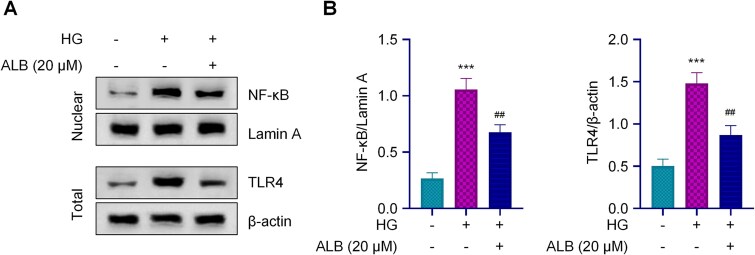
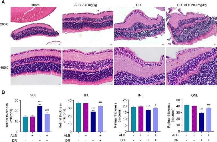
This study was to investigate the effects of Albiflorin (ALB) on oxidative stress and inflammation in diabetic retinopathy (DR) and explore its potential mechanism involving the Toll-like receptor 4 (TLR4)/nuclear factor kappa B (NF-κB) signaling pathway. Human retinal microvascular endothelial cells (HRMECs) were treated with high glucose (HG) and ALB. Cell viability was assessed by MTT assay. Oxidative stress markers and inflammatory cytokines were measured by ELISA. TLR4/NF-κB pathway proteins were analyzed by Western blot. A streptozotocin (STZ)-induced diabetic rat model was established to examine retinal histological changes. Serum metabolic parameters, oxidative stress markers, and inflammatory cytokines were evaluated in the DR model and ALB intervention groups. Results showed that ALB improved HRMEC viability under HG induction and reduced oxidative stress and inflammation. ALB inhibited the TLR4/NF-κB pathway in HG-induced HRMECs. Overexpression of TLR4 partially reversed the protective effects of ALB. In diabetic rats, ALB ameliorated metabolic disorders, improved retinal histological structure, and reduced oxidative stress and inflammation. ALB also suppressed the TLR4/NF-κB signaling pathway in vivo. In conclusion, ALB improves DR by resolving oxidative stress and inflammation through inhibiting the TLR4/NF-κB signaling pathway. These findings suggest ALB as a potential therapeutic agent for DR.

Keywords: Albiflorin; TLR-4/NF-κB signaling; diabetic retinopathy; inflammation; oxidative stress.


Conflict of interest statement

The authors declared that there was no conflict of interest associated with the manuscript.

Figures

Figure 1
Figure 1
ALB mitigates high glucose-induced oxidative stress and inflammation in HRMECs. (A). Cell viability of HRMECs treated with varying concentrations of ALB (0-80 μM) for 24 hours, as determined by MTT assay. B. Cell viability of HRMECs exposed to normal glucose (5.5 mM), high glucose (30 mM), or high glucose with ALB treatment (5, 10, and 20 μM). C. Levels of oxidative stress markers (SOD, MDA, and CAT) in HRMECs under different treatment conditions, as measured by ELISA. D. Expression levels of inflammatory cytokines (TNF-α, IL-6, and IL-1β) in HRMECs, quantified by ELISA. ***p<0.001 compared to control group, #p<0.05, ##p<0.01, and ###p<0.001 compared to HG group.
Figure 2
Figure 2
ALB inhibits the TLR4/NF-κB signaling pathway in high glucose-induced HRMEC injury. A. A. Western blot analysis of TLR4 and nuclear NF-κB expression in HRMECs treated with normal glucose (5.5 mM), high glucose (30 mM), or HG + ALB (20 μM). B. Quantitative analysis of TLR4 and nuclear NF-κB protein levels. Lamin A and β-actin were used as internal controls for nuclear and total protein, respectively. ***p<0.001 compared to control group, and ##p<0.01 compared to HG group.
Figure 3
Figure 3
TLR4 overexpression attenuates ALB-mediated protection against high glucose-induced oxidative stress and inflammation in HRMECs. A. Western blot analysis of TLR4 and nuclear NF-κB expression in HRMECs under different treatment conditions, including TLR4 overexpression. B. Levels of oxidative stress markers (SOD, MDA, and CAT) in HRMECs under various treatment conditions, as measured by ELISA. C. Expression levels of inflammatory cytokines (TNF-α, IL-6, and IL-1β) in HRMECs under different treatments, quantified by ELISA. ***p<0.001 compared to control group, ##p<0.01 compared to HG group, and ^p<0.05; ^^p<0.01 and ^^^p<0.001 compared with ALB treatment group.
Figure 4
Figure 4
ALB ameliorates diabetes-induced metabolic disorders in a rat model of diabetic retinopathy. Rats were randomly divided into six groups and treated with different drugs for 6 weeks: (1) Sham group; (2) ALB 200 mg/kg group; (3) DR group; (4) DR+ALB 50mg/kg; (5) DR+ALB 100 mg/kg; (6) DR+ALB 200 mg/kg (n=6). A. Body weight changes in rats over 6 weeks of treatment. B. Blood glucose levels in rats over the 6-week treatment period. C-F: Serum levels of glucose and lipid metabolism indicators (TC, TG, LDL-C, and HDL-C) as measured by ELISA.***p<0.001 compared with sham group, and #p<0.05 and ##p<0.01 compared to DR group.
Figure 5
Figure 5
ALB improves retinal histopathology in diabetic rats. A. Representative H&E stained images of retinal sections from different treatment groups. Scale bar = 200 μm. B. Quantitative analysis of retinal layer thickness (GCL, IPL, INL, and ONL) across treatment groups. ***p<0.001 compared to sham group, and #p<0.05 and ##p<0.01 compared with DR group.
Figure 6
Figure 6
ALB reduces oxidative stress and inflammation by modulating the TLR4/NF-κB signaling pathway in diabetic rat retinas. A. Levels of oxidative stress markers (SOD, MDA, and CAT) in rat retinas, as measured by ELISA. B. Expression levels of inflammatory cytokines (TNF-α, IL-6, and IL-1β) in rat retinas, quantified by ELISA. C. Western blot analysis and quantification of TLR4 and nuclear NF-κB protein levels in rat retinas. ***p<0.001 compared to sham group, and ###p<0.001 compared to DR group.

Similar articles

References

    1. Sen S et al. Comparative proteomics of proliferative diabetic retinopathy in people with Type 2 diabetes highlights the role of inflammation, visual transduction, and extracellular matrix pathways. Indian J Ophthalmol. 2023:71:3069–3079. 10.4103/IJO.IJO_276_23 - DOI - PMC - PubMed
    1. Tarr JM, Kaul K, Chopra M, Kohner EM, Chibber R. Pathophysiology of diabetic retinopathy. Pathophysiology of diabetic retinopathy. ISRN Ophthalmol. 2013:2013:343560. 10.1155/2013/343560 - DOI - PMC - PubMed
    1. Rodríguez ML, Pérez S, Mena-Mollá S, Desco MC, Ortega ÁL. Oxidative Stress and Microvascular Alterations in Diabetic Retinopathy: Future Therapies. Oxid Med Cell Longev. 2019:2019:1–18. 10.1155/2019/4940825 - DOI - PMC - PubMed
    1. Tang Q et al. Diabetic Retinopathy: New Treatment Approaches Targeting Redox and Immune Mechanisms. Antioxidants (Basel). 2024:13:594. 10.3390/antiox13050594 - DOI - PMC - PubMed
    1. Fung TH, Patel B, Wilmot EG, Amoaku WM. Diabetic retinopathy for the non-ophthalmologist. Clin Med (Lond). 2022;22(2):112–116. doi: 10.7861/clinmed.2021-0792. - DOI - PMC - PubMed

LinkOut - more resources