. 2025 Jul 18:16:1613879.
doi: 10.3389/fimmu.2025.1613879. eCollection 2025.

Mouse B cells engineered to express an anti-HPV antibody elicit anti-tumor T cell responses

Affiliations

Mouse B cells engineered to express an anti-HPV antibody elicit anti-tumor T cell responses

Michal Guberman Bracha et al. Front Immunol. .

Abstract

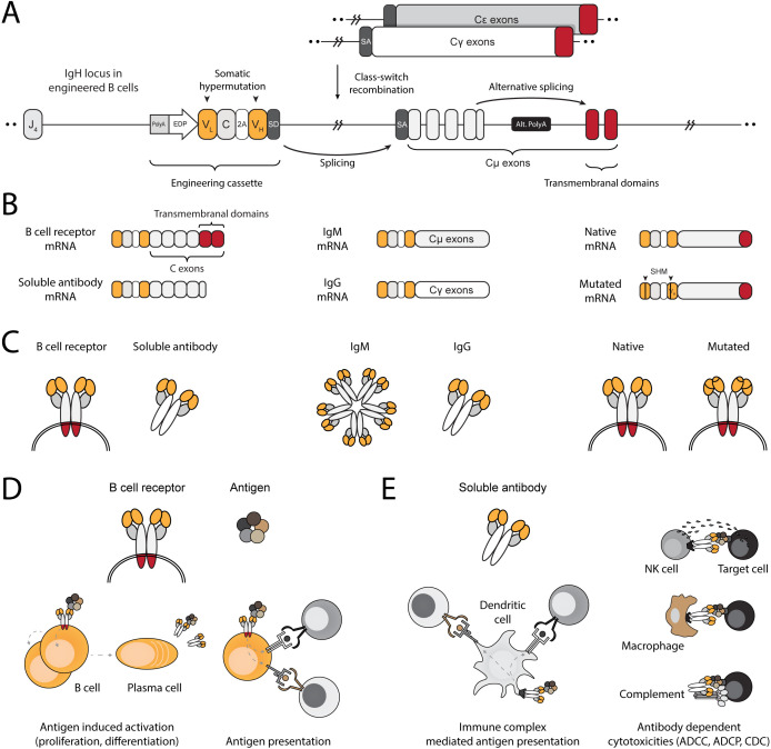
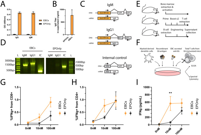
Transplantation of engineered B cells has demonstrated efficacy in HIV disease models. B cell engineering may also be utilized for the treatment of cancer. Recent studies have highlighted that B cell activity is associated with favorable clinical outcomes in oncology. In mice, polyclonal B cells have been shown to elicit anti-cancer responses. As a potential novel cell therapy, we demonstrate that engineering B cells to target a tumor-associated antigen enhances polyclonal anti-tumor responses. We observe that engineered B cells expressing an anti-HPV B cell receptor internalize the antigen, enabling subsequent activation of oncoantigen-specific T cells. Secreted antibodies from engineered B cells form immune complexes, which are taken up by antigen-presenting cells to further promote T cell activation. Engineered B cells hold promise as novel, multi-modal cell therapies and open new avenues in solid tumor targeting.

Keywords: B cell; antibody; cancer; cell engineering; cell therapy (CT); genome editing; tertiary lymphoid structures.


Conflict of interest statement

Authors MG, GB, SS, RA, YV, DB, YK, LK, SH, and AN were employed by the company Tabby Therapeutics Ltd, Israel. The remaining authors declare that the research was conducted in the absence of any commercial or financial relationships that could be construed as a potential conflict of interest.

Figures

Figure 1
Figure 1
Engineering B cells at the IgH locus to enable multi-modal adaptive immune responses. (A) Schematic of the engineered IgH locus in EBCs. The cassette is integrated downstream of the final J segment and upstream of the constant segments. To downregulate endogenous expression, a PolyA signal is added to the 5’ end the cassette, followed by an enhancer dependent promoter (EDP) to upregulate expression upon on-target integration. The coding segment includes a full light chain (VL–C) separated by a 2A peptide from a variable fragment of the heavy chain (VH). A splice donor (SD) at the 3’ end of the cassette enables splicing with the splice acceptor (SA) of the endogenous constant segments, regardless of the isotype expressed in the EBC. (B) mRNA-level expression of the engineering cassette in EBCs. Since it does not include constant segments, the resulting therapeutic antibody expressed in EBCs may be in the form of a BCR when alternative splicing enables incorporation of the membranal exons, or as a soluble antibody when the alternative polyA (Alt. PolyA) is activated in antibody-secreting cells (left). Splicing with endogenous constant segments further enables EBCs to express the antibody as virtually any available isotype (middle). Finally, since the engineering cassette is introduced into the native IgH gene, it can undergo somatic hypermutation (SHM), potentially improving antigen-binding affinity through clonal selection (right). (C) Same as (B), but represented at the protein level. (D) Schematic representation of antibody functions when expressed on the membrane as a BCR. Effector functions of the BCR in EBCs enable antigen-induced activation, leading to EBC proliferation and differentiation into antibody-secreting cells or antigen-presenting cells. The BCR internalizes antigens for processing into peptides, which are then loaded onto MHC complexes. These are subsequently presented to T cells, enabling mutually beneficial interactions. (E) Schematic representation of the antibody after secretion. The soluble antibody binds to soluble antigens, forming immune complexes that facilitate T cell activation via Fc receptor-expressing cells, such as dendritic cells (left). The soluble antibody can bind to membrane-expressed antigens, enabling a range of cytotoxic reactions such as antibody-dependent cell cytotoxicity (ADCC), antibody-dependent cell phagocytosis (ADCP), and complement-dependent cytotoxicity (CDC).
Figure 2
Figure 2
Engineering primary mouse B cells to target HPV-E6. (A) TIDE analysis of mouse splenic lymphocytes edited at the IgH locus. CRISPR-Cas9 activity is detected by insertions and deletions (InDels). Cultured lymphocytes (UT) are compared to cells electroporated with CRISPR-Cas9 ribonucleoproteins (RNPs). Each dot represents an independent experiment, n = 2–9. **pv<0.01, unpaired two-tailed t-test. (B) Representative flow cytometry of mouse splenic lymphocytes engineered to express either the 6F4 (middle column) or C1P5 (right column) anti-HPV-E6 antibodies, compared to cells electroporated but not transduced with AAVs (EPOnly, left column). Engineering rates were assessed using peptide containing either the C1P5 target epitope in the E6 protein (top row) or the 6F4 target epitope (bottom row), both derived from the E6 protein. Pre-gated on live, singlet, CD19+ cells. (C) Quantification of (B), showing engineering rates of primary mouse splenic lymphocytes expressing either the C1P5 or 6F4 anti-HPV-E6 antibodies compared to control EPOnly B cells. To assess specificity, cells were stained using either the C1P5 target epitope (Navajo white) or the 6F4 target epitope (orange), both derived from the E6 protein. Each dot represents an independent experiment, n = 4–8. ns pv>0.05, *pv<0.05, ***pv<0.001, ****pv<0.0001, two-way ANOVA with Tukey’s multiple comparisons test.
Figure 3
Figure 3
EBCs activate CD4 and CD8 T cell responses (A) Schematic representation of the ovalbumin system used in these experiments. B cells were extracted from immunocompetent mice for the production of OVA-specific EBCs. T cells were isolated from OT-I or OT-II mice, which are transgenic for Class I- or Class II-restricted TCRs specific to OVA, respectively. (B) Representative flow cytometry of OVA-specific engineering. EBCs were compared to cells electroporated with RNPs but not transduced with AAVs (EPOnly). Cells were then with or without the OVA antigen. Binding was detected only in electroporated and transduced cells in the presence of the OVA antigen. Pre-gated on live, singlet, CD19+ cells. (C) Quantification of (B) for the +OVA samples. Each dot represents an independent experiment, n = 2–4. *pv<0.05, unpaired two-tailed t-test. (D) Representative flow cytometry of OT-I and OT-II T cells extracted from mice and incubated with APCs pulsed with either the OVA257–264 or the OVA323–339 peptides, recognized by OT-I and OT-II T cells, respectively, but not reciprocally. Pre-gated on singlets, live cells, CD19, CD4+ CD8 (for OT-II co-cultures) and CD8+ CD4 (for OT-I co-cultures). (E) Schematic of the co-culture setup. Engineered B cells were loaded with the OVA antigen and co-cultured with either OT-I or OT-II T cells. (F) Representative flow cytometry of activation markers CD25 and CD69 in OT-I (left) and OT-II (right) co-cultures with EPOnly control cells (top) or EBCs (bottom), pre-loaded with either 0 nM or 100 nM of the OVA antigen. Pre-gated on singlets, live cells, CD4+ CD8 (for OT-II co-cultures) and CD8+ CD4- (for OT-I co-cultures). Numbers in the plots indicate the percentage of CD69+ CD25+ cells. (G) Quantification of (F) for OT-I co-cultures, including data with concentrations of OVA ranging from 0 nM to 1,000 nM. (H) Quantification of (F) for OT-II co-cultures, including data with concentrations of OVA ranging from 0 nM to 1,000 nM. For (G, H), bars represent the mean, error bars indicate SD, n = 2–4, pooled data from two independent experiments. **pv<0.01, ***pv<0.001, two-way ANOVA with Tukey’s multiple comparisons test. (I) IFNg concentrations in the supernatants of OT-I co-cultures. (J) IFNg concentrations in the supernatants of OT-II co-cultures. (K) TFNa concentrations in the supernatants of OT-I co-cultures. (L) TFNa concentrations in the supernatants of OT-II co-cultures. For (I–L), bars represent the mean, error bars indicate SD, n = 2–4, pooled from two independent experiments. ns pv >0.05, **pv<0.01, ***pv<0.001, two-way ANOVA with uncorrected Fisher’s LSD.
Figure 4
Figure 4
EBCs elicit polyclonal anti-tumor T cell responses. (A) Schematic representation of the E6 co-cultures experiments. Mice were immunized three times to generate polyclonal anti-E6 T cells. Independent mice were used as a source for B cell engineering. B cells internalize antigens for presentation to CD4+ and CD8+ T cells (gray arrow). In co-cultures containing CD4+ and CD8+ T cells along with B cells, CD8+ T cell activation may be further enhanced by cytokine-mediated help from CD4+ T cells (orange arrow). (B) Experimental scheme of the co-cultures. EBCs or EPOnly control cells were pre-incubated with the E6 antigen and then added to total T cells from immunized mice. (C) Representative flow cytometry of EBCs engineered to target E6. EBCs were detected with an E6 peptide containing the target epitope of the 6F4 binder. Pre-gated on live, singlet, CD19+ cells. (D) Quantification of (C). Each dot represents an independent experiment, n = 9–10. ****pv<0.0001, unpaired two-tailed t-test. (E) Representative intracellular flow cytometry for IFNg in CD4+ (left) or CD8+ (right) cells from polyclonal co-cultures with EPOnly control cells (above) or EBCs (below) at either 0 nM or 10 nM E6. Pre-gated on singlets, cells, alive, CD4+ CD8 (left) or CD8+ CD4 (right). (F) Quantification of (E) for CD4+ (top) or CD8+ (bottom) T cells. Each dot represents an independent co-culture using either EBC (orange) or EPOnly control cells (black), n = 11. Pooled data from five independent experiments. ns pv >0.05, *pv<0.05, ****pv<0.0001, two-way ANOVA with Šidák’s multiple comparisons tests. (G) ELISA of IFNg secretion from supernatants of T cells from immunized mice co-cultured with EPOnly control B cells (black) or E6-specific EBCs (orange). ns pv >0.05, ****pv<0.0001, two-way ANOVA with Šidák’s multiple comparisons tests. Each dot represents an independent co-culture; data pooled from three individual experiments, n = 7. (H) Degranulation of cytotoxic T cells in co-cultures [as in (B)], monitored by CD107a. Pre-gated on singlets, live cells, CD4 CD8+. ns pv >0.05, **pv<0.01, two-way ANOVA with Šidák’s multiple comparisons tests. Each dot represents an independent co-culture, n = 4. (I) ELISA of Granzyme B in cells from immunized mice co-cultured with either EPOnly control B cells (black) or E6-specific EBCs (orange) as in (B) at 0 nM or 10 nM antigen. ns pv >0.05, **pv<0.01, two-way ANOVA with Tukey’s multiple comparison. Each dot represents an independent co-culture, n = 4.
Figure 5
Figure 5
Immune complexes derived from antibodies secreted by EBCs activate T cells. (A) Isotype-specific ELISA for antibodies against E6 in the supernatants of B cells engineered as in Figure 2A . Mean OD values and SD are shown for IgG (left) or IgM (right) E6-specific antibodies from EBC (orange) or EPOnly (black) cells, n = 2–3. (B) Quantification of IgG data from (A) using a recombinant IgG1 antibody. Mean and SD values are represented. Pooled data from two independent experiments, n = 2. (C) Representative PCR on reverse-transcribed mRNA from EBCs. Primer binding sites are indicated with arrows. The anti-E6 engineering cassette are shown in orange, and endogenous segments in white or gray. For each amplicon, the DNA (top) or mRNA (bottom) are illustrated. Only spliced fragments derived from mRNA are amplified due to polymerization length constraints. (D) Gel electrophoresis of amplicons from (C), using RNA extracted from EBCS (left) or mRNA from EPOnly control B cells (right). Molecular weight ladders with relative sizes are shown beside each gel. (E) Representative schematic of cell sources used in (F–I). Bone marrow from naïve mice was used to generate myeloid-derived dendritic cells. T cells were obtained from mice immunized with E6. B cells for engineering were isolated from separate naïve mice. (F) Representative experimental scheme for (G–I). Myeloid-derived dendritic cells were seeded and loaded with immune complexes formed by incubating E6 antigen with EBC-derived supernatant. T cells from E6-immunized mice were then added to the culture. (G) Intracellular flow cytometry for IFNg in CD8+ T cells from cultures as in (F), incubated with supernatants from either EBCs (orange) or EPOnly control B cells (black). Pre-gated on singlets, live cells, CD4 CD8+. (H) Same as (G), but for CD4+ CD8 T cells. (I) ELISA for IFNg in supernatants from cocultures as described in (F). For (G–I), mean and SEM are represented, n = 5–7, pooled from three independent experiments. **pv<0.01 for two-way ANOVA with Šidák’s multiple comparisons tests.

Similar articles

Cited by

References

    1. Joura EA, Giuliano AR, Iversen OE, Bouchard C, Mao C, Mehlsen J, et al. A 9-valent HPV vaccine against infection and intraepithelial neoplasia in women. N Engl J Med. (2015) 372(8):711–23. doi: 10.1056/NEJMoa1405044, PMID: - DOI - PubMed
    1. Marur S, D’Souza G, Westra WH, Forastiere AA. HPV-associated head and neck cancer: A virus-related cancer epidemic. Lancet Oncol. (2010) 11(8):781–9. doi: 10.1016/S1470-2045(10)70017-6, PMID: - DOI - PMC - PubMed
    1. Näsman A, Du J, Dalianis T. A global epidemic increase of an HPV-induced tonsil and tongue base cancer – potential benefit from a pan-gender use of HPV vaccine. J Intern Med. (2020) 287(2):134–52., PMID: - PubMed
    1. Blagovic K, Smith CK, Ramakrishnan A, Moore L, Soto DR, Thompson Z, et al. Engineered red blood cells (activating antigen carriers) drive potent T cell responses and tumor regression in mice. Front Immunol. (2022) 13:1–21. doi: 10.3389/fimmu.2022.1015585, PMID: - DOI - PMC - PubMed
    1. Ramos da Silva J, Bitencourt Rodrigues K, Formoso Pelegrin G, Silva Sales N, Muramatsu H, de Oliveira Silva M, et al. Single immunizations of self-amplifying or non-replicating mRNA-LNP vaccines control HPV-associated tumors in mice. Sci Transl Med. (2023) 15(686):eabn3464. doi: 10.1126/scitranslmed.abn3464, PMID: - DOI - PubMed

LinkOut - more resources