Case Reports
. 2025 Jul 18:16:1634661.
doi: 10.3389/fimmu.2025.1634661. eCollection 2025.

Uncovering hidden immune defects in childhood granulomatous disorders: a case report

Affiliations
Case Reports

Uncovering hidden immune defects in childhood granulomatous disorders: a case report

Walter Maria Sarli et al. Front Immunol. .

Abstract

Granulomatous diseases in childhood present a complex diagnostic landscape, particularly when histological and clinical findings overlap with those of systemic inflammatory or histiocytic disorders. A subset of these conditions may represent the clinical onset of inborn errors of immunity (IEI), such as Mendelian Susceptibility to Mycobacterial Disease (MSMD), where atypical or sterile granulomas may obscure the underlying infectious or genetic etiology. Recognition of IEI behind granulomatous diseases can radically alter patient's prognosis and therapeutic management. This report describes the case of a 11-years-old with an initial diagnosis of Rosai-Dorfman disease based on clinical and and histological findings. Following relapse after steroid tapering the diagnosis was revised to sarcoidosis, supported by non-caseating granulomas and compatible laboratory findings. Only after cultures from biopsy specimens revealed Mycobacterium avium complex (MAC), immunological investigations were undertaken, revealing a STAT1 dominant negative deficiency, consistent with MSMD. This report underscores the need of considering IEI in pediatric patients presenting with granulomatous inflammation, especially when clinical course is atypical or refractory to standard immunosuppressive therapies. Early microbiological and immunogenetic assessment is essential to avoid diagnostic delay, prevent inappropriate treatment, and guide targeted antimicrobial therapy.

Keywords: Mendelian susceptibility to mycobacterial diseases; STAT1; innate immunity; interferon-gamma (IFNg); mycobacterium avium complex (MAC); mycobacterium avium complex Mendelian susceptibility to mycobacterial diseases (MSMD); nontuberculous mycobacteria (NTM); sarcoidosis.


Conflict of interest statement

The authors declare that the research was conducted in the absence of any commercial or financial relationships that could be construed as a potential conflict of interest. The author(s) declared that they were an editorial board member of Frontiers, at the time of submission. This had no impact on the peer review process and the final decision.

Figures

Figure 1
Figure 1
Multiple follicular papules on the trunk (a) in a 9 years-old female affected by Mycobacterium avium complex (MAC) infection. Subtle and nonspecific histopathological alterations. Hematoxylin and eosin staining, original magnification: (b) 4x, (c) 20x. Erythematous plaque on the abdominal region of the same patient at the age of 11 years (d). Granulomatous inflammatory infiltrate with histiocytic-macrophagic cells (e, f) exhibiting emperipolesis (g), extending throughout the entire dermis (e). Hematoxylin and eosin staining, original magnification: (e) 4x, (f) 10x, (g) 60x. Multiple non-necrotizing epithelioid granulomas. Hematoxylin and eosin staining, original magnification: (h) 2x, (i) 10x. Large erythematous plaque on the trunk with annular shape, four months after antibacterial treatment and during steroid tapering (j). The lesion shows peripheral scaling and central clearing, giving a targetoid appearance.
Figure 2
Figure 2
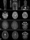
Detail of the initial whole-body MRI of a 11 years-old female patient with Mycobacterium avium complex (MAC) infection and sarcoid-like lesions, showing multiple bone lesions in the tibial diaphyses and epiphyses, left femoral diaphysis, ischial punctate lesions, and left cervical lymph node involvement (a). Brain MRI performed during disease exacerbation, 9 months after the initial whole-body MRI, revealed multiple areas of signal alteration in the right cerebellar hemisphere, left temporal and frontal subcortical white matter, splenium, and left thalamus (b). Overall reduction of the previously noted areas of signal alteration, apart from a small new hyperintense lesion in the right parietal region (c), observed 4 months after the initiation of triple antimycobacterial therapy. Further reduction of the known alterations was noted, with stable size of the left pontine lesion (d), assessed 3 months later.
Figure 3
Figure 3
Clinical timeline of the patient’s medical history. A chronological overview of the patient’s clinical progression, starting from the onset of dermatological symptoms at age 7. ACE, angiotensin converting enzyme; ANA, anti-nuclear antibodies; AZM, azithromycin; CLR, clarithromycin; EMB, ethambutol; IFN, interferon; IgG, immunoglobulin G; m, months; MSMD, Mendelian Susceptibility to Mycobacterial Diseases; MXF, moxifloxacin; NGS, next generation sequencing; PDN, prednisone; RIF, rifampicin; STAT1, signal transducer and activator of transcription 1; y, years.
Figure 4
Figure 4
Schematic comparison of granuloma architecture and immune signaling in normal versus STAT1-deficient conditions. Comparison of granulomas in normal versus STAT1-deficient immunity. Functional STAT1 supports IFNγ signaling, leading to strong Th1 responses, macrophage activation, and caseating necrosis. In STAT1 deficiency, impaired signaling reduces iNOS, IRF1, CXCL9/10, and IL12Rβ1 expression, resulting in non-necrotizing granulomas with poor immune containment of mycobacteria and possible overalapping histological features with other granulomatous diseases such as sarcoidosis. Created in BioRender. Ricci, S. (2025) https://BioRender.com/urn1gx7.

Similar articles

References

    1. Rose CD, Neven B, Wouters C. Granulomatous inflammation: The overlap of immune deficiency and inflammation. Best Pract Res Clin Rheumatol. (2014) 28:191–212. doi: 10.1016/j.berh.2014.03.006, PMID: - DOI - PubMed
    1. Abla O, Jacobsen E, Picarsic J, Krenova Z, Jaffe R, Emile JF, et al. Consensus recommendations for the diagnosis and clinical management of Rosai-Dorfman-Destombes disease. Blood. (2018) 131:2877–90. doi: 10.1182/blood-2018-03-839753, PMID: - DOI - PMC - PubMed
    1. Drent M, Crouser ED, Grunewald J. Challenges of sarcoidosis and its management. New Engl J Med. (2021) 385:1018–32. doi: 10.1056/NEJMra2101555, PMID: - DOI - PubMed
    1. Heinle R, Chang C. Diagnostic criteria for sarcoidosis. Autoimmun Rev. (2014) 13:383–7. doi: 10.1016/j.autrev.2014.01.035, PMID: - DOI - PubMed
    1. Valeyre D, Bernaudin JF, Uzunhan Y, Kambouchner M, Brillet PY, Soussan M, et al. Clinical presentation of sarcoidosis and diagnostic work-up. Semin Respir Crit Care Med. (2014) 35:336–51. doi: 10.1055/s-0034-1381229, PMID: - DOI - PubMed

Publication types

MeSH terms

Substances

LinkOut - more resources