Review
. 2025 Jul 2:12:rbaf068.
doi: 10.1093/rb/rbaf068. eCollection 2025.

Strategic incorporation of metal ions in bone regenerative scaffolds: multifunctional platforms for advancing osteogenesis

Affiliations
Review

Strategic incorporation of metal ions in bone regenerative scaffolds: multifunctional platforms for advancing osteogenesis

Yunnong Luo et al. Regen Biomater. .

Abstract

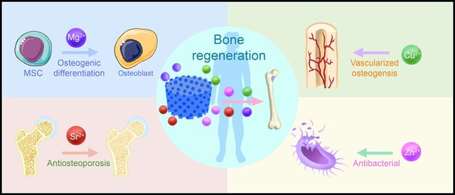
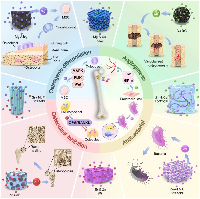
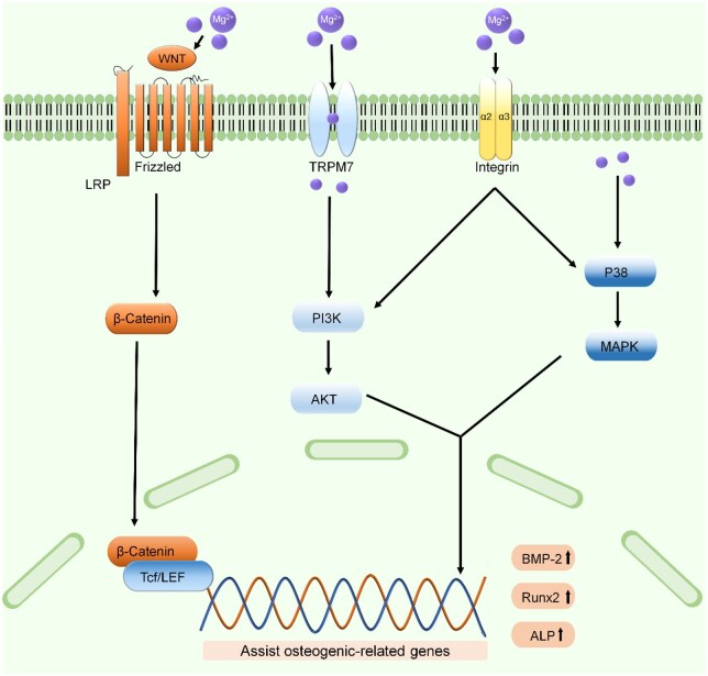
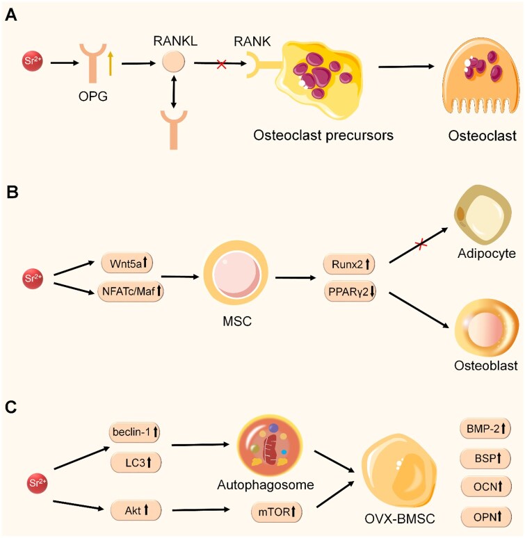
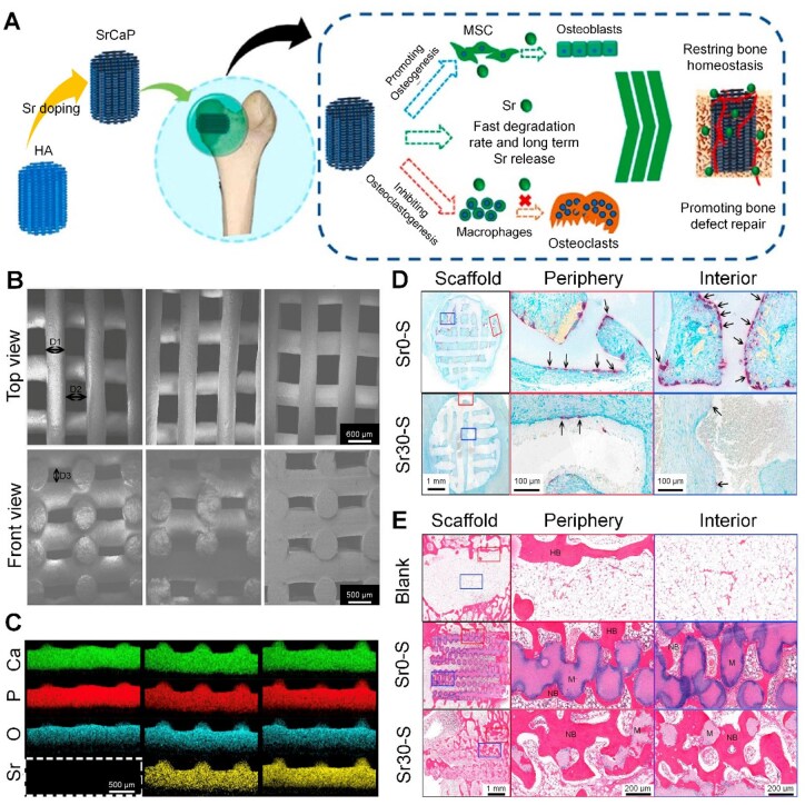
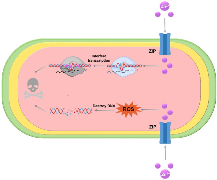
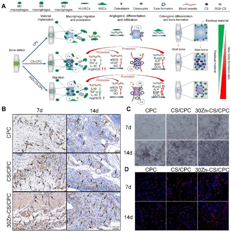
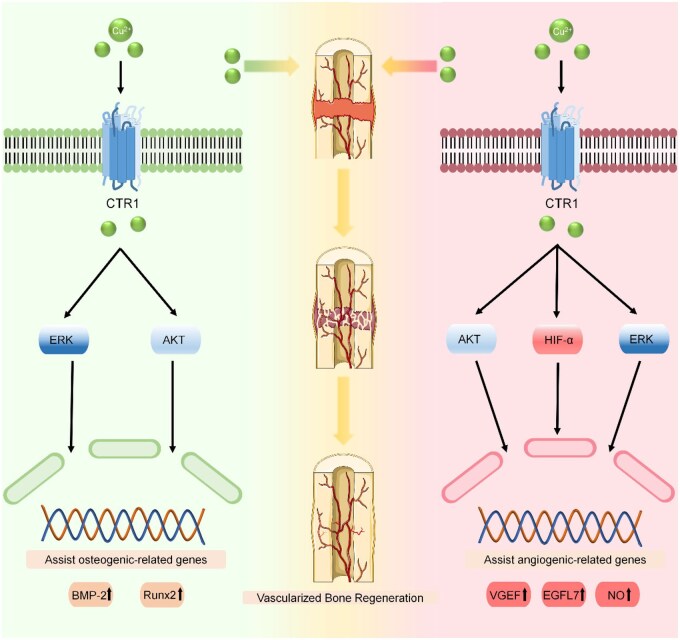
Bone serves as a critical structural framework, enabling movement and protecting internal organs. Consequently, maintaining skeletal health is a pivotal objective in bone tissue engineering. Bioactive metal ions, such as magnesium, strontium, zinc and copper, play essential roles in bone metabolism by participating in key physiological processes that sustain bone health and support regeneration. Recent studies indicate that these ions enhance the physicochemical properties and biological performance of bone tissue engineering materials, thereby facilitating osseointegration through diverse mechanisms. Specifically, magnesium promotes osteogenic differentiation; strontium inhibits osteoclast activity; zinc exhibits antibacterial properties; and copper facilitates vascularization for osteogenesis. Therefore, incorporating bioactive metal ions has emerged as a prevalent strategy in bone tissue engineering to address orthopedic disorders. This review systematically summarizes the roles of magnesium, strontium, zinc and copper in bone repair and regeneration. It provides an in-depth analysis of engineered materials incorporating these ions, with a focus on their applications and modifications across various material types. Furthermore, we explore the synergistic effects of combining these metal ions in bone tissue engineering, emphasizing their enhanced biological properties. By synthesizing recent research findings, this review aims to provide new insights and potential breakthroughs in leveraging bioactive metal ions for advancing treatments of orthopedic diseases.

Keywords: bone regeneration; bone tissue engineering materials; metal ions; osteopororsis.


Figures

None
Graphical abstract
Figure 1.
Figure 1.
Metal active ions possess the capacity to facilitate bone regeneration. Magnesium enhances the osteogenic differentiation of stem cells via the Wnt, PI3K and MAPK signaling pathways. Strontium inhibits osteoclast activity and mitigates osteoporosis via the OPG/RANKL pathway. Zinc demonstrates antibacterial properties by promoting the generation of reactive oxygen species and interfering with transcriptional processes, while copper facilitates vascularized osteogenesis through HIF-α and ERK pathways. The incorporation of these metal ions into various bone tissue engineering materials markedly enhances their capacity for bone integration.
Figure 2.
Figure 2.
Schematic diagram illustrates the mechanism by which Mg²⁺ promotes osteogenic differentiation.
Figure 3.
Figure 3.
Amorphous magnesium phosphate/polylactic acid macroporous biocomposite scaffolds. (A) Schematic representation of the fabrication process for the scaffold, employing fused filament fabrication (FFF)-based three-dimensional printing technology (B) MC3T3 adhesion after a 7-day culture on the 3D printed PLA scaffold and AMP-PLA scaffold. (C) OD540 reading of MC3T3 on control, PLA and AMP-PLA scaffolds [the asterisk (*) is significant concerning the control, and the plus symbol (+) is the significance between PLA and AMP-PLA]. Reproduced from Ref. [79] with permission of American Chemical Society, © 2021.
Figure 4.
Figure 4.
Schematic diagram of Sr²⁺ against osteoporosis in the treatment of bone defect. (A) Sr²⁺ suppresses osteoclast activity by modulating the OPG/RANKL/RANK pathway. (B) Sr²⁺ inhibits the differentiation of MSCs into adipocytes by activating the Wnt 5a and NFATc/Maf pathways to combat osteoporosis. (C) Sr²⁺ reduces ROS production by promoting autophagy and activates the Akt/mTOR pathway to combat osteoporosis.
Figure 5.
Figure 5.
Sr-doped calcium phosphate ceramic scaffolds. (A) Schematic diagram of the production of the scaffolds (B) SEM images of Sr-doped calcium phosphate ceramic scaffolds. (C) Element distribution of Sr-doped CaP ceramic scaffolds (D1: fiber diameter, D2: fibers distance in X and Y direction, D3: fibers distance in Z direction). (D) TRAP staining of the scaffolds after implanted in SD rats for 7 days (black arrows pointed to TRAP-positive cells). (E) HE staining images of the scaffolds after implantation for 12 weeks. (NB: new bone; HB: host bone; M: materials). reproduced from Ref. [118] with permission of Elsevier, © 2024.
Figure 6.
Figure 6.
Schematic diagram of Zn²⁺-mediated resistance to infection.
Figure 7.
Figure 7.
Calcium phosphate cement containing zinc-doped calcium silicate. (A) Schematic illustration. (B) CD31 immunohistochemical staining in tissues surrounding samples implanted in rat subcutaneous tissues at 7 and 14 days post-surgery. (Black arrows point to blood vessels. Statistical analysis of the amount). (C) The ALP activity of mBMSCs cultured in macrophage-conditioned medium derived from cement samples over periods of 7 and 14 days. (D) CD163 immunofluorescent staining images of the surroundings of CPC, CS/CPC and 30Zn-CS/CPC following implantation for 7 and 14 days. Reproduced from Ref. [170] with permission of Elsevier, © 2022.
Figure 8.
Figure 8.
Schematic diagram of Cu²⁺ promoting vascularized bone regeneration.
Figure 9.
Figure 9.
A novel Cu²⁺ doped calcium phosphate cement (Cu-CPC). (A) schematic diagram of the role of Cu²⁺ in Cu-CPC. (B) ALP activity of mBMSCs cultured on Cu-CPCs. (C) In vitro angiogenesis of HUVECs in Cu-CPC extracts. Reproduced from Ref. [198] with permission of Elsevier, © 2020.
Figure 10.
Figure 10.
Strontium-doped MgP-PCL scaffold based on extrusion-based 3D printing system. (A) Schematic illustration of the low-temperature printing process and the composition of the ink. (B) The plastic deformation of the scaffolds commences at an initial strain of 10% and subsequently progresses to buckling and barrel-like deformations. (C) Elastic modulus, yield stress and toughness from compressive loading profile for various concentrations of PCL. (D) ALP images of the scaffold in basal media (a), and ALP images of the scaffold in osteogenic media (b), alizarin red S images of the printed samples in basal media (c) and in osteogenic media (d). Reproduced from Ref. [245] with permission of IOP Publishing, © 2023.
Figure 11.
Figure 11.
Cu-MSNs and ZnO nanoparticles incorporated poly (ethylene glycol) diacrylate/sodium alginate double network hydrogel. (A) Schematic diagram of the preparation of PS@ZnO/Cu-MSNs composite hydrogel. (B) Stress–strain curve of the hydrogels. (C) Antibacterial performance of hydrogels against Staphylococcus aureus. (D) Optical microscopic images of ALP-stained hydrogel. (E) Optical microscopic images of ARS-stained hydrogel. Reproduced from Ref. [268] with permission of Elsevier, © 2024.

Similar articles

References

    1. Srivastava RK, Sapra L, Mishra PK. Osteometabolism: metabolic alterations in bone pathologies. Cells 2022;11:3943. - PMC - PubMed
    1. Šalandová M, Van Hengel IAJ, Apachitei I, Zadpoor AA, Van Der Eerden BCJ, Fratila‐Apachitei LE. Inorganic agents for enhanced angiogenesis of orthopedic biomaterials. Adv Healthc Mater 2021;10:2002254. - PMC - PubMed
    1. Ciosek Ż, Kot K, Kosik-Bogacka D, Łanocha-Arendarczyk N, Rotter I. The effects of calcium, magnesium, phosphorus, fluoride, and lead on bone tissue. Biomolecules 2021;11:506. - PMC - PubMed
    1. Kraft MD. Phosphorus and calcium: a review for the adult nutrition support clinician. Nutr Clin Pract 2015;30:21–33. - PubMed
    1. Kim S-G. Multiple ways for the same destination: bone regeneration. Maxillofac Plast Reconstr Surg 2022;44:9. - PMC - PubMed

LinkOut - more resources