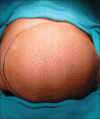
Giant Lipofibromatosis Over Back Mimicking Thoracic Lipomyelomeningocele in Paediatric Age Group: Case Report and Review of Literature
- PMID: 40756045
- PMCID: PMC12316414
- DOI: 10.4103/jiaps.jiaps_3_25
Giant Lipofibromatosis Over Back Mimicking Thoracic Lipomyelomeningocele in Paediatric Age Group: Case Report and Review of Literature
Abstract
Lipofibromatosis is a rare and benign soft-tissue tumor predominantly affecting children. It commonly presents as a slow-growing, painless mass, often misdiagnosed due to its rarity and variable presentation. We report the unusual case of an 8-month-old male with a congenital upper thoracic mass initially suspected to be a lipomyelomeningocele. Clinical examination and ultrasound supported this diagnosis, but magnetic resonance imaging findings suggested a soft-tissue tumor. The child underwent excisional biopsy, and intraoperative findings revealed a highly vascular, well-defined mass without spinal cord involvement. Histopathological analysis confirmed lipofibromatosis. The postoperative course was uneventful, and no recurrence was observed after 1 year of follow-up. This case highlights the diagnostic challenges associated with lipofibromatosis and its potential for misdiagnosis, and the importance of histopathology in establishing a definitive diagnosis. Early complete surgical excision remains the preferred treatment to prevent recurrence.
Keywords: Lipofibromatosis; liposarcoma; pediatric; soft-tissue mass.
Copyright: © 2025 Journal of Indian Association of Pediatric Surgeons.
Conflict of interest statement
There are no conflicts of interest.
Figures
References
-
- Vogel D, Righi A, Kreshak J, Dei Tos AP, Merlino B, Brunocilla E, et al. Lipofibromatosis: Magnetic resonance imaging features and pathological correlation in three cases. Skeletal Radiol. 2014;43:633–9. - PubMed
-
- Fetsch JF, Miettinen M, Laskin WB, Michal M, Enzinger FM. A clinicopathologic study of 45 pediatric soft tissue tumors with an admixture of adipose tissue and fibroblastic elements, and a proposal for classification as lipofibromatosis. Am J Surg Pathol. 2000;24:1491–500. - PubMed
-
- Taran K, Woszczyk M, Kobos J. Lipofibromatosis presenting as a neck mass in eight-years old boy –A case report. Pol J Pathol. 2008;59:217–20. - PubMed
-
- Lao QY, Sun M, Yu L, Wang J. Lipofibromatosis: A clinicopathological analysis of eight cases. Zhonghua Bing Li Xue Za Zhi. 2018;47:186–91. - PubMed
-
- Al-Ibraheemi A, Folpe AL, Perez-Atayde AR, Perry K, Hofvander J, Arbajian E, et al. Aberrant receptor tyrosine kinase signaling in lipofibromatosis: A clinicopathological and molecular genetic study of 20 cases. Mod Pathol. 2019;32:423–34. - PubMed
Publication types
LinkOut - more resources
Full Text Sources


