. 2025 Aug 1;13(8):e70753.
doi: 10.1002/ccr3.70753. eCollection 2025 Aug.

Obstructive Ureteral Metastasis From Prostate Cancer

Affiliations

Obstructive Ureteral Metastasis From Prostate Cancer

Ahmad Al-Bitar et al. Clin Case Rep. .

Abstract

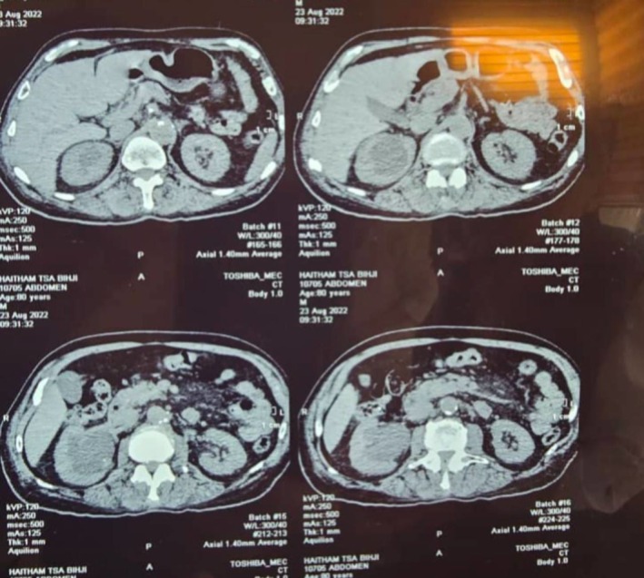
Prostate cancer is the second most common malignancy in males, primarily associated with risk factors such as age, ethnicity, obesity, and family history. While metastases commonly involve lymph nodes, bones, and the liver, ureteral metastasis is exceedingly rare. Ureteral obstruction due to metastatic prostate cancer can lead to hydronephrosis, often requiring ureteroscopy for diagnosis and management. However, persistent symptoms may necessitate exploratory surgery. An 82-year-old Arab male with a history of Gleason 4 + 4 = 8 prostate adenocarcinoma (initially managed with hormonal therapy and surgical castration) presented with right back pain. Imaging revealed stage 4 hydronephrosis, initially attributed to a ureteral stone. Ureteroscopy identified obstructing ureteral tumors instead, with biopsy confirming well-differentiated adenocarcinoma of prostatic origin. Due to persistent obstruction and confirmed malignancy, a right nephroureterectomy was performed. Histopathology demonstrated extensive ureteral involvement (16 cm) with vascular and perineural invasion but no distant metastasis on postoperative PET-CT. This case underscores the diagnostic challenge of ureteral metastasis in prostate cancer, often mimicking benign conditions like ureteral stones. Despite successful local surgical control, the tumor's aggressive features necessitate long-term surveillance and continued hormonal therapy. High clinical suspicion for metastasis is crucial in high-risk prostate cancer patients presenting with obstructive uropathy, even in the absence of significantly elevated PSA. This report highlights the importance of a stepwise diagnostic approach and the need for prompt intervention in such rare presentations.

Keywords: hydronephrosis; metastasis; prostate cancer; ureter stones; ureteral metastasis; ureteral obstruction.


Conflict of interest statement

The authors declare no conflicts of interest.

Figures

FIGURE 1
FIGURE 1
Coronal views from a computed tomography (CT) scan of the abdomen and pelvis demonstrate severe left‐sided hydronephrosis, characterized by marked dilatation of the renal pelvis and calyces. There is associated significant thinning of the renal parenchyma, suggesting a chronic obstructive process. The proximal and mid‐left ureter is also dilated (hydroureter). In contrast, the right kidney and ureter appear unremarkable with no signs of obstruction.
FIGURE 2
FIGURE 2
Axial views from the same CT scan further delineate the abdominal pathology. The images confirm the presence of severe left‐sided hydronephrosis, with massive dilatation of the pelvicalyceal system. The overlying renal parenchyma is severely atrophied and appears as a thin, nonfunctional cortical rim. In contrast, the right kidney demonstrates normal size, enhancement, and a nondilated collecting system.
FIGURE 3
FIGURE 3
Ureteroscopy of the left ureter was performed to identify the cause of the obstruction. The endoscopic view (Figure 3) revealed a large, intraluminal, fungating mass in the distal ureter. The lesion appeared dark, necrotic, and pigmented, with a highly irregular and friable surface. The mass was seen to completely obstruct the ureteric lumen, which directly corresponds to the site of obstruction inferred from the CT imaging. The adjacent ureteric mucosa showed evidence of inflammation.
FIGURE 4
FIGURE 4
(a, b) Postoperative images showing the pathological kidney and ureter.
FIGURE 5
FIGURE 5
(a) (Low‐power view) At low magnification, the section shows extensive infiltration of the ureteral wall by a malignant neoplasm. The tumor is composed of numerous, small, and crowded glands arranged in a back‐to‐back architectural pattern with minimal intervening stroma. This pattern is characteristic of an acinar adenocarcinoma. (b) (High‐power view) On high power magnification, the malignant glands are lined by cells showing nuclear atypia. Critically, this view clearly demonstrates the presence of perineural invasion, where tumor cells are seen infiltrating and surrounding a small nerve bundle.

References

    1. Zuur L. G., de Barros H. A., van der Mijn K. J. C., et al., “Treating Primary Node‐Positive Prostate Cancer: A Scoping Review of Available Treatment Options,” Cancers (Basel) 15, no. 11 (2023): 2962, 10.3390/cancers15112962. - DOI - PMC - PubMed
    1. Pernar C. H., Ebot E. M., Wilson K. M., and Mucci L. A., “The Epidemiology of Prostate Cancer,” Cold Spring Harbor Perspectives in Medicine 8, no. 12 (2018): a030361, 10.1101/cshperspect.a030361. - DOI - PMC - PubMed
    1. Zhang T., Wang Q., Min J., et al., “Metastasis to the Proximal Ureter From Prostatic Adenocarcinoma: A Rare Metastatic Pattern,” Canadian Urological Association Journal 8, no. 11–12 (2014): 859–861, 10.5489/cuaj.2169. - DOI - PMC - PubMed
    1. Tsutsumi S., Kawahara T., Hattori Y., et al., “Ureter Metastatic Castration‐Resistant Prostate Cancer: A Case Report,” Journal of Medical Case Reports 11, no. 1 (2017): 215, 10.1186/s13256-017-1379-z. - DOI - PMC - PubMed
    1. Geavlete P., Multescu R., and Geavlete B., “Pushing the Boundaries of Ureteroscopy: Current Status and Future Perspectives,” Nature Reviews. Urology 11, no. 7 (2014): 373–382, 10.1038/nrurol.2014.118. - DOI - PubMed

LinkOut - more resources