

doi: 10.31138/mjr.310724.iia.
eCollection 2025 Jun.
Psoriatic Onycho-Pachydermo Periostitis (POPP)
Affiliations
- PMID: 40757118
- PMCID: PMC12312468
- DOI: 10.31138/mjr.310724.iia
Item in Clipboard
Psoriatic Onycho-Pachydermo Periostitis (POPP)
Mediterr J Rheumatol.
.
No abstract available
Keywords: MRI; POPP; onychodystrophy; psoriatic arthritis; ultrasonography.
Conflict of interest statement
The authors declare no conflict of interest.
Figures
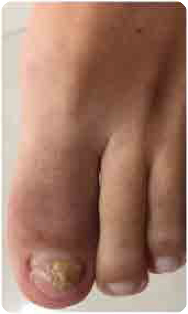
(left). Photograph of the patient’s left forefoot showing nail dystrophy, oedema, and erythema of the interphalangeal joint of the hallux.
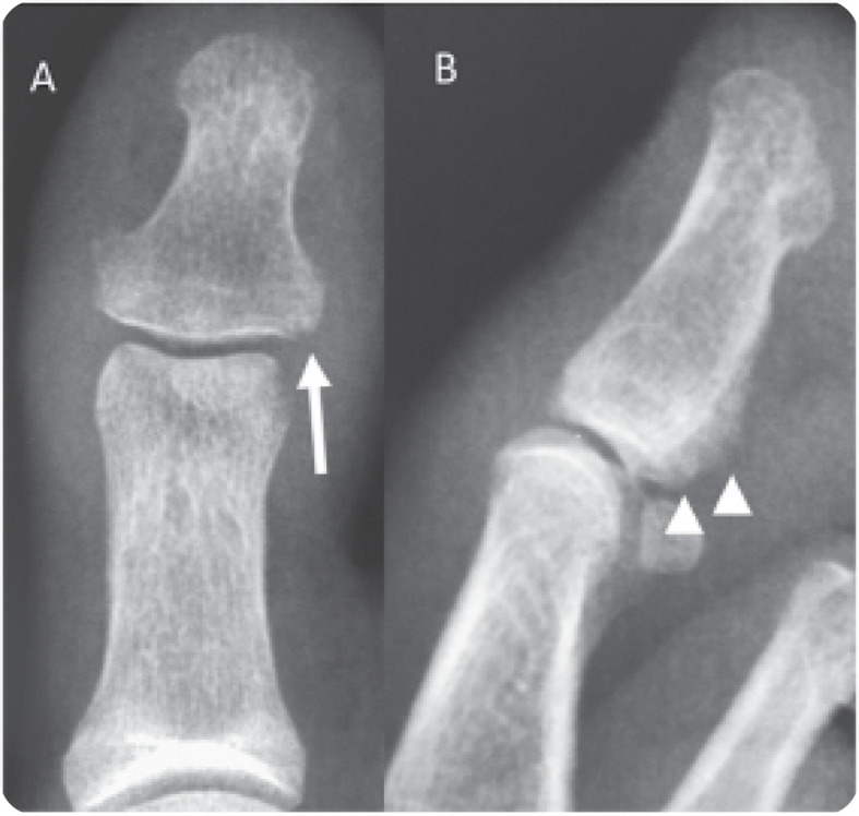
(right). Conventional radiography of the hallux with anteroposterior (A) and lateral (B) views showing periarticular erosions (arrows in A) and periosteal reaction (arrowheads in B) of the distal phalanx.

(left). Ultrasonography of the hallux revealing enthesitis characterised by hyperostosis of the proximal and distal phalanges (arrows in A), and increased thickness and hypoechogenicity of the extensor hallucis longus (arrowheads in B) associated with peritendinous positive Doppler findings adjacent to the tendon’s insertion at the base of the distal phalanx (C). Synovitis of the interphalangeal joint of the hallux (asterisk in B) is also visible.

(right). Magnetic resonance imaging of the hallux. (A) Coronal T1-weighted image showing periarticular erosions (arrow) and soft-tissue swelling with intermediate to low signal intensity (arrowhead). Coronal (B) and sagittal (C) fat-suppressed proton density-weighted images depicting extensive bone-marrow oedema in the proximal and distal phalanges of the hallux (asterisks) related to the extensor and flexor enthesis, and oedema and thickening of the nail bed (open arrow in C).
References
LinkOut - more resources
Full Text Sources