Case Reports
. 2025 Aug 1;14(8):8.
doi: 10.1167/tvst.14.8.8.

Short-Term Outcomes of Pediatric Patients With Mild Autosomal Recessive RPE65-Associated Retinal Dystrophy Treated With Voretigene Neparvovec

Affiliations
Case Reports

Short-Term Outcomes of Pediatric Patients With Mild Autosomal Recessive RPE65-Associated Retinal Dystrophy Treated With Voretigene Neparvovec

David A Merle et al. Transl Vis Sci Technol. .

Abstract

Purpose: Voretigene neparvovec is approved for RPE65-associated inherited retinal degeneration (RPE65-IRD) in the United States and the European Union. According to current knowledge, early treatment benefits efficacy. However, consensus on treating mild cases is lacking due to ambiguity in balancing clinical benefits with potential side effects. Therefore, we present short-term outcomes of four pediatric patients with milder types of RPE65-IRD.

Methods: Two unrelated pediatric patients were unilaterally treated at the University Eye Hospital in Tübingen, Germany. Another two unrelated pediatric patients were bilaterally treated at Ghent University Hospital, Belgium. Examinations were performed before and until at least 6 months after treatment, including best-corrected visual acuity, slit-lamp examination, fundus photography, short-wavelength fundus autofluorescence, optical coherence tomography, 90° kinetic perimetry, dark-adapted chromatic perimetry, and full-field stimulus threshold measurements.

Results: Despite surgical challenges, treatment with voretigene neparvovec was successful in all four patients. All patients showed rod functional rescue with stable best-corrected visual acuity. Three patients suffered chorioretinal atrophy at the retinotomy site but none developed signs of fast-growing CRA. One case developed limited CRA in the bleb area, potentially related to inflammation in the subretinal space.

Conclusions: Treatment with voretigene neparvovec was safe and effective in patients with mild RPE65-IRD. Early treatment showed good functional outcomes. Also, treatment at stages without profound retinal degeneration might lower the risk of fast-growing CRA.

Translational relevance: This study aids clinical decision-making in unclear cases by demonstrating that early treatment with voretigene neparvovec in mild RPE65-IRD provides functional benefits while minimizing the risk of fast-growing chorioretinal atrophy.


Conflict of interest statement

Disclosure: D.A. Merle, None; L. Hertens, None; S. Dimopoulos, None; S. Kohl, None; M. Van Haute, None; E. De Baere, None; M. De Bruyne, None; B. Janssens, None; K. Rüther, None; C. Huchzermeyer, None; P. Mazzola, None; F. Nerinckx, None; T. Haack, None; L. Wolfram, None; M. Kempf, None; L. Kühlewein, Novartis (F), Rhythm (F), Tistou and Charlotte Kerstan Foundation (F); K. Stingl, Johnson & Johnson (C), Lundbeck (C), Novartis (C), ProQR Therapeutics (C), Santen (C), ViGeneron (C), THEA (C), RhyGaze (C), Ascidian (C); B.P. Leroy, 4DMT (C), AAVantgardeBio (C), Akouos (C), Alia Therapeutics (C), Bayer (C), Belite Bio (C), Biogen (C), Coave Therapeutics (C), GenSight Biologics (C), Gyroscope (C), IVERIC Bio (C), Johnson & Johnson (Janssen Pharmaceuticals) (C), MeiraGTx (C), Novartis (C), ProQR Therapeutics (C), Ray Therapeutics (C), REGENXBIO (C), SalioGen (C), Santen (C), Spark Therapeutics (C), SparingVision (C), SpliceBio (C), Stoke Therapeutics (C), Transine Therapeutics (C), Vedere Bio I and II (C), ViGeneron (C); K. Stingl, None

Figures

Figure 1.
Figure 1.
Pre- and post-treatment follow-up for case 1. The left column presents findings immediately prior to surgery (baseline). The middle and right columns show findings 3 months and 6 months after surgery, respectively. (AC) Left-eye kinetic 90° perimetry using Goldmann object III4e. At all time-points, a concentric constriction to approximately 10° to 15° was measured, which was considered stable despite some variations in the measurement. Solid red lines represent the borders of the measured visual field; the broader, pale red lines represent reference values of healthy eyes. (DF) Macular OCT scan of the left eye showed an irregular appearance of the EZ nasally at baseline, with a seemingly decreased granular appearance of the EZ at 3 and 6 months relative to baseline that was not explained by differences in image quality. A small hyperreflective accumulation superior to the retinal nerve fiber layer was detected at 3 months but was not detectable thereafter. (GI) CFP at baseline and at 3 and 6 months after surgery. All pictures show a diffuse retinal atrophy typical for RPE65-associated IRD without spicular intraretinal pigment migration. The inferonasal retinal hole surrounded by laser scars along with some individual laser scars can be seen in panels (H) and (I). Limited, non-progressive CRA can be identified at the retinotomy site around the inferior vascular arcade in panels (H) and (I).
Figure 2.
Figure 2.
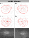
Initial presentation and pre-treatment follow-up of case 2. (AD) The 90° Goldmann kinetic visual fields using object III4e showed clinically significant deterioration in both eyes within 3 years after initial presentation to our clinic: left eye at initial presentation (A), right eye at initial presentation (B), left eye at 3-year follow-up (baseline) (C), right eye at 3-year follow-up (baseline) (D). (E, F) FAF images of both eyes at baseline showing globally reduced but visible diffuse hyperautofluorescence at the posterior pole: left eye (E), right eye (F).
Figure 3.
Figure 3.
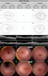
Pre- and post-treatment follow-up of case 2. (AC) Left eye 90° kinetic perimetry using Goldmann object III4e. At baseline and 6 months after treatment, a remaining central visual field with concentric restriction to approximately 10° to 15° was observed with additional large inferior residual islands. At the 3-month follow-up, no kinetic perimetry was performed. No clinically relevant differences were observed between baseline and the 6-month follow-up. (DF) Macular OCT scans of the left eye at baseline (D), 3 months after surgery (E), and 6 months after surgery (F). The residual island remained stable at 3 months after surgery but showed a slight reduction at 6 months. (GI) CFP at baseline (G), 3 months after surgery (H), and 6 months after surgery (I). All pictures show a diffuse retinal atrophy typical of RPE65-IRD. At 3 months post-surgery, a newly developed area of CRA was observed at the lower vascular arcade and a smaller one around the upper vascular arcades, corresponding to the retinotomy sites. However, these areas remained stable (H, I).
Figure 4.
Figure 4.
DACP for case 2. (AC) Local dark-adapted thresholds using cyan stimulus with the DACP were overlaid on the fundus image of the patient at baseline (A), 3 months after surgery (B), and 6 months after surgery (C) for better visibility. The color-coding scale shows the local values in decibels (for comparison, a healthy eye has values around 60 dB in all locations). An improvement in local dark adaptation thresholds in the treated region is obvious and remained relatively stable in both postoperative visits. Of note, at baseline a shorter protocol with fewer stimulus locations designed specifically for children was used, whereas at the follow-up visits the patient was able to follow a standard protocol.
Figure 5.
Figure 5.
Pre- and post-treatment follow-up for case 3. (AD) Right eye (A, B) and left eye (C, D) 90° kinetic perimetry using Goldmann objects V4e, III4e, and I4e. No kinetic perimetry was scheduled for the 3-month follow-up. No clinically relevant differences were observed between baseline and the 6-month follow-up for objects V4e and I4e; however, an improvement of visual fields using Goldmann object III4e was observed in both eyes. (EG) Macular OCT scans of the right eye at baseline (E), 3-month follow-up (F), and 6-month follow-up (G). (HJ) Macular OCT scans of the left eye at baseline (H), 3-month follow-up (I) and 6-month follow-up (J). The residual island remained stable over time, and no other clinically relevant alterations were evident in both eyes. (KM) CFP of the right eye at baseline (K), 1 month after surgery (L), and 6 months after surgery (M). No clinically relevant differences were observed between baseline and 6-month follow-up, except for the development of a small atrophic area within the vascular arcades at the retinotomy site superior to the macula. (NP) CFP of the left eye at baseline (N), 1 month after surgery (O), and 6 months after surgery (P). No CFP images are available for the 3-month follow-up; instead, images from the 1-month follow-up are shown. All CFP images show a diffuse retinal atrophy typical for RPE65-associated IRD. No clinically relevant differences were observed between baseline and the 6-month follow-up.
Figure 6.
Figure 6.
Pre- and post-treatment follow-up for case 4. (AC) Macular OCT scans of the right eye at baseline (A), 3-month follow-up (B), and 6-month follow-up (C). (DF) Macular OCT scans of the left eye at baseline (D), 3-month follow-up (E), and 6-month follow-up (F). The residual island remained stable over time, and no other clinically relevant alterations were evident in both eyes. (GI) CFP of the right eye at baseline (G), 3 months after surgery (H), and 18 months after surgery (I). After surgery, a small area of CRA developed at the injection site at the superior vascular arcade. (JL) CFP of the left eye at baseline (J), 3 months after surgery (K), and 18 months after surgery (L). After surgery, a small area of CRA developed at the injection site at the superior vascular arcade and small atrophic spots emerged temporally to the optic disc, which since have coalesced into a small area of CRA but remained stable until the last follow-up. No CFP images are available for the 6-month follow-up; instead, images from the 18-month follow-up are shown. *FST measurements at 6 months were unreliable; therefore, 12-month values are shown instead.
Figure 7.
Figure 7.
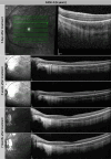
Subretinal deposits and development of CRA in case 4. (A) Macular OCT scan of the left eye 5 days after treatment. A small subretinal deposit near the optic disc and a decrease in choroidal thickness can be seen. (B, C) Follow-up examination at 2 weeks after treatment. Two distinct B-scans are shown, and both scans show enlargement of subretinal deposits. (D, E) Follow-up examination at 7 months after treatment. Both B-scans show extensive CRA in the area corresponding to the previously observed subretinal deposits.

References

    1. Wu W, Takahashi Y, Shin HY, Ma X, Moiseyev G, Ma JX.. The interplay of environmental luminance and genetics in the retinal dystrophy induced by the dominant RPE65 mutation. Proc Natl Acad Sci U S A. 2022; 119: e2115202119. - PMC - PubMed
    1. Hull S, Mukherjee R, Holder GE, Moore AT, Webster AR.. The clinical features of retinal disease due to a dominant mutation in RPE65. Mol Vis. 2016; 22: 626–635. - PMC - PubMed
    1. Jauregui R, Park KS, Tsang SH.. Two-year progression analysis of RPE65 autosomal dominant retinitis pigmentosa. Ophthalmic Genet. 2018; 39: 544–549. - PMC - PubMed
    1. Gu SM, Thompson DA, Srikumari CR, et al.. Mutations in RPE65 cause autosomal recessive childhood-onset severe retinal dystrophy. Nat Genet. 1997; 17: 194–197. - PubMed
    1. Kumaran N, Moore AT, Weleber RG, Michaelides M.. Leber congenital amaurosis/early-onset severe retinal dystrophy: clinical features, molecular genetics and therapeutic interventions. Br J Ophthalmol. 2017; 101: 1147–1154. - PMC - PubMed

Publication types

Substances