Histological Analysis of the Prostate During the Human Fetal Period (12 to 22 Weeks Post-Conception)
- PMID: 40758283
- PMCID: PMC12539898
- DOI: 10.1590/S1677-5538.IBJU.2025.9915
Histological Analysis of the Prostate During the Human Fetal Period (12 to 22 Weeks Post-Conception)
Abstract
Purpose: The aim of this study was to quantitatively analyze the stromal components of the prostate (collagen, muscle fibers and elastic system fibers) during prostate development in normal human fetuses, in order to establish normal growth patterns.
Materials and methods: Twenty-one normal human fetuses, aged between 12- and 22-weeks post-conception (WPC), were studied. The fetuses were divided into three groups: G1, n = 7, 12 to 16 WPC; G2, n = 6, 17 to 19 WPC, and G3, n = 8, 20 to 22 WPC. The samples were fixed in 4% buffered formalin and processed for paraffin embedding for histochemical analysis. Five 5-micrometer-thick sections were taken from each sample and stained using histochemical techniques to identify the prostate stromal components. Histomorphometric analyses were carried out by evaluating the area density of the parameters analyzed, in percentage, using ImageJ software.
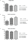
Results: The quantitative analysis of collagen fibers (p = 0.0616) and elastic system fibers (p = 0.6049) showed no significant difference between groups. Smooth muscle fibers were higher in G2 (+82.38%) and G3 (+108.81%) when compared to G1, p = 0.0020. Linear regression of the entire fetal period studied (12 to 22 WPC) showed a proportional increase in muscle fibers in relation to gestational age (r2 = 0.3398; p = 0.0056). Linear regression revealed a strong negative correlation between collagen fibers and smooth muscle fibers in G3 (20 to 22 WPC) (r2 = 0.8209; p = 0.0019).
Conclusions: Changes in the stromal components of the prostate are more significant from the end of the second trimester of pregnancy. Collagen fibers and elastic system fibers showed stable growth throughout the fetal period studied. The density of smooth muscle fibers increased about twofold with increasing fetal age.
Keywords: Fetus; Growth and Development; Prostate.
Copyright® by the International Brazilian Journal of Urology.
Conflict of interest statement
None declared.
Figures


References
-
- Srougi M, Dall’Oglio M, Antunes AA. In: Hiperplasia Prostática Benigna. Srougi M, Dall’Oglio M, Antunes AA, editors. São Paulo: Atheneu; 2010. Hiperplasia prostática benigna (HPB): aspectos morfológicos e fisiopatológicos; pp. 25–35.
-
- Chung BI, Sommer G, Brooks JD. In: Campbell-Walsh Urology. 10ª ed. Wein AJ, Kavoussi LR, Novick AC, Partin AW, Peters CA, editors. Philadelphia: Saunders Elsevier; 2012. Anatomy of the lower urinary tract and male genitalia; pp. 33–70.
-
- Berman DM, Rodriguez R, Veltri RW. In: Campbell-Walsh Urology. 10ª ed. Wein AJ, Kavoussi LR, Novick AC, Partin AW, Peters CA, editors. Philadelphia: Saunders Elsevier; 2012. Development, molecular biology, and physiology of the prostate; pp. 2533–2569. - DOI
MeSH terms
Substances
LinkOut - more resources
Full Text Sources
