. 2025 Aug 4;20(8):e0329272.
doi: 10.1371/journal.pone.0329272. eCollection 2025.

Premature termination of DNA Damage Repair by 3-Methyladenine potentiates cisplatin cytotoxicity in nasopharyngeal carcinoma cells

Affiliations

Premature termination of DNA Damage Repair by 3-Methyladenine potentiates cisplatin cytotoxicity in nasopharyngeal carcinoma cells

Jie Zhou et al. PLoS One. .

Abstract

3-Methyladenine (3-MA) is widely recognized as a PI3K inhibitor involved in autophagy regulation. However, it is also a byproduct of DNA damage repair, and its role in modulating DNA damage response (DDR) mechanisms remains largely unexplored. Cisplatin (CDDP), a cornerstone chemotherapeutic agent for nasopharyngeal carcinoma (NPC), exerts its cytotoxic effects by inducing DNA damage in tumor cells. This study investigates the combined effects of CDDP and 3-MA on NPC cells. Cell viability and the half-maximal inhibitory concentration (IC50) were assessed using the Cell Counting Kit-8 (CCK-8) assay. Flow cytometry was employed to analyze cell cycle distribution, mitochondrial membrane potential (MMP) alterations, and apoptosis. γ-H2AX foci formation and morphological changes were examined via fluorescence microscopy, while Western blotting was used to evaluate proteins associated with the DNA damage response. The combination treatment significantly reduced cell viability and lowered the IC50 compared to CDDP alone. While both treatments induced Sub-G1 phase arrest, the combination resulted in greater MMP loss and apoptosis. Western blot analysis further revealed that 3-MA enhanced CDDP cytotoxicity by suppressing ATM/ATR/p53-mediated DNA damage repair and promoting apoptotic signaling. These findings suggest that 3-MA sensitizes NPC cells to CDDP by disrupting DNA repair processes, offering a promising therapeutic strategy.


Conflict of interest statement

The authors have declared that no competing interests exist.

Figures

Fig 1
Fig 1. Enhancement of CDDP-induced proliferation inhibition in nasopharyngeal cancer cells by 3-MA.
(A, C) CCK8 assay results showed that after 24 hours of treatment with a combination of 3 mM 3-MA and various concentrations of CDDP, the cell viability of nasopharyngeal cancer cells was lower than that of the CDDP-only treatment group. The data are presented as the mean ± SD (n = 3), and the Mann-Whitney U test was used for comparisons between two groups. * present vs. CDDP (20 μM) group at 24h, *P < 0.05, ns means not significant. (B, D) The combination of 3-MA and CDDP reduced the IC50 of CDDP against 5-8F and 6-10B nasopharyngeal cancer cells within 24 hours. (E) Schematic timeline of experimental design. (F) Colony elimination assay results indicated that the combination of 3-MA (3 mM) and CDDP (20 μM) significantly reduced the clonogenic survival rate of nasopharyngeal cancer cells compared to CDDP (20 μM) treatment alone. The data are presented as the mean ± SD (n = 3) and Kruskal-Wallis test followed by Bonferroni correction was employed for multiple group comparisons. * present vs. CDDP (20 μM), *P < 0.05, **P < 0.01, ns means not significant.
Fig 2
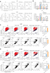
Fig 2. The combination of 3-MA and CDDP significantly increased CDDP-induced cell death.
(A, B) The distribution of the Sub-G1 phase in nasopharyngeal cancer cells treated with 3-MA and CDDP for 24 hours was significantly higher compared to both the CDDP-only treatment group and the control group. The data are presented as the mean ± SD (n = 3) and Kruskal-Wallis test followed by Bonferroni correction was employed for multiple group comparisons. * present vs. NC group, # present vs. CDDP (20 μM), *P < 0.05, **P < 0.01, ns means not significant. (C, D) After 24 hours of treatment with 3-MA and CDDP, nasopharyngeal cancer cells showed a significant increase in JC-1 monomer fluorescence compared to both the CDDP-only treatment group and the control group (C, D, top rows). Additionally, the proportion of dead cells was also significantly higher (C, D, bottom rows). The data are presented as the mean ± SD (n = 3) and Kruskal-Wallis test followed by Bonferroni correction was employed for multiple group comparisons. * present vs. NC group, # present vs. CDDP (20 μM), *P < 0.05, **P < 0.01, ns means not significant.
Fig 3
Fig 3. The combination of 3-MA and cisplatin enhances the initiation of apoptotic signaling and induces DNA damage in nasopharyngeal cancer cells.
(A-C) After 24 hours of treatment with 3-MA and CDDP, nasopharyngeal cancer cells exhibited significantly increased ratio of Bax/Bcl2, cleaved caspase-9/full caspase-9, cleaved caspase-3/full caspase-3, and cleaved-PARP/full PARP compared to both the CDDP-only treatment group and the control group. The data are presented as the mean ± SD (n = 3) and Kruskal-Wallis test followed by Bonferroni correction was employed for multiple group comparisons. * present vs. NC group, # present vs. CDDP (20 μM), *P < 0.05, **P < 0.01, ns means not significant. (D, E) Immunostaining results demonstrated that after 16 hours of treatment with CDDP, Compared to the 3-MA alone and control groups, cells treated with CDDP alone or in combination with 3-MA exhibited stronger γ-H2AX (green) fluorescence signals and a greater number of morphologically abnormal nuclei (blue). Scale bar = 200 μm.
Fig 4
Fig 4. 3-MA accelerates the termination of DNA damage repair and activates p53-mediated apoptosis in nasopharyngeal carcinoma cells.
(A, C) Expression levels and quantification of DNA damage repair-related proteins in 5-8F NPC cells following drug treatment for 8, 16, and 24 hours. (D, E) Expression levels and quantification of DNA damage repair-related proteins in 6-10B NPC cells following drug treatment for 8, 16, and 24 hours. Representative western blot results were analyzed using Kruskal-Wallis test followed by Bonferroni correction for multiple group comparisons. * present vs. NC group, # present vs. CDDP (20 μM), *P < 0.05, **P < 0.01, ns means not significant.
Fig 5
Fig 5. Schematic representation of 3-MA-induced premature termination of DNA damage repair and activation of apoptotic signaling.

References

    1. Bossi P, Chan AT, Licitra L, Trama A, Orlandi E, Hui EP, et al. Nasopharyngeal carcinoma: ESMO-EURACAN Clinical Practice Guidelines for diagnosis, treatment and follow-up(dagger ). Ann Oncol. 2021;32(4):452–65. doi: 10.1016/j.annonc.2020.12.007 - DOI - PubMed
    1. Su ZY, Siak PY, Lwin YY, Cheah SC. Epidemiology of nasopharyngeal carcinoma: current insights and future outlook. Cancer Metastasis Rev. 2024;43(3):919–39. doi: 10.1007/s10555-024-10176-9 - DOI - PubMed
    1. Lugones Y, Loren P, Salazar LA. Cisplatin Resistance: Genetic and Epigenetic Factors Involved. Biomolecules. 2022;12(10). doi: 10.3390/biom12101365 - DOI - PMC - PubMed
    1. Nagasawa S, Takahashi J, Suzuki G, Hideya Y, Yamada K. Why Concurrent CDDP and Radiotherapy Has Synergistic Antitumor Effects: A Review of In Vitro Experimental and Clinical-Based Studies. Int J Mol Sci. 2021;22(6). doi: 10.3390/ijms22063140 - DOI - PMC - PubMed
    1. Forgie BN, Prakash R, Telleria CM. Revisiting the Anti-Cancer Toxicity of Clinically Approved Platinating Derivatives. Int J Mol Sci. 2022;23(23). doi: 10.3390/ijms232315410 - DOI - PMC - PubMed

MeSH terms