. 2025 Aug 5;11(1):67.
doi: 10.1038/s41421-025-00817-w.

AR to GR switch modulates differential TDO2-Kyn-AhR signalling to promote the survival and recurrence of treatment-induced dormant cells in prostate cancer

Affiliations

AR to GR switch modulates differential TDO2-Kyn-AhR signalling to promote the survival and recurrence of treatment-induced dormant cells in prostate cancer

Sangsang Li et al. Cell Discov. .

Abstract

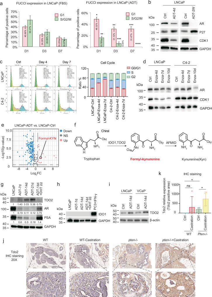
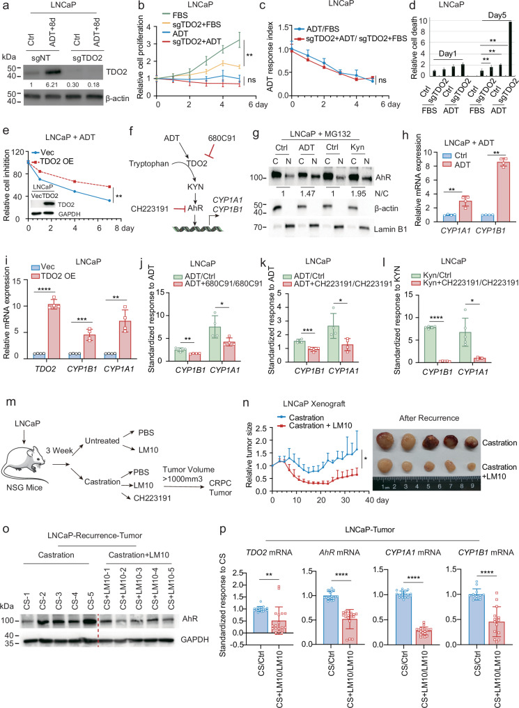
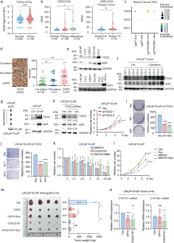
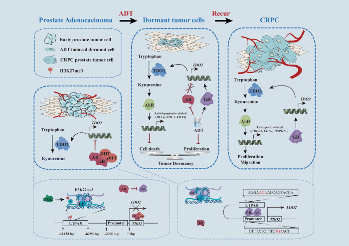
Cancer cells can be induced to dormancy initially by specific cancer therapies, but can be reactivated for subsequent relapse as therapy-resistant cancer cells. Although the treatment-induced dormancy-to-reactivation switch is an important process in tumour spread and recurrence, little is known about the underlying molecular mechanisms, particularly the metabolic underpinnings. In this study, we demonstrated that the tryptophan catabolism-related tryptophan 2,3-dioxygenase (TDO2) -kynurenine (Kyn) -aryl hydrocarbon receptor (AhR) signalling axis was responsible for both sustaining the survival of dormant prostate cancer cells induced by androgen deprivation therapy (ADT) and promoting the reactivation of dormant cells and their recurrent outgrowth, which facilitated the development of therapeutic resistance by allowing the dormancy-to-reactivation switch. Mechanistically, we found that ADT upregulated the expression of TDO2 to produce Kyn, which activated AhR and maintained the survival of ADT-induced dormant cells. Interestingly, the switch of transcription factors from the androgen receptor (AR) to the glucocorticoid receptor (GR) modulated the persistent expression of TDO2 and promoted the reactivation of dormant cells through the same TDO2-Kyn-AhR signalling axis. Additionally, tumour recurrence following ADT was delayed by pharmacological suppression of TDO2-Kyn-AhR signalling with a TDO2 inhibitor or an AhR inhibitor. In summary, we describe a signalling circuit mediated by tryptophan metabolism for regulating tumour cell dormancy and recurrence and propose TDO2 as a new target for the treatment of androgen-sensitive prostate cancer patients in combination with ADT.


Conflict of interest statement

Conflict of interest: The authors declare no competing interests. Ethics: This study was carried out in accordance with the recommendations of the Southern University of Science and Technology Laboratory Animal Welfare Ethics Review Committee.

Figures

Fig. 1
Fig. 1. ADT induces TDO2 upregulation in androgen-dependent prostate cancer.
a FUCCI system was used to show that ADT for 7 days led most cells to enter the G1 phase of the cell cycle. b The protein expression of AR, P27 and CDK1 in LNCaP cells treated with ADT for 4 days or 2 weeks was assessed by Western blotting. c Cell cycle distribution of LNCaP and C4-2 cells treated with enzalutamide for 4 and 7 days. d The protein expression of AR and CDK1 in LNCaP and C4-2 cells after 4, 7 and 10 days of enzalutamide treatment was examined by western blotting assay. e Volcano map (day 14) showing that ADT significantly decreased the levels of most metabolites but increased the levels of a few metabolites. f Formyl-kynurenine is the precursor of Kyn, the first product catalysed by IDO1/2 and/or TDO2 during tryptophan catabolism. g The protein expression of TDO2, AR and PSA in LNCaP cell lines was examined by western blotting assay at different time points after ADT treatment (days 7, 14, and 21) and DHT supplementation. h The protein expression of IDO1 in LNCaP cell lines was assessed at different time points after ADT (days 7 and 14) and DHT supplementation. PC3 cells treated with IFN-γserved as a positive control for IDO1 protein expression. i TDO2 protein expression in LNCaP and VCaP cell lines subjected to ADT for 14 days was examined by western blotting assay. j, k Immunohistochemical staining for TDO2 was performed on prostate tissues from wild-type and Pten/ cKO mice, either castrated or non-castrated (n = 4). *P < 0.05; **P < 0.01; ***P < 0.001; ****P < 0.0001.
Fig. 2
Fig. 2. ADT-induced TDO2 upregulation prevents the death of dormant cells via the Kyn-AhR pathway.
a CRISPR-Cas9 was used to knock out TDO2 in LNCaP cells, and western blotting assay was used to assess the protein expression of TDO2 under ADT. b Cell proliferation was tested on days 0, 1, 2, 3, 4, and 5 for LNCaP/Ctrl and LNCaP sgTDO2 cells with or without ADT. c The cell proliferation rate was normalized for LNCaP/Ctrl and LNCaP sgTDO2 cells. d Cell death was assessed on days 1 and 5 in LNCaP/Ctrl and LNCaP sgTDO2 cells with or without ADT. e LNCaP/Vec and LNCaP/TDO2 overexpression cells with or without ADT were tested for cell growth on days 0, 1, 4, and 7, and the cell numbers were normalised to show the ADT response. f Schematic diagram of the TDO2-Kyn-AhR pathway. g After ADT or Kyn treatment of LNCaP cells for 6 h, isolation of nuclear and cytoplasmic fractions was conducted, followed by WB analysis of the distribution of AhR in the cytoplasm and nucleus. h The mRNA levels of CYP1A1 and CYP1B1 were measured in LNCaP cells with or without ADT (48 h). i CYP1A1 and CYP1B1 mRNA levels were measured in LNCaP/Vec and LNCaP/TDO2 OE cells. j CYP1A1 and CYP1B1 mRNA levels were measured 48 h after the addition of the TDO2 inhibitor 680C91, ADT or both to LNCaP cells. The data were normalised to show the relative effect of 680C91 on ADT-induced AhR activation. k CYP1A1 and CYP1B1 mRNA levels were measured in LNCaP cells 24 h after the addition of the AhR inhibitor CH223191, ADT or their combination. The data were normalised to show the relative effect of CH223191 on ADT-induced AhR activation. l CYP1A1 and CYP1B1 mRNA levels were measured 24 h after the addition of the AhR inhibitor CH223191, Kyn or their combination to LNCaP cells. The data were normalized to show the relative rescue effect of Kyn on AhR activation. m Experimental model of LNCaP subcutaneous tumour formation in mice. n LNCaP cells were subcutaneously seeded into the axillary subcutaneous tissue of NSG mice and allowed to grow for 3 weeks. The mice were treated with a placebo or the TDO2 inhibitor LM10 after castration surgery (n = 5). o Protein was extracted from LNCaP xenograft tumours, and the expression of AhR was assessed by western blotting assay (n = 5). p RNA from LNCaP xenograft tumours was extracted, and the mRNA expression levels of TDO2, AhR, CYP1A1 and CYP1B1 were assessed by RT‒qPCR (n = 5). *P < 0.05; ** P < 0.01; *** P < 0.001; **** P < 0.0001.
Fig. 3
Fig. 3. Persistent TDO2 expression in CRPC also promotes tumour progression and drug resistance via the Kyn-AhR pathway.
a TDO2 mRNA levels were analyzed in normal prostate tissues (n = 495) and prostate cancer tissues (n = 152) from The Cancer Genome Atlas (TCGA) and the Genotype-Tissue Expression (GTEx) databases. b TDO2 mRNA expression levels in normal prostate tissues (n = 29), primary prostate cancer tissues (n = 128) and metastatic prostate cancer tissues from the GEO database (GSE21034) were analysed. The primary prostate cancer tissue was further analysed according to Gleason grade. c Analysis of TDO2 mRNA levels in CRPC cell lines from published single-cell sequencing results. d Immunohistochemical staining analysis was performed on the tissues of patients at low-grade (LG), high-grade (HG) and CRPC stages. The Q score was used to semi-quantify the TDO2 staining. e Western blotting was used to assess the protein expression levels of TDO2 and AhR in the prostate cancer cell lines LNCaP, C4-2, 22RV1, DU145 and PC3. f The protein expression levels of IDO1 in the prostate cancer cell lines LNCaP, C4-2, 22RV1, DU145 and PC3 were examined by Western blot, and PC3 + IFN-γ was used as the positive control for IDO1 expression. g An enzalutamide-resistant LNCaP cell line was established, and the protein levels of TDO2 and AhR were examined by western blotting assay. hj TDO2 was knocked down by shRNA in LNCaP-EnzR cells, the protein levels of TDO2 and AhR were assessed (h), and cell proliferation, colony formation (i), and cell migration (j) were examined. k LNCaP-EnzR cells were treated with the TDO2 inhibitor 680C91 and the AhR inhibitor CH223191/BAY2416964 to assess cell inhibition. l The proliferation of LNCaP-EnzR cells treated with the TDO2 inhibitors 680C91 and 680C91+Kyn was examined on days 0, 1, 3, 5 and 7. m LNCaP-EnzR xenograft tumours were treated separately with a placebo, the TDO2 inhibitor LM10, the TDO2 inhibitor LM10 + Kyn, the AhR inhibitor CH223191, or the AhR inhibitor CH223191 + Kyn. The tumour weights were recorded (n = 4). n RNA from LNCaP-EnzR xenograft tumours was extracted, and the mRNA expression levels of CYP1A1 and CYP1B1 were assessed by RT‒qPCR (n = 6). *P < 0.05; ** P < 0.01; *** P < 0.001; **** P < 0.0001.
Fig. 4
Fig. 4. The AR-to-GR switch mediates the sustained expression of TDO2 in CRPC.
a LNCaP cells were treated with ADT, enzalutamide, bicalutamide, apalutamide, or darolutamide, and the mRNA levels of TDO2 were assessed by RT‒qPCR. b The binding of AR to the TDO2 gene in LNCaP, LNCaP/DHT and LNCaP/enzalutamide cells was analysed by AR-ChIP sequencing data. c ChIP‒qPCR was used to verify the binding of AR to the TDO2 gene in LNCaP, LNCaP + DHT and LNCaP + DHT + enzalutamide cells. d The protein expression levels of AR and GR in the prostate cancer cell lines LNCaP, C4-2, 22RV1, DU145, PC3 and LNCaP-EnzR were examined by western blotting assay. e The protein expression levels of GR and AR in LNCaP cells subjected to ADT (2 weeks) were assessed by western blotting assay. f The mRNA levels of Gr were assessed by RT‒qPCR in LNCaP cells subjected to ADT for different durations (0, 4, 7, 10, and 14 days). g GR was silenced by siRNA in LNCaP cells with or without ADT for 48 h, and the mRNA levels of AR, GR, TDO2, CYP1A1 and CYP1B1 were examined by RT‒qPCR. h GR was knocked down by siRNA in the LNCaP-EnzR cell line, and the protein levels of GR, TDO2 and AhR were measured by western blotting assay. i The mRNA levels of GR, TDO2, CYP1A1 and CYP1B1 were measured RT‒qPCR after GR was knocked down by siRNA in LNCaP-EnzR cells. j, k GR was knocked down by siRNA in LNCaP-EnzR cells, the protein level of GR was measured by western blotting assay, and cell proliferation (j) and cell migration (k) were examined. l GR was knocked down with siRNA in LNCaP-EnzR cells, and cell proliferation was measured at 0, 1, 2, 3, 4, and 5 days in the presence or absence of Kyn. m H3K27me3 binding to the TDO2 genome in LNCaP, LNCaP-abl, DU145 and PC3 cell lines was analyzed by H3K27me3 ChIP sequencing data (data from http://cistrome.org/db). n The protein expression levels of H3K27me3 and H3 in the prostate cancer cell lines LNCaP, C4-2, 22RV1, DU145, PC3 and LNCaP-EnzR were measured by western blotting assay. o LNCaP and LNCaP-EnzR cells were treated with GSK126 for 48 h, and the protein expression levels of AR, GR, TDO2 and AhR were measured by western blotting assay. p LNCaP cells were treated with GSK126 alone or in combination with ADT for 48 h, and the mRNA levels of TDO2, CYP1A1 and CYP1B1 were measured by RT‒qPCR. q The TDO2 binding sequence regulated by GR was predicted on the regulatory element of TDO2 on the basis of the GR motif (binding site prediction website: https://jaspar.elixir.no/), which predicted three glucocorticoid response elements (GRE1–3) in the TDO2 promoter and one in the L1PA5 transposon. r The binding of GR to the TDO2 regulatory element was verified by ChIP‒qPCR. *P < 0.05; ** P < 0.01; *** P < 0.001; **** P < 0.0001.
Fig. 5
Fig. 5. Differential AhR signalling in ADT-induced dormant cells and recurrent cells.
a Transcription start state (TSS) average map and heatmap for CUT-Tag sequencing analysis. b CUT-Tag sequencing analysis was carried out on the transcription factor AhR in LNCaP cells, LNCaP+ADT cells and LNCaP-EnzR cells. After normalization, three sets of co-analyses and pairwise comparisons were performed to compare the intersection of genes with peaks greater than 100. c Kyoto Encyclopedia of Genes and Genomes (KEGG) enrichment analysis of 2214 differential AhR-regulated genes when the ADT-treated and LNCaP groups were compared. d Average map and heatmap of apoptosis-related gene sets. e The mRNA levels of AhR, CYP1A1, BCL6, SHC3 and KRAS were measured after siRNA interference of AhR in LNCaP+ADT cells. f KEGG enrichment analysis of 234 differentially expressed AhR-regulated genes from comparison of the LNCaP-EnzR and LNCaP groups. g The mRNA levels of CCND1, CREB3L2, NFKB1, CREB5, PPP2R3A, EDN1, HSPG2, REL, and ANAPC13 were measured after siRNA interference of AhR in LNCaP-EnzR cells. KEGG enrichment analysis tool: https://david.ncifcrf.gov/. *P < 0.05; ** P < 0.01; *** P < 0.001; **** P < 0.0001.
Fig. 6
Fig. 6. A working model.
In androgen-dependent prostate adenocarcinoma, AR inhibits TDO2 transcription by binding to the TDO2 intron and suppresses GR expression. EZH2-mediated H3K27me3 on the TDO2 transposon (L1PA5) blocks GR binding and suppresses its transcriptional activity for TDO2 in androgen-sensitive prostate cancer. ADT results in dormant prostate cancer, in which AR expression is suppressed to alleviate the suppression of TDO2 transcription and concurrently increases GR expression to form a transcriptional loop between GR and GREs in the L1PA5 and TDO2 promoter, activating TDO2 transcription, which persists into the recurrent CRPC stage. TDO2 upregulation activates the Kyn-AhR pathway, which functions to maintain the survival of dormant tumour cells and encourages the onset of recurrence and the progression of castration-resistant prostate cancer.

Similar articles

References

    1. Zaorsky, N. G. et al. Causes of death among cancer patients. Ann. Oncol.28, 400–407 (2017). - PMC - PubMed
    1. Aguirre-Ghiso, J. A. Models, mechanisms and clinical evidence for cancer dormancy. Nat. Rev. Cancer7, 834–846 (2007). - PMC - PubMed
    1. Endo, H. & Inoue, M. Dormancy in cancer. Cancer Sci.110, 474–480 (2019). - PMC - PubMed
    1. Phan, T. G. & Croucher, P. I. The dormant cancer cell life cycle. Nat. Rev. Cancer20, 398–411 (2020). - PubMed
    1. Kurppa, K. J. et al. Treatment-induced tumor dormancy through YAP-mediated transcriptional reprogramming of the apoptotic pathway. Cancer Cell37, 104–122.e112 (2020). - PMC - PubMed

LinkOut - more resources