. 2025 Aug 4;16(1):7156.
doi: 10.1038/s41467-025-62566-4.

Nucleus-translocated glucokinase functions as a protein kinase to phosphorylate TAZ and promote tumour growth

Affiliations

Nucleus-translocated glucokinase functions as a protein kinase to phosphorylate TAZ and promote tumour growth

Gaoxiang Zhao et al. Nat Commun. .

Abstract

Hypoxia frequently occurs during rapid tumour growth. However, how tumour cells adapt to hypoxic stress by remodeling central cellular pathways remains largely unclear. Here, we show that hypoxia induces casein kinase 2 (CK2)-mediated glucokinase (GCK) S398 phosphorylation, which exposes its nuclear localization signal (NLS) for importin α1 binding and nuclear translocation. Importantly, nuclear GCK interacts with the transcriptional coactivator with PDZ-binding motif (TAZ) and functions as a protein kinase that phosphorylates TAZ T346. Phosphorylated TAZ recruits peptidyl-prolyl cis-trans isomerase NIMA-interacting 1 (PIN1) for cis-trans isomerization of TAZ, which inhibits the binding of β-TrCP to TAZ and β-TrCP-mediated TAZ degradation. Activated TAZ-TEAD induces the expression of downstream target genes to promote tumour growth. These findings reveal an instrumental mechanism by which a glycolytic enzyme regulates the Hippo pathway under hypoxic conditions and highlight the moonlighting function of GCK as a protein kinase in modulating TAZ activity and tumour growth.


Conflict of interest statement

Competing interests: Z.L. owns shares in Signalway Biotechnology (Pearland, TX), which supplied the rabbit antibodies that recognize GCK pS398 and TAZ pT346. Z.L.’s interest in this company had no bearing on its being chosen to supply these reagents. The remaining authors declare no competing interests.

Figures

Fig. 1
Fig. 1. GCK S398 phosphorylation by CK2 induces nuclear translocation of GCK.
a MCF7 and BT549 cells were stimulated with or without hypoxia for 8 h. Immunofluorescence analyses were performed with an anti-GCK antibody. DAPI (blue) was used to stain the nuclei. b MCF7 (luminal A) and BT549 (basal) cells were treated with or without hypoxia for 8 h. Whole-cell lysates (WCLs) were collected and cytosolic (Cyto) and nuclear (Nuc) fractions were prepared. WB, western blot. c MCF7 cells were pretreated with or without the indicated inhibitors for 30 min and then treated with or without hypoxia and the indicated inhibitors for 8 h. Cytoplasm and nucleus were prepared. Whole-cell lysates were collected. d MCF7 cells with or without CK2α knockout were subjected to hypoxia for 8 h. Cytoplasm and nucleus were prepared. WCLs were collected. e MCF7 cells with or without CK2α knockout were subjected to hypoxia for 8 h. Immunoprecipitation (IP) with an anti-GCK antibody was performed. f A GST pulldown assay was performed by mixing purified His-CK2α with purified GST or GST-GCK. Immunoblotting analyses were performed as indicated. g An in vitro kinase assay was performed by incubating GST-CK2α WT or GST-CK2α K68M with His-GCK WT or His-GCK S398A in the presence of active ERK2 and ATP-γ-S. The samples were then alkylated with p-nitrophenyl mesylate (PNBM). h MCF7 cells expressing Flag-GCK WT or Flag-GCK S398A were treated with or without hypoxia for 8 h. IP analyses were performed with an anti-Flag antibody. i MCF7 cells pretreated with or without TBB (10 µM) for 30 min were subjected to hypoxia or/and TBB for the indicated times. Total cell lysates were prepared. j MCF7 and BT549 cells transfected with the indicated plasmids were treated with or without hypoxia for 8 h. Cytoplasm and nucleus were prepared. WCL was collected. k MCF7 and BT549 cells transfected with the indicated plasmids were stimulated with or without hypoxia for 8 h. Immunofluorescence analyses were performed with an anti-Flag antibody. DAPI (blue) was used to stain the nuclei. All immunoblotting and immunofluorescence experiments were performed in at least three independent biological replicates, with consistent results observed across repetitions. Representative data are shown. Source data are provided as a Source Data file.
Fig. 2
Fig. 2. GCK binds to importin α1 for nuclear translocation.
a MCF7 cells were treated with or without hypoxia for 8 h. Immunoprecipitation with an anti-GCK antibody was performed. b MCF7 cells with or without importin α1 (encoded by KPNA2) depletion were treated with or without hypoxia for 8 h. WCL and cytoplasm and nucleus were prepared. c MCF7 cells expressing the indicated Flag-GCK proteins were treated with or without hypoxia for 8 h. IP with an anti-Flag antibody was performed. d MCF7 and BT549 cells expressing the indicated Flag-GCK proteins were treated with or without hypoxia for 8 h. WCL and cytoplasm and nucleus were prepared. e The indicated Flag-GCK proteins were expressed in MCF7 and BT549 cells. Immunofluorescence analyses were performed with an anti-Flag antibody. DAPI (blue) was used to stain the nuclei. All immunoblotting and immunofluorescence experiments were performed in at least three independent biological replicates, with consistent results observed across repetitions. Representative data are shown. Source data are provided as a Source Data file.
Fig. 3
Fig. 3. Nucleus-translocated GCK acts as a protein kinase to phosphorylate TAZ.
a MCF7 cells were treated with or without hypoxia for 8 h. Cytosolic and nuclear fractions were prepared. IP with an anti-TAZ antibody was performed. b A GST pull-down assay was performed by mixing purified His-TAZ protein with purified GST or GST-GCK in the presence or absence of purified 14-3-3. c An in vitro kinase assay was performed by mixing GST-GCK WT/S398D with His-TAZ WT/T346A proteins in the presence of ATP-γ-S. The samples were then alkylated with PNBM. d MCF7 cells transfected with the indicated plasmids were treated with or without hypoxia for 8 h, after which IP and WB analyses were performed with the indicated antibodies. e Parental MCF7 and BT549 cells and the indicated clones with knocked-in expression of TAZ T346A were stimulated with or without hypoxia for 8 h. IP with an anti-TAZ antibody was performed. f MCF7 cells with or without GCK knockout were treated with or without hypoxia for 8 h. IP with an anti-TAZ antibody was performed. All immunoblotting and immunofluorescence experiments were performed in at least three independent biological replicates, with consistent results observed across repetitions. Representative data are shown. Source data are provided as a Source Data file.
Fig. 4
Fig. 4. Nuclear GCK-mediated TAZ phosphorylation recruits PIN1 and increases TAZ stability and transcriptional activity.
a The indicated cells were subjected to hypoxia for the indicated periods. WB analyses with the indicated antibodies were performed. b Parental MCF7 and BT549 cells and the indicated clones with knock-in expression of TAZ T346A mutants were stimulated with or without hypoxia for 8 h. WB analyses were performed with the indicated antibodies. c, d MCF7 and BT549 cells were transfected with the indicated plasmids (c). The indicated clones of these cells with knock-in expression of GCK S398D were constructed (d). These cells were treated with 100 μg/mL cycloheximide (CHX) prior to WB analysis. The quantification of TAZ protein levels relative to initial protein levels is shown. The data are the means ± SD, n = 3 independent experiments, by two-tailed Student’s t tests (c, d). e MCF7 cells with or without CK2α knockout were treated with or without hypoxia for 8 h. IP and WB analyses were performed with the indicated antibodies. f MCF7 cells transfected with the indicated plasmids were treated with or without hypoxia for 8 h before IP and WB analyses. g Purified GST-PIN1 WT or GST-PIN1 WW mutant was mixed with the indicated purified His-TAZ proteins in the presence or absence of purified His-GCK S398D and ATP. A GST pull-down assay was subsequently performed. h Cis–trans isomerization assays were carried out by mixing synthesized phosphorylated or nonphosphorylated oligopeptide of TAZ containing the T346P motif with purified WT GST-PIN1 or GST-PIN1 C113A mutant. Data represent the means ± SD of three independent experiments. i MCF7 and BT549 cells with or without PIN1 shRNA expression were treated with CHX (100 μg/mL) and harvested at the indicated times. WB analyses were performed with the indicated antibodies. The quantification of TAZ protein levels relative to initial protein levels is shown. The data are the means ± SD, n = 3 independent experiments, by two-tailed Student’s t tests. j MCF7 cells transfected with the indicated plasmids were treated with or without hypoxia for 8 h before WB analysis. k MCF7 cells transfected with the indicated plasmids were treated with 10 μM MG132 for 10 h before IP and WB analyses. l MCF7 cells transfected with the indicated plasmids were treated with or without hypoxia for 8 h before IP and WB analyses. m MCF7 cells were transfected with the indicated plasmids. IP and WB analyses were then performed. n MCF7 cells were transfected with the indicated plasmids. WB analyses were then performed. o Parental MCF7 and BT549 cells and the indicated clones with knock-in expression of TAZ T346A mutant stably expressing a luciferase reporter, which was driven by the CTGF promoter, were stimulated with or without hypoxia for 8 h or transfected with the indicated plasmids. A luciferase reporter assay was then performed. The data are the means ± SD, n = 3 independent experiments, by two-tailed Student’s t tests. All immunoblotting and immunofluorescence experiments were performed in at least three independent biological replicates, with consistent results observed across repetitions. Representative data are shown. Source data are provided as a Source Data file.
Fig. 5
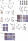
Fig. 5. Nuclear GCK-mediated TAZ phosphorylation and activation promotes tumour cell proliferation and tumour growth.
a Proliferation of MCF7 and BT549 cells with knocked-in expression of TAZ T346A and the indicated proteins was examined. The data are presented as the means ± SD, n = 3 independent experiments, two-tailed Student’s t test. b Parental MCF7 and BT549 cells and the indicated clones with knock-in expression of TAZ T346A were transfected with the indicated plasmids. The colony formation ability of the MCF7 and BT549 cells was assessed. The data are presented as the means ± SD, n = 3 independent experiments, two-tailed Student’s t test. c, d The parental MCF7 cells and the indicated clones with knock-in expression of GCK S398D or GCK S398A mutants were orthotopically injected into the fourth mammary fat pad of female BALB/c nude mice. The mice were sacrificed 30 days after cell injection. Tumour volume was calculated (n = 5 mice per group). Tumour weights were calculated (n = 5 mice per group). The data are presented as the means ± SDs; two-tailed Student’s t test. e IHC analyses of mouse mammary tumours were performed with the indicated antibodies. Representative staining images are shown. f, g The parental MCF7 cells and the indicated clones with knock-in expression of TAZ T346E or TAZ T346A mutants were orthotopically injected into the fourth mammary fat pad of female BALB/c nude mice. The mice were sacrificed 30 days after cell injection. Tumour volume was calculated (n = 5 mice per group). Tumour weights were calculated (n = 5 mice per group). The data are presented as the means ± SDs; two-tailed Student’s t test. h IHC analyses of mouse mammary tumours were performed with the indicated antibodies. Representative staining images are shown. i, j Parental MCF7 cells and the indicated clones with knock-in expression of TAZ T346A mutants were transfected with WT GCK or GCK S398D and orthotopically injected into the fourth mammary fat pad of female BALB/c nude mice. The mice were sacrificed 30 days after cell injection. The tumour volume was calculated (n = 5 mice per group). Tumour weights were calculated (n = 5 mice per group). The data are presented as the means ± SDs; one-way ANOVA. k IHC analyses of mouse mammary tumours were performed with the indicated antibodies. Representative staining images are shown. Source data are provided as a Source Data file.
Fig. 6
Fig. 6
A schematic model showing that nucleus-translocated GCK acts as a protein kinase and phosphorylates TAZ to promote TAZ-TEAD transcriptional activity and tumour growth.

References

    1. Li, X., Egervari, G., Wang, Y., Berger, S. L. & Lu, Z. Regulation of chromatin and gene expression by metabolic enzymes and metabolites. Nat. Rev. Mol. Cell Biol.19, 563–578 (2018). - PMC - PubMed
    1. Guo, D., Meng, Y., Jiang, X. & Lu, Z. Hexokinases in cancer and other pathologies. Cell Insight2, 100077 (2023). - PMC - PubMed
    1. Lu, B., Munoz-Gomez, M. & Ikeda, Y. The two major glucokinase isoforms show conserved functionality in beta-cells despite different subcellular distribution. Biol. Chem.399, 565–576 (2018). - PubMed
    1. Bian, X. et al. Regulation of gene expression by glycolytic and gluconeogenic enzymes. Trends Cell Biol.32, 786–799 (2022). - PubMed
    1. Li, X. et al. Mitochondria-translocated PGK1 functions as a protein kinase to coordinate glycolysis and the TCA cycle in tumorigenesis. Mol. Cell61, 705–719 (2016). - PMC - PubMed

MeSH terms

Substances