. 2025 Aug 4;16(1):7130.
doi: 10.1038/s41467-025-62512-4.

Precise mapping of single-stranded DNA breaks by sequence-templated erroneous DNA polymerase end-labelling

Affiliations

Precise mapping of single-stranded DNA breaks by sequence-templated erroneous DNA polymerase end-labelling

Leonie Wenson et al. Nat Commun. .

Abstract

The ability to analyze whether DNA contains lesions is essential in identifying mutagenic substances. Currently, the detection of single-stranded DNA breaks (SSBs) lacks precision. To address this limitation, we develop a method for sequence-templated erroneous end-labelling sequencing (STEEL-seq), which enables the mapping of SSBs. The method requires a highly error-prone DNA polymerase, so we engineer a chimeric DNA polymerase, Sloppymerase, capable of replicating DNA in the absence of one nucleotide. Following the omission of a specific nucleotide (e.g., dATP) from the reaction mixture, Sloppymerase introduces mismatches directly downstream of SSBs at positions where deoxyadenosine should occur. This mismatch pattern, coupled with the retention of sequence information flanking these sites, ensures that the identified hits are bona fide SSBs. STEEL-seq is compatible with a variety of sequencing technologies, as demonstrated using Sanger, Illumina, PacBio, and Nanopore systems. Using STEEL-seq, we determine the SSB/base pair frequency in the human genome to range between 0.7 and 3.8 × 10⁻⁶ with an enrichment in active promoter regions.


Conflict of interest statement

Competing interests: L.W., J.H., and O.Sö. are inventors on the patent application (WO Patent 2022/093091 A1) covering the design and use of Sloppymerase. The remaining authors declare no competing interests.

Figures

Fig. 1
Fig. 1. Overview of the STEEL-seq method.
Sloppymerase binds to a single-strand break (SSB) on a DNA strand. The strand downstream of the nick is degraded in the 5′–3′ direction by the exonuclease activity of Sloppymerase, while the polymerase activity enables the incorporation of three dNTPs, with dATP omitted from the dNTP mixture. This creates mismatches (N) in positions opposite of dT.
Fig. 2
Fig. 2. Determination of Sloppymerase characteristics.
A Time lapse experiment to assess Sloppymerase processivity. A nicked hairpin (HP) was incubated with either all dNTPs present (4) or with dATP, dGTP, and dTTP present (3), with DNA synthesis followed at different time points (0–120 min). When subjected to denaturing PAGE, the hairpin dissociates into two fragments: a longer 5′ fragment (A) and a shorter 3′ fragment (C). Extension of the longer fragment (B) indicates DNA polymerase activity, while degradation of fragment C indicates 5′–3′ exonuclease activity. A quantification of the bands (A and B) in the gel image are presented, which do not take in account the smear in between that indicate partially extended hairpins. The actual efficiency is hence even higher. B Processivity of Sloppymerase is dependent on the combination of provided dNTPs. A nicked hairpin (HP) was incubated with Sloppymerase and different combinations (either one or two dNTPs were omitted) and concentrations of dNTPs. Bands in position (B) imply full extension of the hairpin, whereas bands in position (A) indicate no elongation. C Structure of Sloppymerase, E. coli DNA polymerase I and DNA polymerase η, as predicted by AlphaFold.
Fig. 3
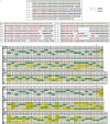
Fig. 3. Sanger and Illumina sequencing results for the application of Sloppymerase to a nicked hairpin.
A Sanger sequencing results when all of the nucleotides were included in the reaction mixture (4 dNTPs) and when either dATP (−dATP) or dCTP (−dCTP) was omitted. The upper row shows the sequence of the hairpin in green letters, with red letters indicating positions where (−dATP or −dCTP) substitutions are expected. Seven clones (1–7, A1–A7, and C1–C7) are shown in the rows below. Substitutions are shown in red, while additional substitutions, insertions, or deletions are marked in blue. B Illumina sequencing results with all dNTP (4 dNTPs) and when dATP was omitted (−dATP) from the dNTP mixture. The table shows the percentages of different nucleotides, along with the share of insertions and deletions. The arrow at positions 24/25 indicate the gap where Sloppymerase bind and alters the sequence downstream, position 25–79.
Fig. 4
Fig. 4. The application of STEEL-seq to identify SSBs.
Testing the utility of STEEL-seq for SSB detection in mitochondrial DNA with different sequencing techniques. STEEL-seq was performed with Nanopore, Illumina, and PacBio whole genome sequencing to detect SSBs at nicking sites. The DNA was nicked with Nt.BsmAI prior to STEEL-seq treatment to create SSBs at GTCTCN*N. The sequencing analysis results show the incorporation of mismatched bases in positions with dA (marked in red) in the reference genome; this was observed downstream of the nicking site at two different loci, A chrM:13020 and B chrM:14960 on the reverse strand, across all three sequencing technologies. The mitochondrial chromosome was chosen for presentation purposes; coverage on other chromosomes is lower on average. Image based on JBrowse screenshot.
Fig. 5
Fig. 5. Detection of SSBs using Nanopore and Illumina sequencing.
A Whole genome analysis of STEEL-seq of HaCaT cells, using Nanopore, and TK6 cells with Illumina. The figure shows SSB/base pair frequency for different regions. The width of the regions is proportional to the regions size in the genome. Lighter hue of the bars represents SSBs on the DNA strand in the forward direction, in relation to gene orientation, and darker hue represents SSBs on the DNA strand in the reverse orientation. B qRT-PCR analysis to confirm that TGFβ-stimulation induced expression of target genes was performed as a control before DNA samples were used for STEEL-seq. HaCaT cells were treated with DMSO or BMH-21 for 3 h, and stimulated or not with 5 ng/ml TGFβ for the last 1.5 h. Data are presented as mean values +/− SD, technical replicate n = 3. TGFβ-induced expression of target genes was confirmed by RNA sequencing (see Supplementary Table 1). C STEEL-seq results, using Illumina, of HaCaT cells treated with DMSO or BMH-21, stimulated or not with TGFβ. Upper row represent whole genome analysis. Second row represent selected genes where no transcript could be found. Third row show genes induced by TGFβ-treatment. The figure shows SSB/base pair frequency for different regions. The width of the regions is proportional to the regions size in the genome. Lighter hue of the bars represents SSBs on the DNA strand in the forward direction, in relation to gene orientation, and darker hue represents SSBs on the DNA strand in the reverse orientation.

References

    1. Caldecott, K. W. DNA single-strand break repair and human genetic disease. Trends Cell Biol.32, 733–745 (2022). - PubMed
    1. Jackson, S. P. et al. The DNA-damage response in human biology and disease. Nature461, 1071–1078 (2009). - PMC - PubMed
    1. Blasiak, J. Single-strand annealing in cancer. Int. J. Mol. Sci.22, 2167 (2021). - PMC - PubMed
    1. Caldecott, K. W. Single-strand break repair and genetic disease. Nat. Rev. Genet.9, 619–631 (2008). - PubMed
    1. Zeng, X. et al. DNA polymerase eta is an A-T mutator in somatic hypermutation of immunoglobulin variable genes. Nat. Immunol.2, 537–541 (2001). - PubMed

MeSH terms

Substances

LinkOut - more resources