Review
. 2025 Aug 4;24(1):211.
doi: 10.1186/s12943-025-02417-4.

Unraveling the glyco-immunity nexus in pancreatic cancer

Affiliations
Review

Unraveling the glyco-immunity nexus in pancreatic cancer

Christabelle Rajesh et al. Mol Cancer. .

Abstract

Pancreatic ductal adenocarcinoma (PDAC) is an extremely aggressive disease, and standard of care therapies have failed to yield significant clinical benefit, with invasive surgery being the only curative treatment for patients with early-stage disease. Tumor-associated glycans in pancreatic cancer have direct effects on the survival and propagation of the tumor proper and contribute to an immunosuppressed tumor microenvironment. The existence of a “tumor glycocode” in PDAC and the role of hypersialylation in this cancer have been hugely underscored. Through this initial understanding, significant strides have been made in the field of glycosylation-mediated immune regulation, uncovering glyco-immune checkpoints that facilitate tumor progression in PDAC and other malignancies. Here, we describe the specific roles of glycan-binding proteins, such as C-type lectin receptors, Siglecs, and Galectins, in generating and promoting immunosuppression, exacerbating survival outcomes, and dampening therapeutic efficacy. We illustrate the scale of glycan-mediated regulation of homeostatic immune responses and how cancer glycans facilitate dampened anti-tumor immunity through the major histocompatibility complex (MHC) and the enhanced expression of immune checkpoints, such as PD-L1 and CTLA-4. A wide array of glycan-targeted therapies against PDAC in the clinic, including monoclonal antibodies, enzymes, and vaccines, has been described. With the help of new glycosylation signatures identified and techniques that allow us to reach single-cell resolution, we can target glycans and generate strategies to activate the immune system against PDAC.

Keywords: Immune signaling; Pancreatic cancer; Therapies; Tumor glycosylation; Tumor microenvironment.


Conflict of interest statement

Declarations. Ethics approval and consent to participate: Not applicable. Consent for publication: Not applicable. Competing interests: The authors declare no competing interests.

Figures

Fig. 1
Fig. 1
Progression of pancreatic cancer in the context of tumor glycosylation. Pancreatic cancer of exocrine origin typically begins with malignant transformation of acinar cells, frequently due to mutations, like those in the (a) KRAS oncogene, followed by mutations in CDKN2A, TP53 and SMAD4 genes, all of which leads to sustained proliferation and formation of a highly dysplastic, metastatic pancreatic tumor. Certain cells that have such high genomic instability are removed via mechanisms like the epithelial defense against cancer (EDAC) or immune surveillance. (b) The cellular events accompanying this begin with acinar to ductal metaplasia (ADM) and progress through pancreatic intraepithelial neoplasms (PanINs) -1, -2 and − 3 prior to the formation of aggressive pancreatic ductal adenocarcinoma. Glycosylation modulating events tie into abnormal metabolic pathways that starts with the Warburg effect, wherein transformed cells switch to aerobic glycolysis to facilitate energy production as well as maintain the synthesis of biosynthetic intermediates for cell proliferation. Such increased glucose uptake also facilitates glucose flux through the hexosamine biosynthetic pathway (HBP), which leads O-GlcNAcylation of the enzyme phosphofructokinase 1 (PFK1), which reduces PFK1 activity and serves as a positive feedback loop for increased HBP flux of glucose, leading to sustained high O-GlcNAcylation of proteins that facilitates oncogenic processes. Several other mechanisms described within this review lead to increasing truncated O-glycans (Tn antigen), increasing tumor sialylation and appearance of glycosylation mediated immune checkpoints, all of which contribute to a marked reduction in anti-tumor immunosurveillance, that aids in immune evasion and tumor progression
Fig. 2
Fig. 2
Overview of key glycosylation pathways affected in pancreatic cancer. O-glycosylation on proteins is initiated in the Golgi with the addition of a N-Acetylgalactosamine (GalNAc) to serine or threonine (Ser/Thr) residues within proteins by polypeptidyl a-GalNAc transferase to yield the Tn antigen or CD175. Next, either ST6GalNAc-1 adds an 𝛼2,6 sialic acid (Neu5Ac) to form the sialyl Tn antigen or CD175s, or T-synthase (properly folded by its molecular chaperone Cosmc) adds Galactose (Gal) to form the T antigen or Core 1 structure (Galβ1-3-GalNAca1-O-Ser/Thr). Core 1 can be extended by ST6GalNAc-2-4 enzymes to 6-sialyl Core 1 (sialyl T antigen) and further via 𝛼3-sialyl transferase (𝛼3ST) to the disialyl core 1 or disialyl T antigen. Core 1 can alternatively also undergo 𝛼3-sialic acid addition by ST3Gal-1 to yield the 3-sialyl Core 1 structure, which can further be extended to the disialyl Core 1 structure (disialyl T) by ST6GalNAc-2-4 enzymes. Blood group Lewis antigens can be found on O- and N-glycans. Type I Lewis antigens start with the β1–3 glycosidic bond (Galβ1-3GlcNAc), also called the type 1 chain. Fucosyltransferase 2 (FUT2) creates the α-1,2-fucosidic linkage on a terminal Gal to yield the H type 1 structure, FUT3 creates the α-1,4-fucosidic linkage on N-Acetylglucosamine (GlcNAc) to yield the Lewis A structure (LeA) or a sialyltransferase (ST) adds an 𝛼3 sialic acid (sialyl Lec), to which FUT3 subsequently adds the α-1,4-fucosidic linkage on N-Acetylglucosamine (GlcNAc) to yield the sialyl Lewis A (sialyl LeA) structure, also called CA19-9. Type II Lewis antigens start with the β1–4 glycosidic bond (Galβ1-4GlcNAc), also called the type 2 chain. FUT1 creates an α-1,2-fucosidic linkage on a terminal Gal to yield the H type 2 structure, FUT3 creates the α-1,3-fucosidic linkage on N-Acetylglucosamine (GlcNAc) to yield the Lewis A structure (LeX) or 𝛼3ST yield a Sialyl LacNAc chain, on which FUT3 catalyzes the α-1,3-fucosidic linkage to yield the sialyl Lewis X structure (sialyl LeX)
Fig. 3
Fig. 3
Siglecs as immune checkpoints in pancreatic cancer. Tumor cell-derived or cancer-associated fibroblast (CAF)-derived sialic acids originate in the nucleus, where N-acetylneuraminic acid (Neu5Ac) is added to cytidine monophosphate (CMP) to yield CMP-Neu5Ac by the enzyme cytidine monophosphate N-acetylneuraminic acid synthetase (CMAS). CMP-Neu5Ac is transported into the Golgi by solute carrier SLC35A1 and is used for subsequent sialylation events. Proteins or lipids bearing O-glycans undergo sialylation using CMP-Neu5Ac via ST6GalNAc1-6 enzymes to yield terminal 𝛼2–6 sialyl GalNAc, or via ST3Gal 1–6 to yield terminal 𝛼2–3 sialyl Gal structures. Simultaneously, proteins or lipids containing N-glycans terminating in mannose are transported from the endoplasmic reticulum (ER) to the Golgi, where they undergo addition of terminal Gal via ST6Gal1-2 enzymes, followed by terminal 𝛼2–8 sialylation via ST8Sia1-5 enzymes. Glycan carrying proteins or lipids are transported to the cell membrane, where their terminal sialic acids can interact with Siglec receptors on cells. Siglec-4 on oligodendrocytes and Schwann cells interacts with disialyl T (carrying both 𝛼2–6 sialyl GalNAc and 𝛼2–3 sialyl Gal) on MUC1, which facilitates perineural invasion of tumor cells; Siglecs − 7, -9, -10 and − 15 on myeloid cells and NK cells bind terminal sialic acid to facilitate immunosuppression through mechanisms of macrophage polarization to the tumor-associated macrophage (TAM) phenotype, upregulation of PD-1 checkpoint on immune cells and disruption of tumor cell killing by immune cells in the microenvironment
Fig. 4
Fig. 4
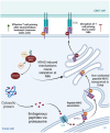
Sialic acids in pancreatic cancer disrupt T cell priming. The major histocompatibility complex 1 (MHC-I) is responsible for presenting endogenous tumor antigens to T cells. Asparagine residue 86 (N86) on the heavy chain of MHC-I is N-glycosylated to ensure proper folding of the MHC-I complex and protein loading into MHC-I followed by membrane presentation. In mutant KRAS-driven cancers like PDAC, improper Ras activation leads to the addition of sialic acid on N86 of the heavy chain of MHC-I, and such sialylated MHC-I is unable to successfully present antigen to the T cell receptor (TCR), which leads to disruption of T cell priming. Treating tumor cells with neuraminidase to remove surface sialic acid prevents this event and potentiates T cell activation
Fig. 5
Fig. 5
Glycosylation directly facilitates membrane expression and stabilization of the PD-L1 immune checkpoint. Programmed death ligand 1 (PD-L1) is an immune checkpoint expressed on tumor cells and other cells of the tumor microenvironment (TME). This figure describes three pathways that regulate PD-L1 expression in cancer. (a). Epidermal growth factor (EGF) binding to its receptor EGFR facilitates transcription factor 4 (TCF4) binding to the beta-1,3-N-acetylglucosaminyltransferase 3 (B3GNT3) gene promoter, and expression of B3GNT3. In the Golgi, the B3GNT3 enzyme adds N-glycans to asparagine residues N192, N200 and N219 of the PD-L1 protein backbone which leads to cell surface retention of PD-L1. Interestingly, glycogen synthase kinase-3 beta (GSK3B) promotes proteasomal degradation of PD-L1, however, EGFR activation disrupts this GSK3B axis. (b). Interleukin-6 (IL-6) binds to and activates its receptor, which leads to recruitment of Janus kinase 1 (JAK1), which phosphorylates tyrosine 112 (Y112) on a non-glycosylated PD-L1 protein. Phospho PD-L1 is transported to the endoplasmic reticulum (ER) where STT3A (the catalytic subunit of an N-oligosaccharyltransferase) adds N-glycans (Glc3Man9GlcNAc2) en bloc, which mediates cell membrane expression of glycosylated PD-L1. (c). Internalization of transiently expressed PD-L1 in pancreatic cancer leads to phosphorylation at threonine (T194/T210) by Never in Mitosis A-Related Kinase 2 (NEK2), which prevents its proteasomal degradation, and facilitates membrane PD-L1 stabilization. T194/T210 phosphorylation also perturbs serine (S195) phosphorylation. Phospho-S195 facilitates displacement of PD-L1 from the cell surface, and thus NEK2 facilitates increased PD-L1 expression in PDAC

References

    1. Levene PA, Alsberg CL. The cleavage products of vitellin. J Biol Chem. 1906;2:127–33. 10.1016/S0021-9258(17)46054-6
    1. Li W, Li F, Zhang X, Lin H, Xu C. Insights into the post-translational modification and its emerging role in shaping the tumor microenvironment. Signal Transduction and Targeted Therapy. 2021;6:1–30. 10.1038/s41392-021-00825-8 - PMC - PubMed
    1. Jiang G, Li C, Lu M, Lu K, Li H. Protein lysine crotonylation: past, present, perspective. Cell Death Disease. 2021;12:7. 10.1038/s41419-021-03987-z - PMC - PubMed
    1. Zhang Z, Tan M, Xie Z, Dai L, Chen Y, Zhao Y. Identification of lysine succinylation as a new post-translational modification. Nat Chem Biology. 2010;7:1. 10.1038/nchembio.495 - PMC - PubMed
    1. Mondal S, Wang S, Zheng Y, Sen S, Chatterjee A, Thompson PR. Site-specific incorporation of citrulline into proteins in mammalian cells. Nat Commun. 2021;12:1. 10.1038/s41467-020-20279-w - PMC - PubMed

LinkOut - more resources