

Early prediction of proton therapy dose distributions and DVHs for hepatocellular carcinoma using contour-based CNN models from diagnostic CT and MRI
- PMID: 40759962
- PMCID: PMC12323130
- DOI: 10.1186/s13014-025-02708-6
Early prediction of proton therapy dose distributions and DVHs for hepatocellular carcinoma using contour-based CNN models from diagnostic CT and MRI
Abstract
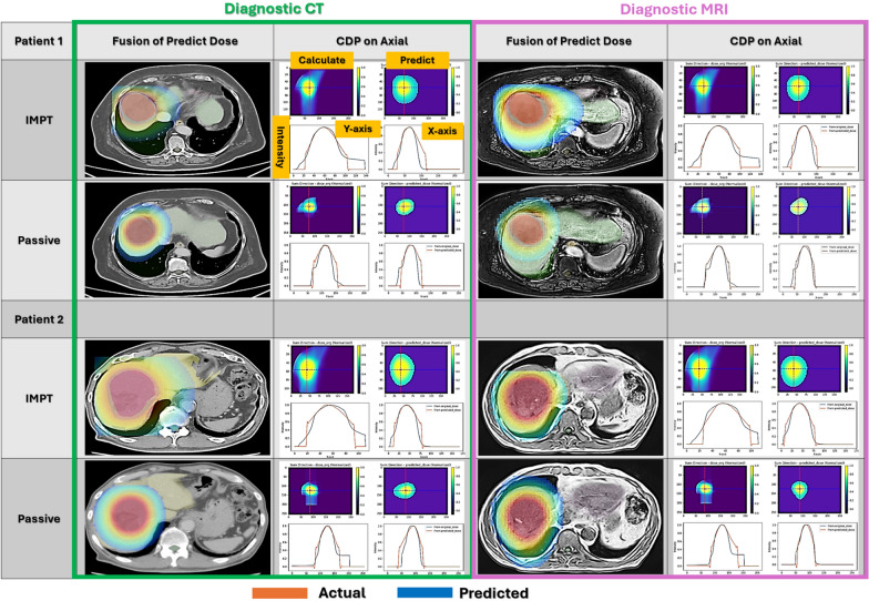
Background: Proton therapy is commonly used for treating hepatocellular carcinoma (HCC); however, its feasibility can be challenging to assess in large tumors or those adjacent to critical organs at risk (OARs), which are typically assessed only after planning computed tomography (CT) acquisition. This study aimed to predict proton dose distributions using diagnostic CT (dCT) and diagnostic MRI (dMRI) with a convolutional neural network (CNN), enabling early treatment feasibility assessments.
Methods: Dose distributions and dose-volume histograms (DVHs) were calculated for 118 patients with HCC using intensity-modulated proton therapy (IMPT) and passive proton therapy. A CPU-based CNN model was used to predict DVHs and 3D dose distributions from diagnostic images. Prediction accuracy was evaluated using mean absolute error (MAE), mean squared error (MSE), peak signal-to-noise ratio (PSNR), structural similarity index (SSIM), and gamma passing rate with a 3 mm/3% criterion.
Results: The predicted DVHs and dose distributions showed high agreement with actual values. MAE remained below 3.0%, with passive techniques achieving 1.2-1.8%. MSE was below 0.004 in all cases. PSNR ranged from 24 to 28 dB, and SSIM exceeded 0.94 in most conditions. Gamma passing rates averaged 82-83% for IMPT and 92-93% for passive techniques. The model achieved comparable accuracy when using dMRI and dCT.
Conclusions: This study demonstrates that early dose distribution prediction from diagnostic imaging is feasible and accurate using a lightweight CNN model. Despite anatomical variability between diagnostic and planning images, this approach provides timely insights into treatment feasibility, potentially supporting insurance pre-authorization, reducing unnecessary imaging, and optimizing clinical workflows for HCC proton therapy.
Keywords: Convolutional neural network; Diagnostic imaging; Dose distribution; Dose volume histogram; Hepatocellular carcinoma; Intensity-modulated proton therapy; Passive methods; Proton therapy.
© 2025. The Author(s).
Conflict of interest statement
Declarations. Ethical approval and consent to participate: This study is a retrospective analysis of radiotherapy outcomes using existing patient data. Ethical approval for the use of these data was granted by the Ethics Committee of the National Cancer Center Hospital, Japan (Approval No. 2020-272, dated 12 October 2020). All procedures were carried out in accordance with the Declaration of Helsinki and relevant institutional guidelines." Consent for publication: Written informed consent was obtained from the patient(s) for the use of personal data and images in this publication. The patient(s) were fully informed about the purpose of the study, the intended use of their data and images, and their rights to privacy. The consent form is retained by the corresponding author and is available upon request, should it be necessary for legal or ethical purposes. Additionally, this information can be referenced on the National Cancer Center Hospital East website: https://www.ncc.go.jp/jp/ncce/index.html . Competing interest: The authors declare no competing interests.
Figures


References
-
- Takeda A, Sanuki N, Tsurugi Y, Iwabuchi S, Matsunaga K, Ebinuma H, et al. Phase 2 study of stereotactic body radiotherapy and optional transarterial chemoembolization for solitary hepatocellular carcinoma not amenable to resection and radiofrequency ablation. Cancer. 2016;13:2041–9. 10.1002/cncr.30008. - DOI - PubMed
-
- Hong TS, Wo JY, Yeap BY, Ben-Josef E, McDonnell EI, Blaszkowsky LS, et al. Multi-institutional phase II study of high-dose hypofractionated proton beam therapy in patients with localized, unresectable hepatocellular carcinoma and intrahepatic cholangiocarcinoma. J Clin Oncol. 2016;34:460–8. 10.1200/JCO.2015.64.2710. - DOI - PMC - PubMed
-
- Korean Liver Cancer Study Group, National Cancer Center Korea. Practice guidelines for management of hepatocellular carcinoma 2009. Korean J Hepatol. 2009;15:391–423. - PubMed
MeSH terms
Grants and funding
LinkOut - more resources
Full Text Sources
Medical
Miscellaneous
