

Decellularised matrices from force loaded periodontal ligament stem cells support osteogenic differentiation
- PMID: 40760151
- PMCID: PMC12322110
- DOI: 10.1038/s41598-025-14343-y
Decellularised matrices from force loaded periodontal ligament stem cells support osteogenic differentiation
Abstract
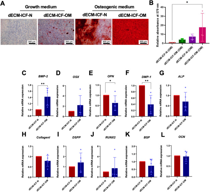
Periodontal ligament stem cells (hPDLSCs) are mechanosensing cells responding to mechanical forces. This study investigates the impact of decellularised extracellular matrix (dECM) derived from intermittent compressive force (ICF)-treated hPDLSCs on osteogenic differentiation. hPDSCLs were subjected to ICF loading at 1.5 g/cm2 for 24 h and then maintained with normal medium (N) or osteogenic induction medium (OM) followed by a decellularisation process. dECMs derived from ICF (dECM-ICF) were characterised using a scanning electron microscope, energy-dispersive X-ray spectroscopy, and proteomic analysis. hPDLSCs were re-seeded on dECM-ICF. Cell proliferation and viability were examined by resazurin and LIVE/DEAD assays. Mineralisation was determined by Alizarin Red S staining. Results demonstrated that dECM-ICF-derived from OM (dECM-ICF-OM) exhibited a fibrillar network structure and showed no cellular component while preserving fibronectin and type I collagen. dECM-ICF exhibited biocompatibility, as indicated by the absence of cytotoxic effects and the ability of hPDLSCs to attach, spread, and proliferate. dECM-ICF-OM significantly enhanced mineral deposition compared to dECM-ICF from normal conditions (dECM-ICF-N). Proteomic analysis of dECM-ICF demonstrated the upregulated proteins in the PI3K-Akt, Ras, MAPK, mTOR, ErbB, TNF, and VEGF signallings. In conclusion, dECM-ICF supports hPDLSCs growth and osteogenic differentiation. dECM-ICF is a promising cell-free natural scaffold to promote periodontal regeneration.
Keywords: Decellularisation; Extracellular matrix; Intermittent compressive force; Periodontal ligament stem cells.
© 2025. The Author(s).
Conflict of interest statement
Declarations. Competing interests: The authors declare no competing interests.
Figures


Similar articles
-
Astragaloside IV Promotes Osteogenic Differentiation of Periodontal Ligament Stem Cells via Activating PI3K/AKT/eNOS/NO Signaling Pathway: In vitro and in vivo Study.Drug Des Devel Ther. 2025 Jul 16;19:6073-6088. doi: 10.2147/DDDT.S514682. eCollection 2025. Drug Des Devel Ther. 2025. PMID: 40687900 Free PMC article.
-
Apoptotic extracellular vesicles derived from human dental pulp stem cells facilitate periodontal tissue regeneration.BMC Oral Health. 2025 Jul 11;25(1):1150. doi: 10.1186/s12903-025-06531-z. BMC Oral Health. 2025. PMID: 40646544 Free PMC article.
-
Human dental follicle cell-derived conditioned media enhance periodontal regeneration by regulating the osteogenic differentiation and inflammation of periodontal ligament stem cells and macrophage polarization.Mol Cell Biochem. 2025 Jul;480(7):4431-4448. doi: 10.1007/s11010-025-05260-9. Epub 2025 Apr 2. Mol Cell Biochem. 2025. PMID: 40175780
-
Advancements in diabetic foot ulcer therapy: The role of exosomes and decellularised extracellular matrix scaffolds.Diabetes Res Clin Pract. 2025 Aug;226:112364. doi: 10.1016/j.diabres.2025.112364. Epub 2025 Jul 10. Diabetes Res Clin Pract. 2025. PMID: 40651532 Review.
-
Periodontal ligament-derived cells for periodontal regeneration in animal models: a systematic review.J Periodontal Res. 2015 Apr;50(2):160-72. doi: 10.1111/jre.12205. Epub 2014 Jun 26. J Periodontal Res. 2015. PMID: 24965968
References
-
- Turker, K. S., Sowman, P. F., Tuncer, M., Tucker, K. J. & Brinkworth, R. S. The role of periodontal mechanoreceptors in mastication. Arch. Oral Biol.52, 361–364. 10.1016/j.archoralbio.2006.11.014 (2007). - PubMed
-
- Li, B., Chen, X., Jin, Y. & Tooth Dent. Pulp Regeneration 367–392 10.1016/b978-0-12-811920-4.00015-x (2019).
-
- Tuli, R. et al. Characterization of multipotential mesenchymal progenitor cells derived from human trabecular bone. Stem Cells. 21, 681–693. 10.1634/stemcells.21-6-681 (2003). - PubMed
-
- Manokawinchoke, J. et al. Mechanical loading and the control of stem cell behavior. Arch. Oral Biol.125, 105092. 10.1016/j.archoralbio.2021.105092 (2021). - PubMed
MeSH terms
Substances
Grants and funding
LinkOut - more resources
Full Text Sources
Medical
Research Materials
Miscellaneous
