. 2025 Aug;27(8):1240-1255.
doi: 10.1038/s41556-025-01708-8. Epub 2025 Aug 4.

A programmed decline in ribosome levels governs human early neurodevelopment

Affiliations

A programmed decline in ribosome levels governs human early neurodevelopment

Chunyang Ni et al. Nat Cell Biol. 2025 Aug.

Abstract

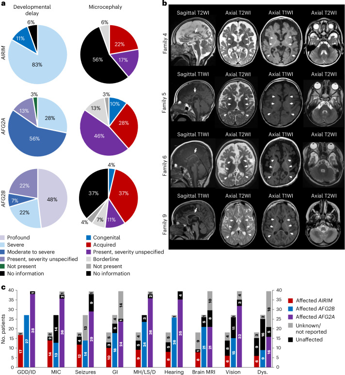
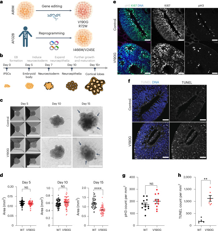
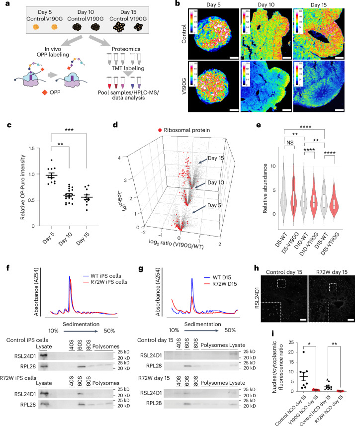
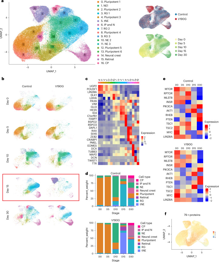
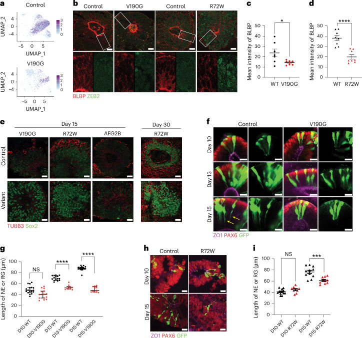
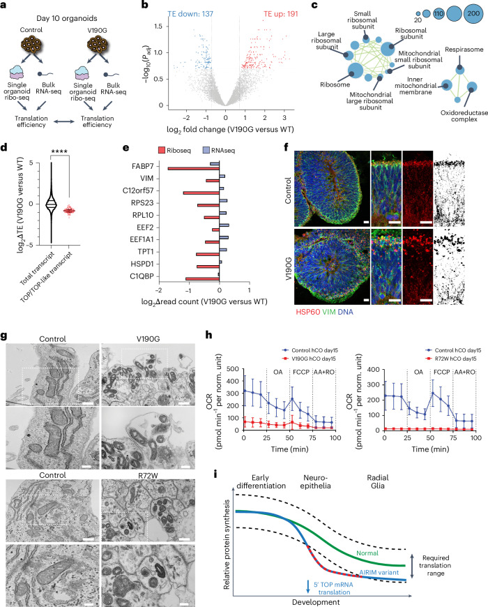
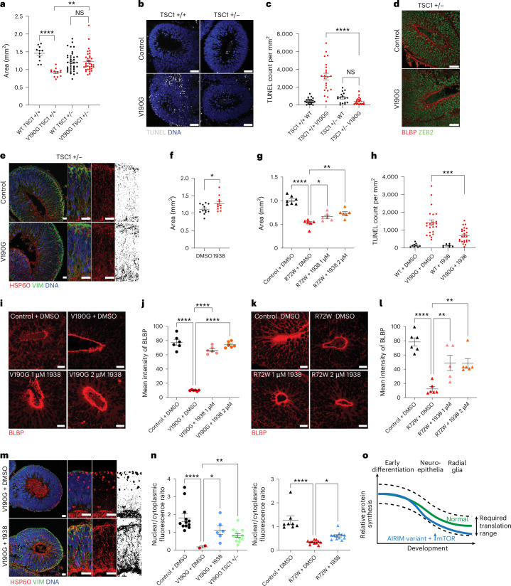
Many neurodevelopmental defects are linked to genes involved in housekeeping functions, such as those encoding ribosome biogenesis factors. How reductions in ribosome biogenesis can result in tissue- and developmental-specific defects remains unclear. Here we describe variants in the ribosome biogenesis factor AIRIM/C1orf109 that are primarily associated with neurodevelopmental disorders. Using human cerebral organoids in combination with proteomic, single-cell RNA sequencing and single-organoid translation analyses, we identify a previously unappreciated drop in protein production during early brain development. We find that ribosome levels decrease during neuroepithelial differentiation, making differentiating cells particularly vulnerable to perturbations in ribosome biogenesis during this time. Reduced ribosome availability more profoundly impacts the translation of specific transcripts, disrupting both survival and cell fate commitment of transitioning neuroepithelia. Enhancing mTOR activity suppresses the growth and developmental defects associated with AIRIM/C1orf109 variants. This work provides evidence for the functional importance of regulated changes in global protein synthesis capacity during cellular differentiation.


Conflict of interest statement

Competing interests: The authors declare no competing interests.

Figures

Fig. 1
Fig. 1. Family pedigrees, variant schematic on gene and protein level and amino acid conservation of AIRIM substitutions.
a, Pedigrees showing segregation of the variants. Affected and unaffected individuals are indicated by filled and open and filled symbols, respectively. Probands are marked with arrows. Double lines indicate consanguinity. Red asterisks represent a nonhomozygous genotype. Segregation results are shown by the presence of a red minus symbol for the variant or a black plus symbol representing the reference allele, where −/− and +/+ represent a homozygous variant or wild-type genotype, respectively, and +/− represents a heterozygous genotype. b, Schematic representation of the gene and protein positions of AIRIM variants. AIRIM is located on chromosome 1 at cytogenetic position p34.3 (top). Below: the genetic variants are mapped to the NM_017850.3 transcript of AIRIM. c, AIRIM variants shown on the protein level. The casein kinase II substrate (CK2S) region is shown in maroon with the corresponding amino acid coordinates of the protein and domain in blue (top). Variants are labelled by family codes. Alignment of multiple AIRIM orthologues with amino acid substitutions marked with arrows (bottom).
Fig. 2
Fig. 2. Clinical features of the affected individuals with biallelic AIRIM variants.
a, Summary of global developmental delay and microcephaly in aggregated published individuals to date. b, Brain MRI of the 1-year and 11-month-old proband in family 4 (II:2). Sagittal T2-weighted image (WI), axial T2WI and axial T1WI showing atrophy in the supratentorial compartment of the brain, including a diffusely thin corpus callosum (arrow) and enlargement of the extra-axial spaces. Abnormal T2WI hyperintensity of the deep and superficial white matter with relatively normal myelination appearance along the anterior and posterior limbs of the internal capsules, as well as the optic tracts (arrowheads). Brain MRI of the sibling of the proband in family 5 (II:3) showing severe atrophy in the supratentorial compartment of the brain, including a diffusely thin corpus callosum (arrow) and enlargement of the extra-axial spaces. Abnormal T2WI hyperintensity of the deep and superficial white matter with relatively normal myelination appearance along the anterior and posterior limbs of the internal capsules, as well as the optic tracts (arrowheads). It is also important to note the relatively normal appearance of the overall structures of the posterior fossa with normal volume, morphology and myelination appearance of the cerebellum. Brain MRI of the proband in family 6 (II:1) at 6 months old; sagittal T1WI, axial T2WI and axial T1WI. Severe atrophy in the supratentorial compartment of the brain, including a diffusely thin corpus callosum (arrows) and diffuse enlargement of the extra-axial spaces with bilateral under-opercularization of Sylvian fissures. Abnormal T2WI hyperintensity of the deep and superficial white matter with relatively normal myelination appearance along the anterior and posterior limbs of the internal capsules, as well as the optic tracts (arrowheads). It is also important to note the relatively normal appearance of the overall structures of the posterior fossa with normal volume, morphology and myelination appearance of the cerebellum. Brain MRI of the proband in family 9 (II:3) showing severe atrophy in the supratentorial compartment of the brain, including a diffusely thin corpus callosum (arrow) and enlargement of the extra-axial spaces. Abnormal T2WI hyperintensity of the deep and superficial white matter with relatively normal myelination appearance along the anterior and posterior limbs of the internal capsules, as well as the optic tracts (arrowheads). It is also important to note the relatively normal appearance of the overall structures of the posterior fossa with normal volume, morphology and myelination appearance of the cerebellum. c, Bar graphs summarizing the comparative proportions of various clinical findings in individuals with AIRIM, AFG2B and AFG2A variants. Dys., dysmorphic facial features; GDD/ID, global developmental delay/intellectual disability; GI, gastrointestinal issues; MH/LS/D, muscular hypotonia/limb spasticity/dystonia; MIC, microcephaly.
Fig. 3
Fig. 3. The NDD-associated AIRIMV190G variant causes growth defects at a specific stage of cerebral organoid development.
a, Schematic explaining the origin of the variant samples used in this study. b, Schematic of the timeline for generating brain organoids from pluripotent stem cells. c, Bright-field images of control and mutant (V190G) organoids at days 5, 10 and 15. Black arrows indicate NE buds, which seem less organized and elongated in the mutant at day 15. Scale bar, 1 mm. d, Quantification of bright-field images at days 5 (left), 10 (middle) and 15 (right) show that control neural tissue is enlarged relative to mutant (V190G); ****P < 0.0001, unpaired two-sided t-test with Welch’s correction, day 5, n = 73 control EBs 62 mutant EBs from three independent batches; day 10, n = 77 control organoids 84 mutant organoids from four independent batches; day 15, n = 46 control organoids 51 mutant organoids from three independent batches. Error bars show s.e.m. P = 0.8776 (day 5), P = 0.1128 (day 10), P = 2.515 × 10−11 (day 15). NS, not significant. e, Representative images showing the proliferation marker pH3 (grey) and Ki67 (green) signal in control (WT) and mutant (V190G) day 15 organoids. Scale bar, 50 μm. f, Representative images showing TUNEL labelling in control and mutant (V190G) day 15 organoids. Mutant organoids exhibit a higher TUNEL signal. Scale bar, 50 μm. g, Quantification of pH3 signal of individual NE buds at day 15 control (WT) and mutant (V190G) organoids. Unpaired two-sided t-test with Welch’s correction. n = 12 control and 11 mutant imaged regions from six organoids from two independent batches. Error bars show s.e.m. h, Quantification of TUNEL signal of individual NE bud showing that mutant organoids exhibit a higher TUNEL signal. TUNEL counts were normalized to the area of the imaged bud. **P < 0.01, unpaired two-sided t-test with Welch’s correction, P = 0.0022, n = 6 control n = 6 mutant imaged regions from two independent batches. Error bars show s.e.m. The image in a was made using BioRender. Source data
Fig. 4
Fig. 4. The AIRIMV190G variant causes stage-specific defects in protein synthesis and ribosome levels.
a, Experimental schemes of OP-puro based translation capacity and TMT assays. b, Representative images showing the OP-puro signal in control and mutant (V190G) organoids at days 5, 10 and 15. Mutant organoids exhibited lower OP-puro signals than controls starting at day 10. Scale bar, 50 μm. c, Quantification of OPP signal of individual NE bud at day 5, 10 and 15 mutant (V190G) organoids relative to their corresponding control. Dunn’s multiple comparisons test (two-sided). n = 11, 17 and 11 individual EB or NE buds from six individual organoids imaged from two independent batches. Error bars show s.e.m. Day 5 versus day 10 adjusted P value (Padj) = 8.013453 × 10−5; day 5 versus day 15 Padj = 4.692448 × 10−5, **P < 0.01, ***P < 0.001. d, Volcano plots of the −log10-transformed P value versus the log2-transformed ratio of mutant/control organoids at days 5, 10 and 15 (n = 3,539, 3,544 and 3,541 proteins, respectively n = 3 biologically independent samples). Discovery was determined using the two-stage linear step-up procedure of Benjamini, Krieger and Yekutieli, a false discovery rate (FDR)-controlling method for multiple comparisons (two-sided test). e, Violin plots showing the distribution of r-proteins as in d (n = 76 r-proteins; n = 3 biologically independent samples). Adjusted P values were calculated from Tukey’s multiple comparisons test (two-sided). **P < 0.01, ****P < 0.0001. Error bars show s.e.m. f, Polysome profiling with the corresponding western blots comparing the distribution of RSL24D1 and RPL28 in control and AIRIMR72W iPS cells. Three independent batches were performed. g, Polysome profiling with corresponding western blots comparing the distribution of RSL24D1 and RPL28 in control and AIRIMR72W day 15 organoids. Three independent batches were performed. h, IF showing the subcellular distribution of RSL24D1 in control and AIRIMR72W day 15 organoids. Scale bar, 20 μm. Three independent batches were performed. i, Quantification of the nuclear/cytoplasm ratio of RSL24D1 immunofluorescence of control, AIRIMV190G and AIRIMR72W variant day 15 cerebral organoids. *P = 0.032, **P = 0.0042. Dunnett’s T3 multiple comparisons test (two-sided). Control for V190G, n = 8; control for V190G, n = 9; control for R72W, n = 10; control for R72W, n = 13 imaged regions from two independent batches. Error bars show s.e.m. Source data
Fig. 5
Fig. 5. scRNA-seq reveals AIRIMV190G mutant organoids display a transient delay of neuroepithelial differentiation.
a, UMAP embedding of scRNA-seq data from one pooled sample per condition (n = 50 day 5 EBs, 3–6 day 10 NE, 3–6 day 15 organoids and 3 day 30 organoids per genotype, 10,000 cells were sequenced per condition). tNE, transitioning NE; IP, intermediate progenitor; N, neuron; CP, choroid plexus. b, Split-view of full integrated dataset, coloured by Louvain clusters. c, Heatmap showing the average expression of cluster marker genes related to brain development from the full integrated dataset. d, Quantification of different cell types identified at each time point in control and V190G samples. e, Heatmap showing the average expression of translation regulator genes across the development of control and V190G organoids. f, Gene module expression scores for a set of all 79 human r-protein genes across developmental time points.
Fig. 6
Fig. 6. The AIRIMV190G cerebral organoids exhibit impaired radial glial cell fate specification.
a, UMAP showing FABP7 expression in control and AIRIM variant cells in day 15 organoids. b, IF image of day 15 control and mutant (V190G and R72W) organoids of transitioning NE marker ZEB2 and committed RG marker BLBP (encoded by FABP7). Note that the mutant exhibits less expression of BLBP and less-organized nuclei (marked by ZEB2). Scale bar, 20 µm. c, Quantification of mean BLBP IF intensity of day 15 control and mutant (V190G) organoids. *P = 0.0463. Unpaired two-tailed t-test with Welch’s correction. WT, n = 7; V190G, n = 8 imaged regions from two independent batches. Error bars show s.e.m. d, Quantification of mean BLBP IF intensity of day 15 control and mutant (R72W) organoids. ****P = 8.65486 × 10−5. Unpaired two-tailed t-test with Welch’s correction. WT, n = 8; R72W, n = 8 imaged regions from two independent batches. Error bars show s.e.m. e, IF image of day 15 control and AIRIMV190G, AIRIMR72W and AFG2BI466M/V245E organoids of the progenitor marker Sox2 and the neuronal differentiation marker TUBB3. Scale bar, 20 μm. Also included are IF images of day 30 control and AIRIMR72W organoids stained for the progenitor marker Sox2 and the neuronal differentiation marker TUBB3. Scale bar, 40 μm. Three independent batches were performed. f, Representative whole-mount organoid IF images showing the morphology of neural progenitor cells (PAX6+), around apical (ZO1+) lumens, revealed by sparse labelling with GFP in control and mutant (V190G) organoids. Day 10 cells are columnar and exhibit typical NE morphology. Day 15 control cells show a thinning of apical processes (yellow arrows), whereas mutant cells still seem to be columnar. Scale bar, 20 μm. g, Quantification of the length of neural progenitor cells in control and mutant (V190G) organoids at days 10, 13 and 15, showing reduced length of the progenitor cells in mutant compared with control organoids. Cells with clear apical and basal labelling were used for quantification. Mann–Whitney U-test, ****P < 0.0001, two-tailed, multiple-comparison corrected. Day 10 control, n = 10 cells; day 10 V190G, n = 8 cells; day 13 control, n = 14 cells; day 13 V190G, n = 16 cells; day 15 control, n = 8 cells; and day 15 V190G, n = 8 cells. Padj = 0.0714, day 10; Padj = 9.177792 × 10−7, day 13; Padj = 1.098352 × 10−7, day 15. Error bars show s.e.m. h, Representative whole-mount organoid IF images showing the morphology of neural progenitor cells revealed by sparse labelling with GFP in control and mutant (R72W) organoids. Day 10 cells are columnar and exhibit typical NE morphology. Day 15 control cells show a thinning of apical processes, whereas mutant cells do not. Scale bar, 20 μm. i, Quantification of the length of neural progenitor cells in control and AIRIMR72W organoids at days 10 and 15. Cells with clear apical and basal labelling were used for quantification. Mann–Whitney U-test, ***P = 0.0009, two-tailed, day 10 control, n = 12 cells; day 10 V190G, n = 9 cells; day 15 control, n = 11 cells; and day 15 V190G, n = 10 cells. Error bars show s.e.m. Source data
Fig. 7
Fig. 7. Reduced ribosome availability more profoundly affects a select subset of transcripts in differentiating neuroepithelia.
a, Schematic of ribo-seq experiment. b, Volcano plot of change in TE of mutant (V190G) relative to control organoids at day 10. Red, log2(fold change) > 0.7 (higher TE in mutant); blue, log2(fold change) < −0.7 (lower TE in mutant); n = 2 independent batches for bulk RNA-seq, n = 2 control organoids and n = 3 mutant organoids for single-organoid ribo-seq; FDR < 0.2. Differential expression analysed by Genewise Negative Binomial GLM with quasi-likelihood tests (two-sided), adjusted for multiple comparisons using the Benjamini–Hochberg FDR method. c, Gene-set enrichment analysis (GO: cellular components) showing enriched terms among transcripts with decreased TE in mutant organoids. All displayed terms showed significant enrichment (FDR < 0.01) in the ‘Down’ direction from CAMERA-PR testing, indicating these cellular components are overrepresented among genes with reduced TE in mutants compared with controls. d, Violin plot of total transcripts and scatter-plot showing the distribution of TE changes of transcripts whose 5′ UTRs contain 5′ TOP-like motifs, shown across FDR thresholds for differential translation. ****P < 0.0001, n = 12,176 total and 105 TOP mRNAs. Unpaired two-sided t-test with Welch’s correction, P = 7.319627 × 10−58. e, Relative change in ribo-seq cpm (red) and RNA-seq cpm (blue) of mutant (V190G) day 10 organoids compared with controls for selected high-confidence transcripts. f, IF image of day 15 control and mutant (V190G) organoids of VIM (green) and mitochondrial matrix HSP60 (red). Note that the mutant exhibits more aggregated mitochondria. Scale bar, 20 μm. Three independent batch were performed. g, TEM micrographs of control and mutant (both V190G and R72W) day 15 organoids. This analysis shows the variant cells exhibit morphologically defective mitochondria, which are often located in autophagic vesicles. Scale bar, 500 nm; zoomed in scale bar, 200 nm. h, OCRs in control, AIRIMV190G and AIRIMR72W day 15 organoids. Control for V190G, n = 5; V190G, n = 6; control for R72W, n = 6; R72W, n = 6 biological replicates. Error bars show s.e.m. i, Model describing how reduced levels of ribosomes in AIRIM variant cerebral organoids results in decreased translation of key mRNAs encoding translation machinery components and neurogenesis factors. Error bars show s.e.m. Source data
Fig. 8
Fig. 8. Enhancing global protein synthesis suppresses cell survival and developmental defects within AIRIMV190G organoids.
a, Quantification of bright-field images at day 15. V190G TSC1+/− neural tissue is enlarged relative to V190G TSC1+/+; **P < 0.01; ****P < 0.0001; Dunnett’s multiple comparison, unpaired two-sided t-test with Welch’s correction. WT TSC1+/+, n = 11; V190G TSC1+/+, n = 12; WT TSC1+/−, n = 37; V190G TSC1+/−, n = 35, from two independent batches; P = 1.734489 × 10−6 (TSC1+/+ WT versus TSC1+/+ V190G); P = 4.751388 × 10−6 (TSC1+/+ V190G versus TSC1 +/− V190G); and P = 0.9354378 (TSC1+/− WT versus TSC1+/− V190G). Error bars show s.e.m. b, Representative images showing the TUNEL signal in control TSC1+/+, V190G TSC1+/+, control TSC1+/− and V190G TSC1+/− organoids at day 15. Scale bar, 50 μm. c, Quantification of TUNEL signal of individual NE bud. TUNEL counts were normalized to the area of the imaged bud. ****P < 0.0001, unpaired, two-sided t-test with Welch’s correction. WT TSC1+/+, n = 21; V190G TSC1+/+, n = 22; WT TSC1+/−, n = 17; V190G TSC1+/−, n = 20 NE buds from two independent batches; P = 2.310684 × 10−7 (TSC1+/+ V190G versus TSC1+/− V190G); and P = 0.1644 (TSC1+/− WT versus TSC1 +/− V190G). Error bars show s.e.m. d, IF image of day 15 control (WT TSC1+/−) and mutant (V190G TSC1+/−) organoids of transitioning NE marker ZEB2 and committed RG marker BLBP. Scale bar, 50 μm. Two independent batch were performed. e, IF image of day 15 control (WT TSC1+/−) and mutant (V190G TSC1+/−) organoids of VIM (green) and mitochondrial matrix HSP60 (red). Scale bar, 20 μm. Two independent batch were performed. f, Quantification of size of day 15 mutant (V190G) organoids treated with vehicle (DMSO) or 1 μM PI3Kα activator UCL-TRO-1938. *P < 0.05. Unpaired two-sided t-test with Welch’s correction. WT + DMSO, n = 7; V190G + DMSO, n = 12; V190G + 1938 1 μM, n = 11, organoids from two independent batches; P = 0.037447. Error bars show s.e.m. g, Quantification of size of day 15 mutant (R72W) organoids treated with vehicle (DMSO) or 1 μM or 2 µM PI3Kα activator UCL-TRO-1938. *P = 0.0373, **P = 0.0015, ****P = 8.67138 × 10−7. Dunnett’s multiple comparison, unpaired t-test with Welch’s correction (two-sided). Control + DMSO, n = 7; R72W + DMSO, n = 6; V190G + 1938 1 μM, n = 6; V190G + 1938 2 μM, n = 6 organoids from two independent batches. Error bars show s.e.m. h, Quantification of TUNEL signal of individual NE bud. TUNEL counts were normalized to the area of the imaged bud. ***P < 0.001, Dunnett’s T3 multiple comparisons test (two-sided). WT + DMSO, n = 9; V190G + DMSO, n = 11; WT + 1938, n = 7; V190G + 1938, n = 13 from two independent batches. P = 0.0008 (V190G + DMSO versus V190G + 1938). Error bars show s.e.m. i, Representative IF images of day 15 control and mutant (V190G, 1938 1 μM and 1938 2 μM) organoids of BLBP (red). Scale bar, 20 μm. j, Quantification of mean BLBP IF intensity of day 15 control and mutant (V190G, 1938 1 μM and 1938 2 μM) organoids. ****V190G versus control, P = 1.60703 × 10−5; ****V190G versus 1938 1 μM, P = 1.39136 × 10−6; ****V190G versus 1938 2 μM, P = 2.35979 × 10−7. Dunnett’s multiple comparison unpaired t-test with Welch’s correction (two-sided). Control + DMSO, n = 6; V190G + DMSO, n = 6; control + 1938, n = 6; V190G + 1938, n = 6 imaged regions from two independent batches, error bars are s.e.m. k, Representative IF images of day 15 control and mutant (R72W, 1938 1 μM and 1938 2 μM) organoids of BLBP (red). Scale bar, 20 μm. l, Quantification of mean BLBP immunofluorescence intensity of day 15 control and mutant (R72W, 1938 1 μM and 1938 2 μM) organoids. **R72W versus 1938 1 μM, P = 0.0027; **R72W versus 1938 2 μM, P = 0.0037; ****R72W versus control, P = 1.49216 × 10−6. Dunnett’s multiple comparison, unpaired two-sided t-test with Welch’s correction. Control + DMSO, n = 6; R72W + DMSO, n = 6; R72W + 1938, n = 5; R72W + 1938, n = 6 imaged regions from two independent batches. Error bars show s.e.m. m, IF images of day 15 control (V190G and DMSO 0.1%) and treated (V190G, UCL-TRO-1938 1 μM) organoids stained for VIM (green) and mitochondrial matrix HSP60 (red). Scale bar, 20 μm. n, Quantification of the nuclear/ cytoplasm ratio of RSL24D1 IF of control + DMSO, AIRIMV190G + DMSO, AIRIMV190G + 1938 2 μM, AIRIMV190G TSC1+/− and control + DMSO, AIRIMR72W + DMSO, AIRIMR72W + 1938 2 μM day 15 cerebral organoids. ****AIRIMV190G versus control, P = 1.26963 × 10−5; *AIRIMV190G versus 1938 2 μM, P = 0.024; **AIRIMV190G versus AIRIMV190G TSC1+/−, P = 0.0018; ****AIRIMR72W versus control, P = 1.0138 × 10−5; *AIRIMR72W versus 1938 2 μM, P = 0.0491. Dunnett’s multiple comparison, unpaired two-sided t-test with Welch’s correction. Control (V190G + DMSO), n = 12; V190G + DMSO, n = 2; V190G + 1938 2 μM, n = 6; V190G TSC1+/−, n = 12; control (R72W + DMSO), n = 8; R72W + DMSO, n = 11; V190G + 1938 2 μM, n = 12 organoids from two independent batches. Error bars show s.e.m. o, Model describing a potential explanation for the observed suppression of AIRIMV190G phenotypes by UCL-TRO-1938. Source data
Extended Data Fig. 1
Extended Data Fig. 1. Structural and functional analysis of AIRIM variants.
a Close-up views and structural details of AIRIM residues mutated in neurodevelopmental syndromes. Wild-type and mutant AIRIM variants predicted by AlphaFold 3 are shown in cartoon representation, and coloured light green or grey respectively. Mutated residues are shown as ball & sticks; N- and C-termini are indicated. AFG2A, AFG2B and CINP subunits have been removed for clarity. b. Top view of the 55LCC structure (PDBid: 8RHN), showing the arrangement of the 55LCC “Lid” unit. Cartoon models of the AIRIM–CINP heterodimer, and of the N-termini of AFG2A and AFG2B are coloured in light green, blue, purple and light orange respectively. Missing loops within each CINP protomer or unstructured regions in the AFG2A N-termini are indicated with dashed lines; superscript numbers refer to the AFG2A and AFG2B subunits. Dashed black rectangles highlight regions where AIRIM mutants and interacting residues are located (left). Close-up views and structural details of AIRIM residues mutated in neurodevelopmental syndromes. Disease-related and interacting residues are labelled in bold and roman respectively and shown as ball & sticks (right) NTD = N-terminal domain. c. Western blot of FLAG-immunoprecipitation (IP) and input samples from HEK293T transiently expressing FLAG–HA-AIRIM variants, empty vector (EV), or wild-type (WT). n = 3 biological replicates. Source data
Extended Data Fig. 2
Extended Data Fig. 2. Proliferation and pluripotency assays of iPS cell lines.
a Growth of parental and AIRIMV190G lines. Error bars are SEM. b. Teratoma assay of SCVI274 (control) and the derived AIRIMV190G mutant (V190G) lines. Scale bar, 200 μm. c. Growth of parental and AIRIMR72W lines. Error bars are SEM. d. Immunofluorescence image of pluripotency markers SOX2 (green) and OCT4 (red) of AIRIMR72W iPS cell clones. Scale bar, 1 mm. e. Growth of parental and AFG2B patient lines. Error bars are SEM. f. Immunofluorescence image of pluripotency markers SOX2 (green) and OCT4 (red) of iPS cell clones derived from the AFG2B patient and parental control fibroblasts. Scale bar, 1 mm. Two-way RM ANOVA. n = 4 individual wells quantified per genotype, per time point for panels a,c,e. Source data
Extended Data Fig. 3
Extended Data Fig. 3. Disease associated AIRIM and AFG2B variants induces stage-specific growth defects of cerebral organoids.
a Bright-field images of SCVI274 (control) and the AIRIMV190G mutant (V190G) organoids at day 30. Note both control and mutant organoids show cortical plate structure. Scale bar, 1 mm. b. Quantification of organoid size in bright-field images at day 30. Control neural tissue is enlarged relative to mutant (V190G); ****P < 0.0001, Unpaired two-sided t-test with Welch’s correction, n = 12 control organoids and 12 mutant organoids from 2 independent batches, P = 7.371387e-05, error bars are SEM. c. Quantification of organoid size in bright-field images at days 2, 5, 7, 10 and15 (R72W). ****P(Day 10) = 2.28472e-11, ****P(Day 15) = 1.99452e-23. Dunettes multiple comparison Unpaired t-test with Welch’s correction (two-sided). n (Day 2) = 23 control EBs 24 mutant organoids from 3 independent batches, n (Day 5) = 23 control organoids 23 mutant organoids from 3 independent batches, n (Day7) = 21 control organoids 22 mutant organoids from 3 independent batches, n (Day 10) = 39 control organoids 37 mutant organoids from 3 independent batches, n (Day 15) = 21 control organoids 23 mutant organoids from 3 independent batches, error bars are SEM. d. Control and AIRIMR72W derived EBs and organoids at days 2, 5, 10, and 15. Scale bar in day 2 and day 5, 130 μm, Scale bar in day 10 and day 15, 320 μm. 3 independent batches were performed. e. Parental control and AFG2B patient derived EBs and organoids at days 5, 10, and 15. Scale bar, 1 mm. Note organoids from patient derived iPS cells exhibit reduced growth and less elongated neuroepithelial buds on day 15. 2 independent batch were performed. f. Representative images showing the proliferation marker phospho-Histone 3 (pH3, grey) and Ki67 (green) signal in control (parent) and mutant (AFG2B patient) day 15 organoids. Scale bar, 50 μm. g. Quantification of pH3 signal of individual neuroepithelial bud at day 15 control (parent) and mutant (AFG2B patient) organoids. Unpaired two-sided t-test with Welch’s correction. n = 10 control and 14 mutant imaged regions from 2 independent batches. P = 0.8875, error bars are SEM. h. Representative images showing the TUNEL signal in control (parent) and mutant (AFG2B patient) day 15 organoids. Note mutant organoids exhibit significantly more TUNEL signal. Scale bar, 50 μm. i. Quantification of TUNEL signal of individual neuroepithelial bud showing mutant organoids exhibit more TUNEL signal. TUNEL counts were normalized to the area of the imaged bud. **P < 0. 01, Unpaired two-sided t-test with Welch’s correction. n = 10 control and 11 mutant imaged regions from 2 independent batches. P = 0.0044, error bars are SEM. Source data
Extended Data Fig. 4
Extended Data Fig. 4. Additional AIRIMV190G variant clone derived from H9 cells exhibits similar growth defect as other 55LCC complex members.
a Schematic illustrating the main stages of the method for generating human dorsal forebrain organoids from H9 hESCs. b. Sanger sequencing of C1orf109 V190G mutation in mutation cell line. c. Bright-field images of brain organoid at day 9 and day 20, with 2500 WT and V190G start cells respectively. d. Bright-field images of brain organoid at day 9 and day 20, with 9000 WT and V190G start cells, respectively. e. Measuring area of brain organoid generated from 2,500 WT and V190G cells (day 9 P = 0.047, day 20 P = 0.385, n = 3 biological replicates, unpaired, 2-tailed Student’s t-test), respectively. Data are presented as mean ± SD. f. Measuring area of brain organoid generated from 9,000 WT and V190G cells (day 9 P = 0.003, day 20 P = 0.150, n = 3 biological replicates, unpaired, 2-tailed Student’s t-test), respectively. Data are presented as mean ± SD. Source data
Extended Data Fig. 5
Extended Data Fig. 5. AIRIM variant organoids exhibit increased cytoplasmic RSL24D1.
Polysome profiles of Control and AIRIMV190G a. iPS cells and b. day 15 organoids. Western blots for RSL24D1 and RPL28 of the fractions corresponding to the indicated regions of the gradient for Control and AIRIMV190G c. iPS cells and d. day 15 organoids. 3 independent batches were performed. e. IF showing the subcellular distribution of RSL24D1 in control and AIRIMR72W day 15 organoids. Scale bar, 20μm. f. Quantification of the nuclear/cytoplasm ratio of RSL24D1 immunofluorescence of day 15 control (WT) and mutant (V190G, R72W) iPS cells. *P(V190G vs Control) = 0.0302, *P(R72W vs Control) = 0.01. Kruskal-Walli’s comparison test (two-sided). n = 6 control and 5 mutant (V190G), n = 6 control and 6 mutant (R72W) imaged regions from 2 independent batches. Error bars are SEM. g. IF showing the subcellular distribution of RSL24D1 in parental and AFG2B variant day 15 organoids. Scale bar, 20μm. h. Quantification of the nuclear/cytoplasm ratio of RSL24D1 immunofluorescence of individual neuroepithelial bud of day 15 Parental and AFG2B variant organoids. **P = 0.0013, Unpaired two-sided test with Welch’s correction. n = 10 parental and 10 AFG2B variant imaged regions from 2 independent batches. error bars are SEM. Source data
Extended Data Fig. 6
Extended Data Fig. 6. Comparison of r-protein genes and p53 target gene expression in control and AIRIM organoids.
a Representative images showing the SOX2 (green) and p53 (red) in control (WT) and mutant (V190G) day 15 organoids. Scale bar, 50μm. b. Quantification of nuclear p53 signal of individual neuroepithelial bud at day 15 control (WT) and mutant (V190G) organoids. Unpaired t-test with Welch’s correction. n = 16 control and 17 mutant imaged regions from 3 independent batches, error bars are SEM. c. UMAP embedding of a subset of the organoid development at days 10 and 15 coloured by time point and genotype. d. Expression of individual p53 target genes in cells from control (WT) and mutant (V190G) samples. Source data
Extended Data Fig. 7
Extended Data Fig. 7. Quality control and analysis of single-organoid ribo-seq analysis.
a The ribo-seq data exhibit high start site coverage across samples based upon metagene analysis of CDS regions. b. The ribo-seq data exhibit high stop site coverage across samples based upon metagene analysis of CDS regions. c. Violin/scatter dot plot quantifying TE changes relative to predicted 5’UTR free energy normalized to UTR length. ****P < 0.0001. Dunnett’s T3 multiple comparisons test(two-sided). n = 7982 (total), 37 (FDR < 0.1), 140 (FDR < 0.2) transcripts, P = 6.016844e-07(total vs FDR < 0.1), P = 2.003477e-19(total vs FDR < 0.2). d. Box dot plot quantifying TE changes relative to log2 transformed 5’UTR length. ****P < 0.0001. Dunnett’s T3 multiple comparisons test(two-sided). n = 7982 (total), 37 (FDR < 0.1), 140 (FDR < 0.2) transcripts, P = 9.487886e-11(total vs FDR < 0.1), P = 2.29069e-15 (total vs FDR < 0.2). In the order of groups “total transcript”, “FDR < 0.1”, “FDR < 0.2”, Minimum=1.58, 4.64, 1.58, Maximum=11.1, 8.03, 8.76, median=7.12, 5.75, 5.99, Q1(25%, lower bound)=6.11, 5.25, 5.29, Q3(75%, upper bound)=7.98, 6.23, 6.69, lower whisker=3.30, 4.64, 3.18, upper whisker=10.79, 7.70, 8.77,. e. Example of TOP-like element containing transcripts identified in this study. f. Motifs enriched in the 5’UTRs of AIRIMV190G sensitive transcripts are shown. P values were calculated based on the hypergeometric distribution (two-sided, multiple-comparison corrected test). Source data
Extended Data Fig. 8
Extended Data Fig. 8. Pharmacological activation of PI3Kα suppresses cerebral organoid growth defects caused by AIRIM and AFG2B variants.
a Sanger sequencing of control and AIRIMV190G TSC1 mutant clones. b. Western blot showing TSC1 expression in control and TSC1 mutant clones. 3 independent batches were performed. c. Representative images showing the OP-puro signal in day 15 control, AIRIMV190G, AIRIMV190G TSC +/-, and AIRIMV190G organoids treated with 2 µM UCL-TRO-1938. Scale bar, 20 μm. d. Quantification of mean OP-puro signal intensity of day 15 control, AIRIMV190G, AIRIMV190G TSC1 +/-, and AIRIMV190G organoids treated with 2 µM UCL-TRO-1938. *P(AIRIMV190G vs Control) = 0.0194, *P(AIRIMV190G vs AIRIMV190G TSC1 +/-) = 0.0188, **P(AIRIMV190G vs AIRIMV190G + 1938) = 0.0014. Dunnett’s multiple comparison Unpaired two-sided t-test with Welch’s correction (two-sided). n (Control) = 6, n (AIRIMV190G) = 10, n (AIRIMV190G TSC1 +/-) = 9, n (AIRIMV190G + 1938) = 9 imaged regions from 2 independent batches, error bars are SEM. e. Representative images showing the OP-puro signal in day 15 control, AIRIMR72W, and AIRIMR72W organoids treated with 2 µM UCL-TRO-1938. Scale bar, 20 μm. f. Quantification of mean OP-puro signal intensity of day 15 control, AIRIMR72W, and AIRIMR72W organoids treated with 2 µM UCL-TRO-1938. ****P(AIRIMR72W vs Control) = 9.59586e-05, ****P(AIRIMR72W vs AIRIMR72W + 1938) = 7.00537e-05. Dunnett’s multiple comparison Unpaired two-sided t-test with Welch’s correction. n (Control) = 13, n (AIRIMR72W) = 13, n (AIRIMR72W + 1938) = 13 imaged regions from 2 independent batches, error bars are SEM. g. Quantification of TUNEL signal of individual neuroepithelial buds. TUNEL counts were normalized to the area of the imaged bud. ****P(AIRIMR72W vs Control) = 2.43186e-07, ****P(AIRIMR72W vs AIRIMR72W + 1938 1 µM) = 2.8307e-06, ****P(AIRIMR72W vs AIRIMR72W + 1938 2 µM) = 1.73774e-05. Dunette’s multiple comparison Unpaired t-test with Welch’s correction (two-sided). n (Control+DMSO) = 7, n (R72W + DMSO) = 8, n (R72W + 1938 1 µM) = 7, n (R72W + 1938 2 µM) = 7 imaged regions from 2 independent batches, error bars are SEM. h. Bright-field images of day 15 control (parent) and patient (AFG2BI466M/V245E) organoids treated with vehicle (DMSO) or 2 μM PI3Kα activator UCL-TRO-1938 (1938). Scale bar, 1 mm. i. Quantification of size of day 15 AFG2BI466M/V245E patient organoids treated with vehicle (DMSO) or 2 μM PI3Kα activator UCL-TRO-1938(1938). ****P<0.0001. Unpaired two-sided t-test with Welch’s correction. n = 28 DMSO and 23 UCL-TRO-1938 treated organoids, P = 6.2856e-06, error bars are SEM. j. Representative whole mount organoid immunofluorescence images showing the morphology of neural progenitor cells revealed by sparse labelling with GFP in control, AIRIMR72W organoids, and AIRIMR72W organoids treated with 2 µM UCL-TRO-1938 (1938). Scale bar, 20 µm. k. Quantification of the length of neural progenitor cells in day 15 control, AIRIMR72W organoids, and AIRIMR72Worganoids treated with 2 µM UCL-TRO-1938 (1938). Cells with clear apical and basal labelling were used for quantification. ****P(AIRIMR72W vs Control) = 8.35763e-21, ****P(AIRIMR72W vs AIRIMR72W + 1938 2 µM) = 1.84064e-15. Dunette’s multiple comparison Unpaired t-test with Welch’s correction (two-sided). n (control) = 20 cells, n (AIRIMR72W) = 20 cells, and n (AIRIMR72W + 1938 2 µM) = 20 cells. Error bars are SEM. l. Quantification of aggregated mitochondrial signal of individual neuroepithelial bud at day 15 organoids. *P < 0. 05; Dunnett’s multiple comparison Unpaired two-sided t-test with Welch’s correction. n(WT) = 6, n(V190G) = 6, n(WT + TSC1 +/-) = 8, n(V190G + TSC1 +/-) = 5 imaged regions from 2 independent batches, P = 0.0002(WT vs V190G), 0.5622(WT vs WT TSC1 +/-), 0.9995(WT vs V190G TSC1 +/-). error bars are SEM. m. Quantification of aggregated mitochondria signal of individual neuroepithelial bud at day 15 V190G and V190G + 1938 1 μM organoids. **P < 0.01. Unpaired two-sided t-test with Welch’s correction. n (V190G + DMSO) = 7, n (V190G + 1938 1 μM) = 3 imaged regions from 2 independent batches, P = 0.002938, error bars are SEM. Source data
Extended Data Fig. 9
Extended Data Fig. 9. Model for how AIRIM and AFG2B variants cause differentiation defects in human cerebral organoids.
(Top) The 55LCC complex promotes the maturation of the pre-60S subunit by fostering RSL24D1 recycling. Normal levels of mature 60S subunits allow for the normal differentiation of cerebral organoids. (Bottom) Allelic variants in AIRIM and AFG2B perturb the function of the 55LCC complex, compromising the maturation of pre-60S subunits. This results in reduced protein synthesis capacity in cerebral organoids, marked by delayed differentiation, reduced size, increased apoptosis, and mitochondrial defects.

References

    1. Zhou, Y., Song, H., & Ming, G. -l. Genetics of human brain development. Nat. Rev. Genet.25, 26–45 (2023). - PMC - PubMed
    1. Camp, J. G. et al. Human cerebral organoids recapitulate gene expression programs of fetal neocortex development. Proc. Natl Acad. Sci. USA112, 15672–15677 (2015). - PMC - PubMed
    1. Eze, U. C., Bhaduri, A., Haeussler, M., Nowakowski, T. J. & Kriegstein, A. R. Single-cell atlas of early human brain development highlights heterogeneity of human neuroepithelial cells and early radial glia. Nat. Neurosci.24, 584–594 (2021). - PMC - PubMed
    1. Francés, L. et al. Current state of knowledge on the prevalence of neurodevelopmental disorders in childhood according to the DSM-5: a systematic review in accordance with the PRISMA criteria. Child Adolesc. Psychiatry Ment. Health16, 27 (2022). - PMC - PubMed
    1. Parenti, I., Rabaneda, L. G., Schoen, H. & Novarino, G. Neurodevelopmental disorders: from genetics to functional pathways. Trends Neurosci.43, 608–621 (2020). - PubMed

MeSH terms

Substances

Grants and funding

LinkOut - more resources