. 2025 Aug 4;42(5):45.
doi: 10.1007/s10585-025-10366-x.

A CSF-1R inhibitor both prevents and treats triple-negative breast cancer brain metastases in hematogenous preclinical models

Affiliations

A CSF-1R inhibitor both prevents and treats triple-negative breast cancer brain metastases in hematogenous preclinical models

Wei Zhang et al. Clin Exp Metastasis. .

Abstract

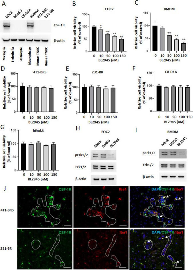
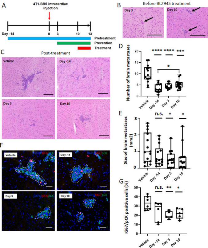
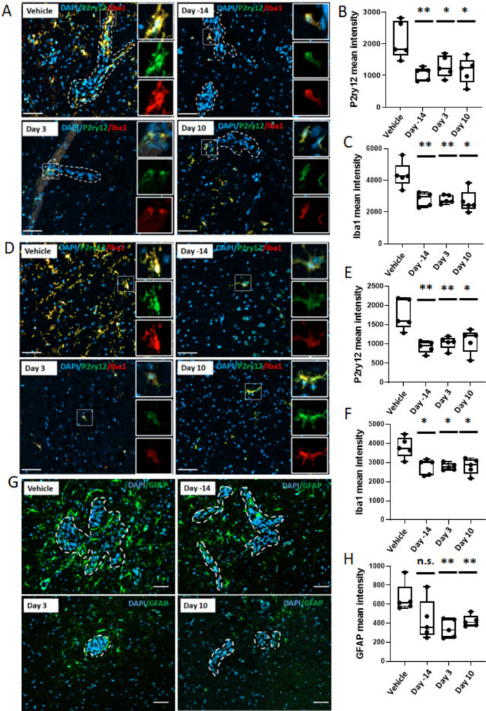
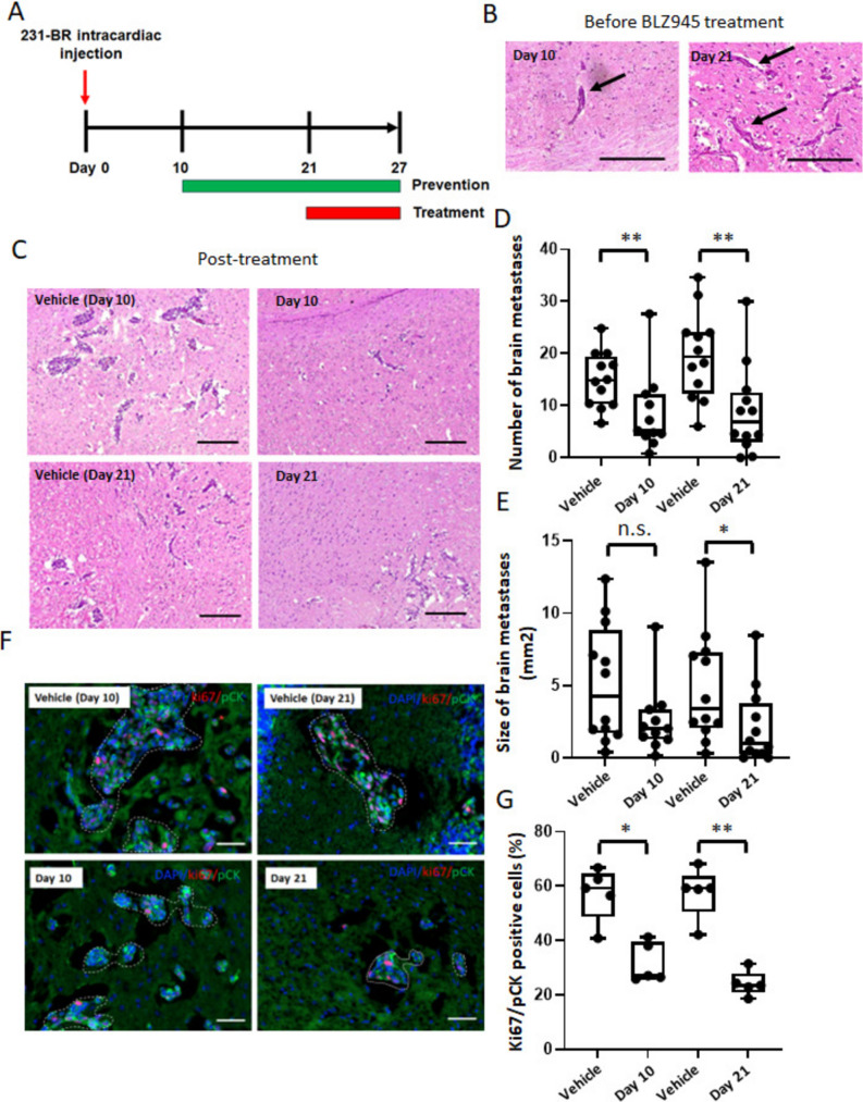
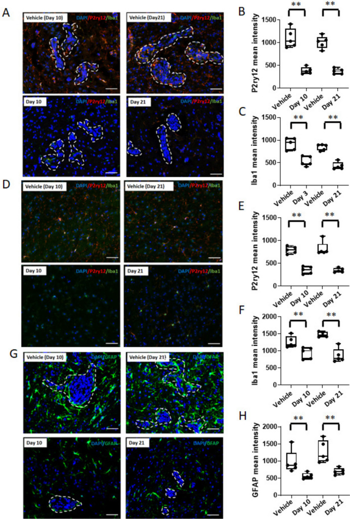
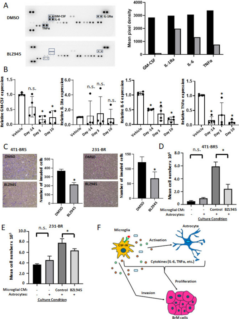
Brain metastasis is a common and serious complication of metastatic triple-negative breast cancer (TNBC) with few effective treatments. Here, we evaluated the effect of targeting the brain tumor microenvironment via the myeloid colony-stimulating factor-1 receptor (CSF-1R) pathway using the small molecule inhibitor BLZ945. Studies were conducted in two TNBC hematogenous brain-tropic models, 4T1-BR5 and 231-BR, with endpoints of prevention of brain metastasis formation and treatment of established brain metastasis. BLZ945 reduced the formation of brain metastases in both models by 57–65% (all p < 0.01) in the prevention setting. In the treatment setting, more analogous to the clinical situation, BLZ945 reduced the number and size of metastases in both models by 44–65% and 61–72%, respectively (all p < 0.05). Treatment with BLZ945 significantly reduced the number of myeloid cells in both the uninvolved brain and metastatic regions, by 15–54% across models as early as three days post-treatment. Efficacy was achieved without the need for complete suppression of brain myeloid cells, suggesting that potential adverse effects of full myeloid suppression can be minimized. Additionally, BLZ945 reduced cancer cell proliferation and astrocyte activation in the tumor microenvironment in vivo. In vitro studies showed that BLZ945 inhibited the secretion of inflammatory cytokines that stimulated cancer cell invasion; BLZ945 also indirectly reduced cancer cell proliferation through astrocyte interaction. Our findings suggest that microglial CSF-1R controls a series of myeloid regulatory pathways, both alone and in concert with other brain microenvironmental cells. The data preclinically credential CSF-1R inhibition as a potential therapeutic strategy for TNBC brain metastases.

Supplementary Information: The online version contains supplementary material available at 10.1007/s10585-025-10366-x.

Keywords: Astrocyte; BLZ945; Brain metastasis; Colony-stimulating factor-1; Colony-stimulating factor-1 receptor; Macrophage; Microglia; Triple-negative breast cancer; Tumor microenvironment.


Conflict of interest statement

Declarations. Conflict of interest: The authors declare no competing interests.

Figures

Fig. 1
Fig. 1
CSF-1R expression is limited to brain microglia/macrophages. A Western blot analysis of CSF-1R in EOC2 (microglia), bEnd.3 (endothelia), C8-D1A (astrocyte), BMDM (primary macrophage), 4T1-BR5 (mouse TNBC) and 231-BR (human TNBC). β-actin is used as a loading control. BG EOC2, BMDM, 4T1-BR5, 231-BR, C8-D1A or bEnd.3 cells were exposed to dimethyl sulfoxide (DMSO, vehicle control) or BLZ945 (10, 50, 100 and150 nM) for 72 h followed by alamarBlue cell viability assay. Values represent the mean fold change ± SEM relative to vehicle control (n = 6). HI Western blot analysis of pErk1/2, Erk and β-actin protein in EOC2 and BMDM cells treated with BLZ945 (EOC2, 150 nM; BMDM, 100 nM) or DMSO for 48 h. *, p < 0.05 vs DMSO control; **, p < 0.01 vs DMSO control by Mann–Whitney test. J CSF-1R (green) co-staining with Iba1 (red) in frozen, formaldehyde-fixed and OCT-embedded metastases and their microenvironments of 4T1-BR5 (upper panel) and 231-BR (lower panel) mouse models of breast cancer brain metastasis. Arrows indicate examples of co-stained cells, and white dotted lines indicate brain metastases. All nuclei are stained with DAPI (blue). Scale bar = 50 μm for all panels
Fig. 2
Fig. 2
CSF-1R suppression in the prevention and treatment settings of 4T1-BR5 TNBC brain metastasis model. A Schematic representation of the pretreatment, prevention and treatment studies. B Representative hematoxylin and eosin (H&E) staining images of 4T1-BR5 cancer cells in the brain after intracardiac injection at the start of the prevention (day 3) and treatment (day 10) arms. Arrows indicate metastatic cancer cells. C Representative H&E staining images of 4T1-BR5 brain metastases on day 13 after vehicle or BLZ945 treatment at the endpoint. Scale bar = 200 μm for panel B & C. D Number of brain metastases per brain section. E Size of brain metastases per animal. n = 14–15. F Representative immunofluorescence staining images of Ki67 (red) and pCK (green) in brain metastases (white dotted line) from vehicle and BLZ945 treated mice. Scale bar = 50 μm. G Quantification of Ki67 and pCK-positive cells in metastases from 5 to 6 biological replicates. Each dot represents one mouse, and the line designates the group median. Statistical significance versus vehicle control was calculated using the Mann–Whitney test (*, p < 0.05; **, p < 0.01; ***, p < 0.001; ****, p < 0.0001 vs vehicle control; n.s., not significant)
Fig. 3
Fig. 3
Effect of CSF-1R suppression on microglia/infiltrating monocytes and astrocyte activation in 4T1-BR5 model. Representative immunofluorescence images and quantification of P2ry12 (green) and Iba1 (red) staining in brain metastases (AC) and uninvolved brain tissues (DF) in control and BLZ945-treated mice at the endpoint. G and H Representative immunofluorescence staining images and quantification of GFAP (green) in the tumor microenvironment in control and BLZ945-treated mice (n = 5). All nuclei are stained with DAPI (blue). Each dot represents one mouse, and the line designates the group median. Scale bar = 50 μm. *, p < 0.05; **, p < 0.01 vs vehicle control; n.s., not significant by the Mann–Whitney test
Fig. 4
Fig. 4
CSF-1R suppression in the prevention and treatment settings in 231-BR TNBC brain metastasis model. A Schematic representation of the prevention and treatment studies. B Representative H&E staining images of 231-BR cancer cells in the brain after intracardiac injection at the start of the prevention (day 10) and treatment (day 21) arms. Arrows indicate metastatic cancer cells. C Representative H&E staining images of 231-BR brain metastases on day 27 after vehicle or BLZ945 treatment at the endpoint. Scale bar = 200 μm for panels B&C. D and E Number of brain metastases per brain section and size of brain metastases per animal. Each dot represents one mouse, and the line designates the group median. n = 11–12. F and G Representative immunofluorescence staining images and quantification of Ki67 and pCK-positive cells in brain metastases (white dotted line) from vehicle and BLZ945 treated mice (n = 5). Scale bar = 50 μm. *, p < 0.05; **, p < 0.01 vs vehicle control; n.s., not significant by the Mann–Whitney test
Fig. 5
Fig. 5
Effect of CSF-1R suppression on microglia/infiltrating monocytes and astrocyte activation in 231-BR model. Representative immunofluorescence images and quantification of P2ry12 (red) and Iba1 (green) staining in brain metastases (AC) and uninvolved brain tissues (DF) in control and BLZ945-treated mice (n = 5) at the endpoint. G and H, Representative immunofluorescence staining images and quantification of GFAP (green) in the tumor microenvironment in control and BLZ945-treated mice (n = 5). All nuclei are stained with DAPI (blue). Each dot represents one mouse, and the line designates the group median. Scale bar = 50 μm. **, p < 0.01 vs vehicle control; n.s., not significant by the Mann–Whitney test
Fig. 6
Fig. 6
CSF-1R suppression alters microglial cytokine secretion patterns with effects on astrocytes and tumor cells. A Mouse cytokine antibody array analysis of EOC2-derived conditioned medium treated with 100 nM BLZ945 or DMSO for 72 h. The images of cytokines with marked GM-CSF, IL-1Rα, IL-6 and TNFα are shown in the left panel. The pixel density of the above cytokines relative to the internal control is shown by the graphs in the right panel. B RT-PCR analysis of GM-CSF, IL-1Rα, IL-6, and TNFα was performed on fixed, frozen specimens from vehicle control and BLZ945 treatment settings of the 4T1-BR5 TNBC brain metastasis model mice on Days -14, 3, and 10. Values represent the fold change ± SD relative to vehicle. Each dot represents one mouse, and the line designates the group median. C 4T1-BR5 and 231-BR cells were allowed to invade through Matrigel for 12 h in the presence of control or BLZ945 treated EOC2-derived conditioned medium. Invaded cells were visualized by Hema 3 staining and counted using ImageJ software. D and E EOC2 conditioned medium promotes the growth of 4T1-BR5 and 231-BR cells under co-culture conditions. 4T1-BR5 and 231-BR cells were labelled with mCherry and EGFP, respectively. Brain trophic cancer cell numbers are quantified and calculated by flow cytometry based on the ratio between cancer cells and quantitative beads. The effect of astrocytes alone on cancer cell growth was measured in low serum medium under co-culture conditions. n ≥ 3 biologically independent experiments. *, p < 0.05 vs DMSO control; n.s., not significant by the Mann–Whitney test. F Schematic representation of microglial action in breast cancer brain metastasis. CSF-1R+ microglia/macrophages regulate the secretion of cytokines that influence the behavior of astrocytes and brain metastatic (BrM) cells within the microenvironment

Similar articles

References

    1. Hackshaw MD et al (2021) Prognostic factors of brain metastasis and survival among HER2-positive metastatic breast cancer patients: a systematic literature review. BMC Cancer 21(1):967 - PMC - PubMed
    1. Kabraji S et al (2023) Preclinical and clinical efficacy of Trastuzumab deruxtecan in breast cancer brain metastases. Clin Cancer Res 29(1):174–182 - PMC - PubMed
    1. Jerusalem G et al (2022) Trastuzumab deruxtecan in HER2-positive metastatic breast cancer patients with brain metastases: a DESTINY-Breast01 subgroup analysis. Cancer Discov 12(12):2754–2762 - PMC - PubMed
    1. Lin NU et al (2020) Intracranial efficacy and survival with tucatinib plus trastuzumab and capecitabine for previously treated HER2-positive breast cancer with brain metastases in the HER2CLIMB trial. J Clin Oncol 38(23):2610–2619 - PMC - PubMed
    1. Morris PG et al (2012) Limited overall survival in patients with brain metastases from triple negative breast cancer. Breast J 18(4):345–350 - PubMed

LinkOut - more resources