Evaluation of calcaneal inclusion angle in the diagnosis of pes planus with pretrained deep learning networks: An observational study
- PMID: 40760538
- PMCID: PMC12324017
- DOI: 10.1097/MD.0000000000043639
Evaluation of calcaneal inclusion angle in the diagnosis of pes planus with pretrained deep learning networks: An observational study
Abstract
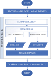
Pes planus is a common postural deformity related to the medial longitudinal arch of the foot. Radiographic examinations are important for reproducibility and objectivity; the most commonly used methods are the calcaneal inclusion angle and Mery angle. However, there may be variations in radiographic measurements due to human error and inexperience. In this study, a deep learning (DL)-based solution is proposed to solve this problem. Lateral radiographs of the right and left foot of 289 patients were taken and saved. The study population is a homogeneous group in terms of age and gender, and does not provide sufficient heterogeneity to represent the general population. These radiography (X-ray) images were measured by 2 different experts and the measurements were recorded. According to these measurements, each X-ray image is labeled as pes planus or non-pes planus. These images were then filtered and resized using Gaussian blurring and median filtering methods. As a result of these processes, 2 separate data sets were created. Generally accepted DL models (AlexNet, GoogleNet, SqueezeNet) were reconstructed to classify these images. The 2-category (pes planus/no pes planus) data in the 2 preprocessed and resized datasets were classified by fine-tuning these reconstructed transfer learning networks. The GoogleNet and SqueezeNet models achieved 100% accuracy, while AlexNet achieved 92.98% accuracy. These results show that the predictions of the models and the measurements of expert radiologists overlap to a large extent. DL-based diagnostic methods can be used as a decision support system in the diagnosis of pes planus. DL algorithms enhance the consistency of the diagnostic process by reducing measurement variations between different observers. DL systems accelerate diagnosis by automatically performing angle measurements from X-ray images, which is particularly beneficial in busy clinical settings by saving time. DL models integrated with smartphone cameras can facilitate the diagnosis of pes planus and serve as a screening tool, especially in regions with limited access to healthcare.
Keywords: calceneal inclusion angle; deep learning; flat foot; pes planus; transfer learning.
Copyright © 2025 the Author(s). Published by Wolters Kluwer Health, Inc.
Conflict of interest statement
The authors have no funding and conflicts of interest to disclose.
Figures


References
-
- Flores DV, Mejía Gómez C, Fernández Hernando M, Davis MA, Pathria MN. Adult acquired flatfoot deformity: anatomy, biomechanics, staging, and imaging findings. Radiographics. 2019;39:1437–60. - PubMed
-
- Berkeley R, Tennant S, Saifuddin A. Multimodality imaging of the paediatric flatfoot. Skeletal Radiol. 2021;50:2133–49. - PubMed
Publication types
MeSH terms
LinkOut - more resources
Full Text Sources
Research Materials

