Case Reports
. 2025 Jul 28;20(10):5196-5204.
doi: 10.1016/j.radcr.2025.06.098. eCollection 2025 Oct.

Cardiac lipoma-induced neonatal hypoxemia: a case report underscoring radiological diagnosis and a literature review

Affiliations
Case Reports

Cardiac lipoma-induced neonatal hypoxemia: a case report underscoring radiological diagnosis and a literature review

Ahmad Al-Bitar et al. Radiol Case Rep. .

Abstract

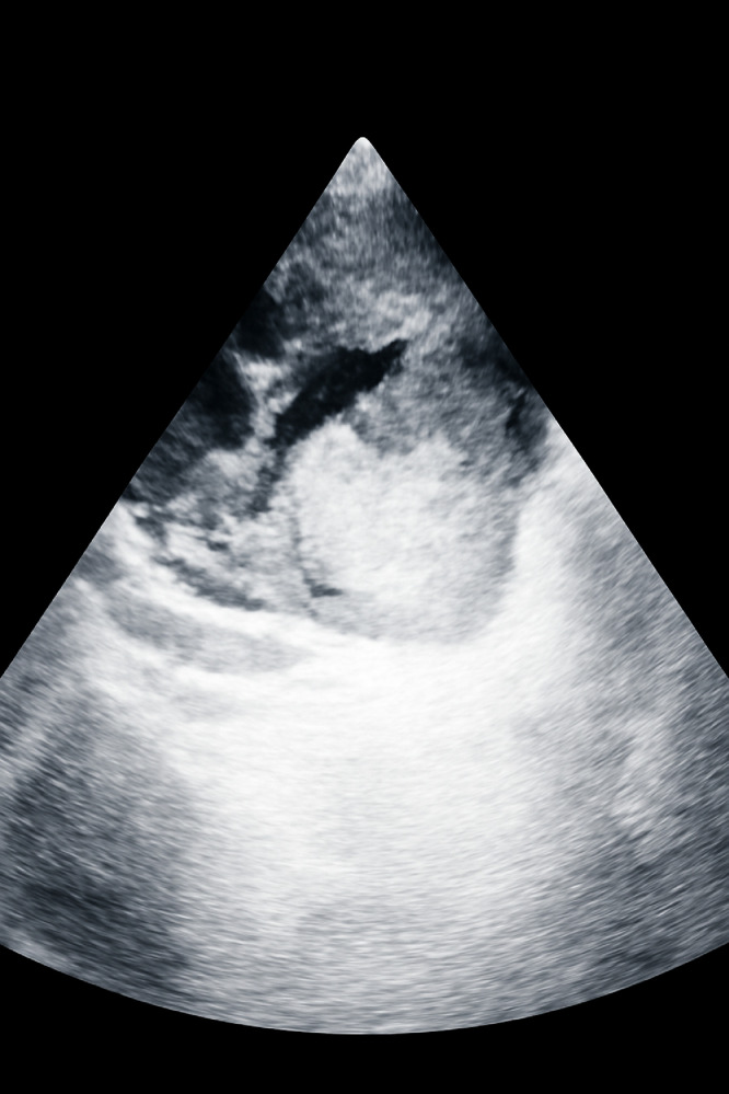
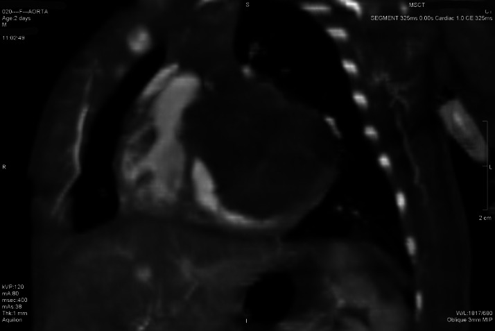
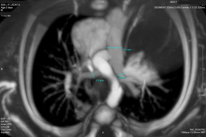
Cardiac tumors in neonates are exceedingly rare, with cardiac lipomas representing an exceptionally uncommon subtype associated with high mortality due to hemodynamic compromise. This report presents a 2-day-old Arab male neonate admitted with acute hypoxemia and cardiomegaly. Echocardiography and CT imaging revealed a large hyperechoic, fatty-density mass (3.8 × 3.5 × 3.3 cm) in the left ventricle, suggestive of a cardiac lipoma, but biopsy or surgical intervention could not be performed due to rapid clinical deterioration culminating in fatal cardiopulmonary failure. This case underscores the diagnostic and therapeutic challenges of neonatal cardiac tumors, highlighting the need for clinical suspicion and early prenatal surveillance to avoid their life-threatening mass effects and enable multidisciplinary planning. Future research should prioritize biomarkers, risk-stratification tools, and improved imaging algorithms to facilitate timely diagnosis and intervention in low-resource contexts.

Keywords: Cardiac lipoma; Fetal cardiac tumors; Multimodality imaging; Neonatal hypoxemia; Prenatal screening; Resource-limited settings.


Figures

Fig 1
Fig. 1
Echocardiogram showing a large, echogenic mass (cardiac lipoma) within the right ventricle, appearing hyperechoic relative to the surrounding myocardium.
Fig 2 –
Fig. 2
Echocardiogram demonstrating the extent of the cardiac lipoma within the right ventricle, highlighting its homogenous echogenicity.
Fig 3 –
Fig. 3
Color Doppler echocardiogram illustrating normal blood flow patterns around the cardiac lipoma, indicating no significant obstruction to valvular flow or intracardiac shunting. The red color likely signifies flow towards the transducer.
Fig 4 –
Fig. 4
Echocardiogram depicting the cardiac lipoma in an alternative view, further confirming its location and size within the cardiac chamber.
Fig 5 –
Fig. 5
Echocardiogram showing the multiple cystic or anechoic components within the cardiac lipoma, a characteristic feature that can be seen in some lipomas.
Fig 6 –
Fig. 6
Axial CT scan showing a lipomatous mass in the left ventricle, measuring 38.0 mm by 33.6 mm. The mass has fatty density, appearing lighter than the surrounding myocardium. Lung tissues, rib cage, and vertebrae are visible for anatomical context. Axial CT scan showing a lipomatous mass in the left ventricle, measuring 38.0 mm by 33.6 mm. The mass has fatty density, appearing lighter than the surrounding myocardium. Lung tissues, rib cage, and vertebrae are visible for anatomical context.
Fig 7 –
Fig. 7
Sagittal MSCT scan showing a lipomatous mass in the left ventricle, attached to the interventricular septum. The mass measures approximately 36.6 mm in height and exhibits fatty density, appearing lighter than the surrounding myocardium. Surrounding anatomical structures, including the ribcage, lung tissue, and spinal column, are visible, providing context for the location of the mass in the thoracic cavity.
Fig 8 –
Fig. 8
Oblique MIP MSCT scan of a lipomatous mass in the left ventricle, attached to the interventricular septum. The mass exhibits fatty density, appearing lighter than the surrounding myocardial tissue. Key anatomical structures, including the ascending aorta, pulmonary artery, ribcage, and vertebral column, are clearly visible, providing detailed context for the mass's position and surrounding cardiovascular and skeletal anatomy.
Fig 9 –
Fig. 9
Oblique MIP MSCT scan of a lipomatous mass in the left ventricle. The mass has fatty density, appearing lighter than the surrounding myocardium. Measurement lines show the mass dimensions: 6.7 mm in diameter, with additional measurements of 3.2 mm and 3.8 mm for surrounding structures.
Fig 10 –
Fig. 10
Oblique MIP MSCT scan of a lipomatous mass in the left ventricle, with fatty density. The mass measures 7.1 mm, with additional dimensions of 5.4 mm and 3.3 mm. The ascending aorta and pulmonary artery are visible, with the ribcage and spinal column providing anatomical context.

Similar articles

  • Prenatal interventions for congenital diaphragmatic hernia for improving outcomes.
    Grivell RM, Andersen C, Dodd JM. Grivell RM, et al. Cochrane Database Syst Rev. 2015 Nov 27;2015(11):CD008925. doi: 10.1002/14651858.CD008925.pub2. Cochrane Database Syst Rev. 2015. PMID: 26611822 Free PMC article.
  • Intraoral Lipoma on the Palate of an 11-Year-Old Patient: A Case Report.
    Zisis V, Charisi C, Poulopoulos K, Papadopoulos P, Poulopoulos A. Zisis V, et al. Reports (MDPI). 2025 Feb 8;8(1):19. doi: 10.3390/reports8010019. Reports (MDPI). 2025. PMID: 40729232 Free PMC article.
  • Ornithine Transcarbamylase Deficiency.
    Lichter-Konecki U, Caldovic L, Morizono H, Simpson K, Ah Mew N, MacLeod E. Lichter-Konecki U, et al. 2013 Aug 29 [updated 2022 May 26]. In: Adam MP, Feldman J, Mirzaa GM, Pagon RA, Wallace SE, Amemiya A, editors. GeneReviews® [Internet]. Seattle (WA): University of Washington, Seattle; 1993–2025. 2013 Aug 29 [updated 2022 May 26]. In: Adam MP, Feldman J, Mirzaa GM, Pagon RA, Wallace SE, Amemiya A, editors. GeneReviews® [Internet]. Seattle (WA): University of Washington, Seattle; 1993–2025. PMID: 24006547 Free Books & Documents. Review.
  • Classic Galactosemia and Clinical Variant Galactosemia.
    Berry GT. Berry GT. 2000 Feb 4 [updated 2021 Mar 11]. In: Adam MP, Feldman J, Mirzaa GM, Pagon RA, Wallace SE, Amemiya A, editors. GeneReviews® [Internet]. Seattle (WA): University of Washington, Seattle; 1993–2025. 2000 Feb 4 [updated 2021 Mar 11]. In: Adam MP, Feldman J, Mirzaa GM, Pagon RA, Wallace SE, Amemiya A, editors. GeneReviews® [Internet]. Seattle (WA): University of Washington, Seattle; 1993–2025. PMID: 20301691 Free Books & Documents. Review.
  • 123I-MIBG scintigraphy and 18F-FDG-PET imaging for diagnosing neuroblastoma.
    Bleeker G, Tytgat GA, Adam JA, Caron HN, Kremer LC, Hooft L, van Dalen EC. Bleeker G, et al. Cochrane Database Syst Rev. 2015 Sep 29;2015(9):CD009263. doi: 10.1002/14651858.CD009263.pub2. Cochrane Database Syst Rev. 2015. PMID: 26417712 Free PMC article.

References

    1. Isaacs H. Fetal and neonatal cardiac tumors. Pediatr Cardiol. 2004;25(3):252–273. - PubMed
    1. Tao T.Y., Yahyavi-Firouz-Abadi N., Singh G.K., Bhalla S. Pediatric cardiac tumors: clinical and imaging features. Radiographics. 2014;34(4):1031–1046. doi: 10.1148/rg.344135163. - DOI - PubMed
    1. Ajami G.H., Cheriki S., Amoozgar H., Borzouee M., Soltani M. Accuracy of Doppler-derived estimation of pulmonary vascular resistance in congenital heart disease: an index of operability. Pediatr Cardiol. 2011;32(8):1168–1174. - PubMed
    1. Taylor A.J., Cerqueira M., Hodgson J.M., Mark D., Min J., O’Gara P. ACCF/SCCT/ACR/AHA/ASE/ASNC/NASCI/SCAI/SCMR 2010 appropriate use criteria for cardiac computed tomography. A report of the American College of Cardiology Foundation Appropriate use Criteria Task Force, the Society of Cardiovascular Computed Tomography, the American College of Radiology, the American Heart Association, the American Society of Echocardiography, the American Society of Nuclear Cardiology, the North American Society for Cardiovascular Imaging, the Society for Cardiovascular Angiography and Interventions, and the Society for Cardiovascular Magnetic Resonance. J Am Coll Cardiol. 2010;23(22):1864–1894. 56. - PubMed
    1. Tyebally S., Chen D., Bhattacharyya S., Mughrabi A., Hussain Z., Manisty C., Westwood M., Ghosh A.K., Guha A. Cardiac Tumors: JACC CardioOncology State-of-the-Art Review. JACC. CardioOncology. 2020;2(2):293–311. doi: 10.1016/j.jaccao.2020.05.009. - DOI - PMC - PubMed

Publication types

LinkOut - more resources