. 2025 Jul 29:12:1661-1676.
doi: 10.2147/JHC.S533777. eCollection 2025.

Single-Cell RNA Sequencing Integrated with Bulk-RNA Sequencing Analysis Reveals Prognostic Signatures Based on PANoptosis in Hepatocellular Carcinoma

Affiliations

Single-Cell RNA Sequencing Integrated with Bulk-RNA Sequencing Analysis Reveals Prognostic Signatures Based on PANoptosis in Hepatocellular Carcinoma

Jiyin Wang et al. J Hepatocell Carcinoma. .

Abstract

Purpose: Drug resistance severely compromises therapeutic efficacy in hepatocellular carcinoma (HCC); however, the selection of precise treatment strategies for patients remains a critical unmet clinical need. This study investigated PANoptosis-related mechanisms underlying HCC progression to identify actionable therapeutic targets and optimize patient-specific treatment outcomes.

Patients and methods: Multi-omics analysis (single-cell/bulk RNA sequencing) combined with machine learning was used to identify the PANoptosis-related prognostic features. The association of PANoptosis-related expression with the tumor immune microenvironment and drugs was explored using bioinformatic analysis and experimental studies.

Results: High PANoptosis risk exhibited immunosuppressive microenvironments and therapeutic resistance. The PANoptosis-related gene YIF1B has emerged as a dual prognostic biomarker and tumor driver that promotes proliferation, and is linked to immune dysfunction and drug resistance.

Conclusion: YIF1B may be a promising therapeutic target. This PANoptosis framework bridges molecular mechanisms to clinical management, offering strategies for personalized HCC therapy and overcoming treatment resistance.

Keywords: PANoptosis; YIF1B; drug resistant; hepatocellular carcinoma.


Conflict of interest statement

The authors declare no competing interests for this work.

Figures
None
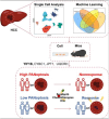
Graphical abstract
Figure 1
Figure 1
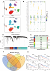
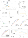
Identification of PANoptosis-Related Genes in HCC from Single-Cell Transcriptomics and TCGA Database. (A) Umap visualization for clustering. (B and C) Different cell types identified by the expression of surface marker genes. (D and E) WGCNA identifies MEtan module as the most strongly correlated with PANoptosis activity. (F) LASSO, SVM, XGBoost and GBM to identify the most diagnostically significant PANoptosis-related genes. (G) Four PANoptosis-related genes associated with survival outcomes in the TCGA cohort.
Figure 2
Figure 2
Validation of a PANoptosis-Related Prognostic Model in HCC. (A) Risk stratification visualization: distribution of risk scores, survival status, and signature gene expression patterns in the training cohort. (B) Time-dependent ROC curves validate predictive accuracy in TCGA cohorts. (C) Kaplan-Meier analysis reveals significantly poorer overall survival in high-risk TCGA-HCC patients. (D) Time-dependent ROC curves validate predictive accuracy in GSE16747 cohort. (E and F) High-risk HCC patients exhibited poorer overall survival and recurrence-free survival in GSE16747 cohort.
Figure 3
Figure 3
High-Risk Groups Demonstrate Impaired Antitumor Immune Response. (A) 28 immune cell subtypes expressed differences between high-risk and low-risk groups evaluated by ssGSEA. (B) The correlation of PANoptosis-associated genes (CYBC1, JPT1, UQCRH and YIF1B) with 28 immune cell subtypes. (C and D) The difference of TIDE scores, Dysfunction, Exclusion, MSI and MDSC in high-risk and low-risk groups. (E) Hepa1-6 cells stably transfected with empty vector or shYIF1B were subcutaneously injected into C57BL/6. Tumor weight was quantitatively analyzed after 3 weeks. (F–H) Tumor-infiltrating immune cells were detected by flow cytometry in empty vector or shYIF1B tumors.
Figure 4
Figure 4
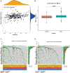
High-Risk Groups Exhibit Elevated Tumor Mutation Burden. (A) The correlation of risk score with TMB. (B) The difference TMB between male and female. (C and D) The high-risk group exhibited a higher mutation frequency overall.
Figure 5
Figure 5
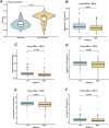
High-Risk Groups Exhibit Poor Response to Sorafenib and Chemotherapies. (A) The correlation of risk score with sorafenib. (BF) The low-risk group had lower IC50 values for Bortezomib_1191, Docetaxel_1007, Fulvestrant_1816, Temozolomide_1375 and Vinblastine_1818.
Figure 6
Figure 6
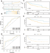
PANoptosis Risk Scores as Prognostic Determinants and Validation of Nomograms. (A and B) A nomogram was developed via integrating clinical data and PANoptosis scores. (C and E) The calibration curves, the C-index and DCA were assessed the accuracy of the nomogram. (FH) The predictive ability of the nomogram confirmed by multivariable ROC curve analysis. (I) The stability of nomogram was validated by survival curves.
Figure 7
Figure 7
YIF1B Promotes the Growth and Tumorigenesis of HCC. (A and B) The protein levels of YIF1B and GAPDH were detected by Western blotting in HepG2 and Huh7 cells transfected with HA-YIF1B or with shYIF1B. (C and D) Proliferation rates of the cells above were measured by CCK-8 assays. (E) The migratory potential of HepG2 and Huh7 cells transfected with HA-YIF1B or with shYIF1B were measured by transwell assay. Scale bar, 200μm. (F and G) HepG2 cells stably transfected with empty vector, HA-YIF1B or shYIF1B were subcutaneously injected into nude mice. Tumor weight was quantitatively analyzed after 4 weeks.

Similar articles

References

    1. Yeo YH, Abdelmalek M, Khan S, et al. Current and emerging strategies for the prevention of hepatocellular carcinoma. Nat Rev Gastroenterol Hepatol. 2025;22(3):173–190. doi: 10.1038/s41575-024-01021-z - DOI - PubMed
    1. Llovet JM, De Baere T, Kulik L, et al. Locoregional therapies in the era of molecular and immune treatments for hepatocellular carcinoma. Nat Rev Gastroenterol Hepatol. 2021;18(5):293–313. doi: 10.1038/s41575-020-00395-0 - DOI - PubMed
    1. Vogel A, Meyer T, Sapisochin G, Salem R, Saborowski A. Hepatocellular carcinoma. Lancet. 2022;400(10360):1345–1362. doi: 10.1016/S0140-6736(22)01200-4 - DOI - PubMed
    1. Yang C, Zhang H, Zhang L, et al. Evolving therapeutic landscape of advanced hepatocellular carcinoma. Nat Rev Gastroenterol Hepatol. 2023;20(4):203–222. doi: 10.1038/s41575-022-00704-9 - DOI - PubMed
    1. Chan YT, Zhang C, Wu J, et al. Biomarkers for diagnosis and therapeutic options in hepatocellular carcinoma. Mol Cancer. 2024;23(1):189. doi: 10.1186/s12943-024-02101-z - DOI - PMC - PubMed

LinkOut - more resources