Artificial intelligence and radiomics applications in adrenal lesions: a systematic review
- PMID: 40761430
- PMCID: PMC12319203
- DOI: 10.1177/17562872251352553
Artificial intelligence and radiomics applications in adrenal lesions: a systematic review
Abstract
Background: Adrenal lesions, often incidentally detected, present diagnostic challenges in distinguishing benign from malignant or hormonally active lesions. Conventional imaging (computed tomography/magnetic resonance imaging (CT/MRI)) has limitations, driving interest in artificial intelligence (AI) and radiomics for enhanced accuracy.
Objectives: To systematically evaluate AI and radiomics applications in adrenal lesion characterization, focusing on diagnostic performance, methodologies, and clinical utility.
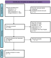
Design: PRISMA-guided systematic review of studies published up to June 2024.
Data sources and methods: PubMed, Scopus, Web of Science, and Google Scholar were searched using the keywords: adrenal lesions, AI, radiomics, and machine learning. Inclusion followed PICO criteria: patients with indeterminate lesions, AI/radiomics interventions, comparisons to standard diagnostics, and diagnostic accuracy. Two reviewers screened studies, resolving discrepancies via consensus. Eleven retrospective studies (996 patients) met eligibility.
Results: CT-based radiomics (eight studies) achieved a mean AUC of 0.88 (range: 0.84-0.94) in differentiating benign/malignant or functional/non-functional lesions. Top-performing models identified aldosterone-producing adenomas (AUC: 0.99). MRI-based radiomics (three studies) yielded mean AUC: 0.82 (0.72-0.92), with test-set performance declines (e.g., AUC: 0.72) suggesting overfitting. Nuclear medicine (four studies) demonstrated that hybrid 18F-FDG PET/CT models (SUVmax + texture features) achieved an AUC of 0.97 for metastatic versus benign lesions. AI applications extended to intraoperative navigation (AUC: 0.93) and prognostic prediction.
Conclusion: CT-based radiomics outperformed MRI, aligning with guidelines favoring CT for adrenal assessment. AI-enhanced models show promise in refining diagnostics and reducing invasive procedures. However, retrospective designs, small cohorts, and protocol variability limit generalizability. Future work requires multicenter collaboration, standardized protocols, and prospective validation to translate AI/radiomics into clinical practice.
Keywords: adrenal tumors; artificial intelligence; computed tomography; magnetic resonance imaging; radiomics.
© The Author(s), 2025.
Conflict of interest statement
The authors declare that there is no conflict of interest.
Figures


References
-
- Boland GWL, Blake MA, Hahn PF, et al. Incidental adrenal lesions: principles, techniques, and algorithms for imaging characterization. Radiology 2008; 249: 756–775. - PubMed
-
- Song JH, Chaudhry FS, Mayo-Smith WW. The incidental indeterminate adrenal mass on CT (>10 H) in patients without cancer: is further imaging necessary? Follow-up of 321 consecutive indeterminate adrenal masses. Am J Roentgenol 2007; 189: 1119–1123. - PubMed
Publication types
LinkOut - more resources
Full Text Sources
Miscellaneous

