. 2025 Aug 3;6(8):e70311.
doi: 10.1002/mco2.70311. eCollection 2025 Aug.

Nanoparticle-Delivered siRNA Targeting NSUN4 Relieves Systemic Lupus Erythematosus through Declining Mitophagy-Mediated CD8+T Cell Exhaustion

Affiliations

Nanoparticle-Delivered siRNA Targeting NSUN4 Relieves Systemic Lupus Erythematosus through Declining Mitophagy-Mediated CD8+T Cell Exhaustion

Bincheng Ren et al. MedComm (2020). .

Abstract

5-Methylcytosine modification (m5C) is an important posttranscriptional regulatory mechanism of gene expression. Exhausted CD8+T cells contribute to the development of many major diseases; however, their exact role and relationship to m5C in systemic lupus erythematosus (SLE) remain unknown. In this study, we identified a CD7highCD74high CD8+T subgroup that were robustly expanded in SLE patients through single-cell transcriptome sequencing (scRNA-seq). CD7highCD74high CD8+T cells displayed exhausted features and exhibited a superior diagnostic value in SLE. Then, we explored the m5C landscape of SLE patients by performing m5C epitranscriptome sequencing (m5C-seq). ScRNA-seq and m5C-seq were conjointly analyzed to screen m5C-related therapeutic targets for SLE, and NOP2/Sun RNA methyltransferase 4 (NSUN4) was identified as a key regulator of SLE pathogenesis. Knockdown of NSUN4 downregulated CD74 expression via reduction of m5C and suppressed CD8+T cell exhaustion by declining CD44/mTOR (mechanistic target of rapamycin kinase)-mediated mitophagy. Finally, we verified that nanoparticle-delivered siRNA against Nusn4 decreased autoimmune reaction kidney damage in both spontaneous and pristane-induced SLE mouse models. In conclusion, we identify an exhausted CD7highCD74high CD8+T cell subset and propose the crucial role of NSUN4/CD74-induced dysregulation of mitophagy in SLE pathogenesis, and targeting NSUN4 is a promising treatment strategy for SLE patients.

Keywords: CD8+T cell mitophagy and exhaustion; NSUN4; nanoparticle‐delivered siRNA; single‐cell RNA sequencing and m5C sequencing; systemic lupus erythematosus.


Conflict of interest statement

The authors declare no conflicts of interest.

Figures
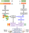
FIGURE 1
FIGURE 1
Over design of this study. C, healthy control; DEG, differentially expressed gene; HD, healthy donor; P, SLE patients; SLE, systemic lupus erythematosus.
FIGURE 2
FIGURE 2
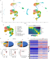
Deep UMAP assay in all clusters and cell–cell communications among all cell types. (A) UMAP assay for cell clusters (left panel) and cell types (right panel) of blood cells in overall samples. (B) Overall interaction numbers between every two cell types in all subjects. (C) Interaction strengths of every cell type between them as an incoming signaling and an outcoming signaling. (D) Total interfered interaction numbers of all cell types between HDs and SLEs. (F) Cross matrices about interaction numbers and interaction strengths between every two cell types respectively in HDs and SLEs.
FIGURE 3
FIGURE 3
An exhausted CD7highCD74high CD8+T cell subgroup was discovered in cluster 16 of SLE blood cells. (A) UMAP assay for cell clusters of blood cells in HDs and SLEs. (B) Annotation for the characteristic surface biomarkers of cell clusters in total T cells based on UMAP assay. (C) Annotation for the subgroups of total T cells based on the surface biomarkers. (D) The percentages of each T cell subgroup in HDs and SLEs, respectively. (E) Heatmap of DEGs in cluster 16 between HDs and SLEs. (F) Flow cytometry assay for the (F) percentages of CD8+ Tex cells/total T cells in the subjects enrolled in the scRNA‐seq assay and (G) CD7highCD74high T cells/CD8+NKG7+ T cells in expanded population of controls (N = 36) and SLEs (N = 52), respectively. (H) Receiver operating characteristic curve (ROC) was used to evaluate the diagnostic sensitivity and specificity in 52 SLE patients. ***p < 0.01.
FIGURE 4
FIGURE 4
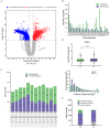
m5C‐seq analysis for the DMGs and their distributions in chromosomes and samples. m5C epitranscriptome assay was performed in six SLE patients and six controls. MACS software was applied to identify the methylation genes in each sample, diffReps software was applied for differential methylation gene (DMG) identification. Peaks located on the exon of mRNA were screened and annotated. (A) Volcano plots of the DMGs. (B) Chromosome distribution of DMGs. (C) The percentages of methylated genes and nonmethylated genes in each sample. (D) The percentages of genes with different number of m5C peaks. (E) Gene numbers of unique m5C‐modified genes, common m5C‐modified genes, and non‐m5C‐modified genes in C2 and P2 groups.
FIGURE 5
FIGURE 5
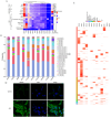
NSUN4 was screened as a key m5C regulator in SLE through conjoint analysis of scRNA‐seq and m5C‐seq. (A) Average expression of m5C regulators in the single cells based on different clinical variables containing cell types by using z‐score. (B) NMF clustering for the differentially expressed m5C regulators in different cell types. (C) Heatmap distribution of differentially expressed m5C regulators in different cell types. (D) Immunofluorescence staining was used to evaluate the NSUN4 levels in CD8+NKG7+ T cells from Controls and SLEs. ×400.
FIGURE 6
FIGURE 6
Knockdown of NSUN4 suppresses exhaustion of CD8+ T cells in vitro. 50 nM NSUN4 siRNAs or scrambled siRNA was transfected into cultured CD8+NKG7+ T cells by using the PEI–PBA (polyvinylimine system modified with phenylboronic acid) system. After 72 h, cells were harvested for functional evaluation. (A) The m5C high‐throughput microarray was used to evaluate the m5C levels of DEGs in cluster 16. (B) RIP and m5C antibody‐based methylated RIP (meRIP) assays were used to verify the binding of NSUN4 with CD74 mRNA. (C) Spot hybridization and meRIP–qPCR were respectively applied to evaluate the effect of silencing NSUN4 on overall level of m5C transcripts and m5C‐modified CD74 mRNA. (D) Actinomycin D was used to incubate CD8+NKG7+ T cells the effect of silencing NSUN4 on the stability of CD74 mRNA. (E) Dual immunofluorescence staining was used to evaluate the CD7 and CD74 levels in CD8+NKG7+ T cells with or without NSUN4 knocked down. DAPI was used to mark the nucleus (blue). ×200. (F) Interference efficiencies of NSUN4 siRNAs detected with qPCR. (G) Western blotting was applied to detect the protein levels NSUN4 and CD8+ T dysfunctional markers, including PD‐1, TIM3, and CTLA‐4. (H) ELISA was used to detect the secretion of landmark immunoactive cytokines of CD8+T cells, including IFN‐γ, IL‐2, and TNF‐α. N = 3, *< 0.05, **< 0.01 compared with scramble.
FIGURE 7
FIGURE 7
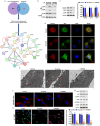
Knockdown of NSUN4 inactivates the CD44/mTOR/S6K pathway and improves mitophagy in CD8+ T cells in vitro. (A) The screening process of the interacting proteins of CD74. (B) Coimmunoprecipitation (co‐IP) was used to verify the binding capacity of CD74 and CD44. 50 nM NSUN4 siRNAs or scrambled siRNA was transfected into cultured CD8+NKG7+ T cells by using the PEI–PBA system. After 72 h, cells were harvested for functional evaluation. (C) Western blotting was used to detect the expression of NSUN4, CD74, and CD44. (D) Autophagic flux assay was used to evaluate the level of gross autophagy. DAPI was used to mark the nucleus (blue). ×1000. (E) Transmission electron microscopy was used to observe damaged mitochondria and mitochondrial autophagosomes in the cells. ×8000. The arrows indicate the mitochondrial autophagosomes. (F) MitoSOXTM Red was used to evaluate the level of mitochondrial oxidative stress. (G) JC‐1 fluorescence probe was used to evaluate the mitochondrial membrane potential. N = 3, *< 0.05, **< 0.01 compared with control and scramble.
FIGURE 8
FIGURE 8
Knockdown of Nsun4 alleviates CD7highCD74high CD8+T cell infiltration, mitophagy, and kidney tissue damage in SLE mice. Spontaneous lupus MRL/lpr mice applied to investigate the role of Nsun4 in SLE progression. Liposome–protamine–hyaluronic acid (LPH) nanoparticle (NP)‐delivered siRNA against Nsun4 was administrated into MRL/lpr mice by the tail vein injection and the NP‐scramble was used as a negative control. Blood was collected from the controls and SLE mice, and CD8+ T subgroups were segregated and counted with flow cytometry. The proportions of (A) NKG7+CD8+ T cells and (B) CD7highCD74high CD8+T cells in each group. (C) After blood collection, the kidney tissues were collected, spliced and stained with HE and immunohistochemistry to observe the tissue morphology and NSUN4 expression. (D) Dual immunofluorescence was used to observe the infiltration of CD7highCD74high T cells in the kidney. (E) Transmission electron microscopy was used to observe damaged mitochondria and mitochondrial autophagosomes in the kidney. (F) PAS staining and Masson staining were to observe the integrity of basement membrane and degree of fibrosis of the kidney tissue. N = 6.

References

    1. Kiriakidou M., Ching C. L., “Systemic Lupus Erythematosus,” Annals of Internal Medicine 172, no. 11 (2020): 81–96. - PubMed
    1. Quintero‐González D. C., Muñoz‐Urbano M., Vásquez G. “Mitochondria as a Key Player in Systemic Lupus Erythematosus,” Autoimmunity 55, no. 8 (2022): 497–505. - PubMed
    1. Bolouri N., Akhtari M., Farhadi E., et al., “Role of the Innate and Adaptive Immune Responses in the Pathogenesis of Systemic Lupus Erythematosus,” Inflammation Research 71, no. 5‐6 (2022): 537–554. - PubMed
    1. Chen P. M., Tsokos G. C., “The Role of CD8+ T‐Cell Systemic Lupus Erythematosus Pathogenesis: An Update,” Current Opinion in Rheumatology 33, no. 6 (2021): 586–591. - PMC - PubMed
    1. Buang N., Tapeng L., Gray V., et al., “Type I Interferons Affect the Metabolic Fitness of CD8(+) T Cells From Patients With Systemic Lupus Erythematosus,” Nature Communications 12, no. 1 (2021): 1980. - PMC - PubMed

LinkOut - more resources